Case description
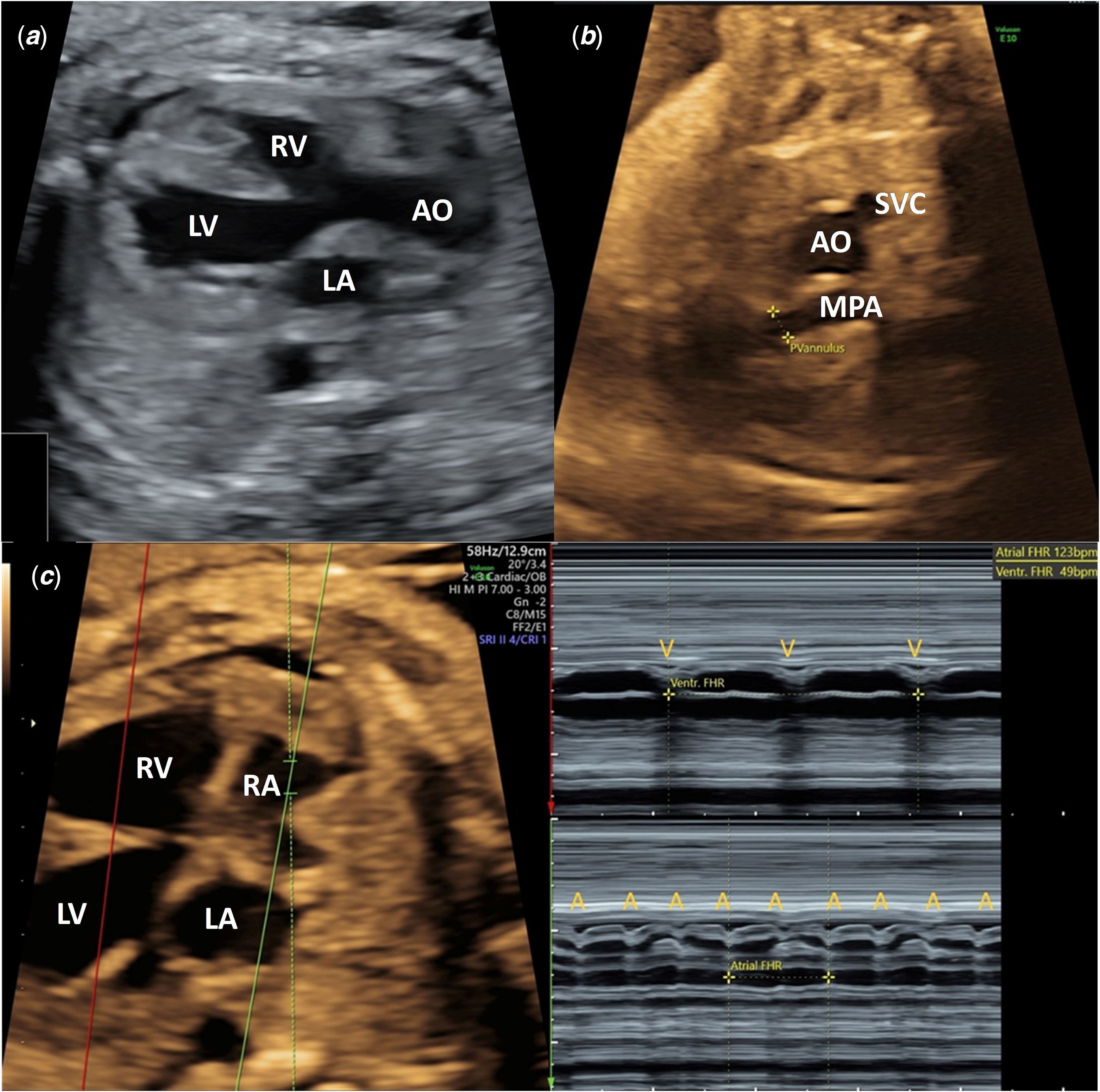
A 33-year-old Burmese woman, gravida 3 para 2 at 27 weeks’ gestation, was referred to our centre due to fetal bradyarrhythmia. Her medical history was unremarkable, and two previous uncomplicated pregnancies resulted in healthy, living children. Ultrasound evaluation showed shortened femur and humerus lengths (−2.5 standard deviations) without additional soft markers for Down syndrome. Umbilical artery Doppler indicated high resistance (pulsatility index >97.5th percentile), and the ductus venosus exhibited a reversed a-wave. Fetal echocardiography demonstrated tetralogy of Fallot characterised by a subaortic malaligned ventricular septal defect, overriding aorta, and moderate pulmonary stenosis with Z-score of −3.75. The main pulmonary artery was hypoplastic; however, the branch PAs were of normal size (right pulmonary artery Z-score: −0.32; left pulmonary artery Z-score: 0.52). Additionally, there was forward flow across the ductus arteriosus. Anatomical M-mode also revealed complete atrioventricular block with fetal atrial and ventricular rates of 123 and 49 beats per minute (bpm), respectively (Figure 1). Fetal cardiomegaly, minimal pericardial effusion, and endocardial fibroelastosis were present. A cardiovascular profile score of 5/10 (holosystolic tricuspid and mitral regurgitation, cardiothoracic area ratio 0.5, pericardial effusion, reversed ductus venosus a-wave) indicated severe cardiac failure without overt hydrops fetalis.
Illustrates fetal tetralogy of Fallot and complete atrioventricular block. (a) Transabdominal ultrasound showing a subaortic malaligned ventricular septal defect and overriding aorta in the left ventricular outflow tract view. (b) Three-vessel view demonstrating a small pulmonic valve and a smaller main pulmonary artery relative to the aorta. (c) Anatomical M-mode depicting complete heart block with a dissociation between atrial and ventricular contractions. The atrial contraction rate is 123 beats/minute, while the slower ventricular rhythm is 49 beats/minute. The green and red lines traverse atriums and ventricles, respectively. A = Atrial Contraction; AO = Ascending Aorta; LA = Left Atrium; LV = Left Ventricle; MPA = Main Pulmonary Artery; RA = Right Atrium; RV = Right Ventricle; SVC = Superior Vena Cava; V = Ventricular Contraction.

Figure 1. Long description
Fetal echocardiography illustrates fetal tetralogy of Fallot and complete atrioventricular block. (A) Transabdominal ultrasound showing a subaortic malaligned ventricular septal defect and overriding aorta in the left ventricular outflow tract view. (B) Three-vessel view demonstrating a small pulmonic valve and a smaller main pulmonary artery relative to the aorta. (C) Anatomical M-mode depicting complete heart block with a dissociation between atrial and ventricular contractions. The atrial contraction rate is 123 beats/minute, while the slower ventricular rhythm is 49 beats/minute. The green and red lines traverse atriums and ventricles, respectively. Abbreviations: A, atrial contraction; AO, ascending aorta; LA, left atrium; LV, left ventricle; MPA, main pulmonary artery; RA, right atrium; RV, right ventricle; SVC, superior vena cava; V, ventricular contraction.
Down syndrome screening was not performed due to the delayed initiation of antenatal care at 26 weeks of gestation. Invasive genetic testing was declined due to advanced gestational age, financial constraints, and maternal concern regarding procedure-related risks to a fetus with significant cardiac compromise. Maternal anti-Ro and anti-La antibodies were screened due to fetal atrioventricular block and were positive for high titres (3+) of anti-Ro52 and negative anti-La antibodies. At 28 weeks’ gestation, the patient was managed by a multidisciplinary team for fetal complete atrioventricular block complicated by severe bradycardia (heart rate < 55 bpm) and endocardial fibroelastosis. A targeted transplacental pharmacological regimen was initiated, comprising hydroxychloroquine (200 mg daily), oral dexamethasone (8 mg daily), and salbutamol (12 mg daily). Despite this in-utero therapy, the cardiovascular profile score deteriorated, decreasing to 3/10 (increased size of cardiomegaly and umbilical vein pulsation) on weekly assessment. Weekly fetal biophysical profile scores were consistently 8/8 (not including a non-stress test).
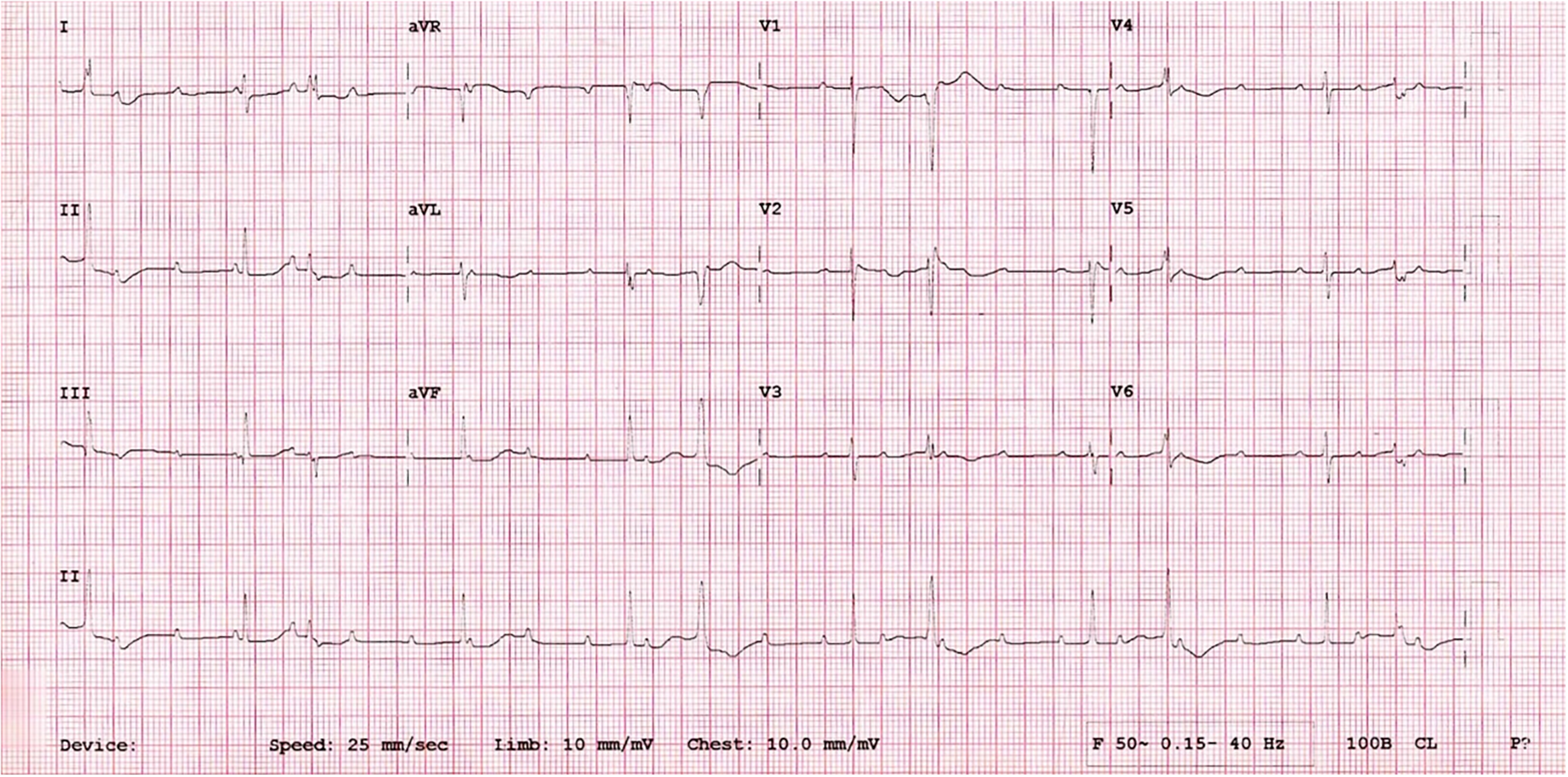
At 37 weeks of gestation, an elective caesarean section was performed, delivering a male newborn weighing 2,680 g, with Apgar scores of 6 and 8 at 1 and 5 minutes, respectively. At birth, the patient’s oxygen saturation was 60%, which improved to approximately 80% following intubation. Postnatal examination revealed phenotypic features indicative of Down syndrome, including generalised hypotonia, upward slanting palpebral fissures, and the characteristic “sandal gap” toes. Subsequent postnatal karyotyping analysis validated the trisomy 21 diagnosis. The neonatal echocardiogram and electrocardiogram confirmed the diagnosis of tetralogy of Fallot (mild to moderate pulmonary stenosis) and complete atrioventricular block (Figure 2). Neonatal lupus erythematosus was suspected based on the presentation of complete atrioventricular block, impaired cardiac function, and thrombocytopenia, prompting treatment with intravenous immunoglobulin and surgical pacing. Following placement of a dual-chamber pacemaker (programmed to VVI mode with a base rate of 90 bpm, a ventricular output of 3.5 V at 0.4 ms, and a sensitivity of 1.2 mV), the patient developed hypotension alongside overall and differential desaturation. Persistent pulmonary hypertension was suspected and successfully managed with inhaled nitric oxide. Despite these interventions, the neonate subsequently experienced ventricular fibrillation and cardiac arrest requiring cardiopulmonary resuscitation. This was attributed to neonatal sepsis, acidosis, concurrent disseminated intravascular coagulation, and subsequent pacemaker malfunction. The patient stabilised following a 14-day antibiotic regimen. Unfortunately, he later succumbed on the 55th day of life due to ventilator-associated pneumonia.
Neonatal electrocardiography, taken shortly after birth, shows third-degree atrioventricular block, a junctional escape rhythm, and ventricular bigeminy.

Discussion
Congenital complete atrioventricular block is an uncommon fetal arrhythmia. It can occur in association with structural cardiac defects or in isolation linked to maternal anti-Ro and anti-La autoantibodies, which cause inflammation in the fetal myocardium and conduction system. Reference Jaeggi, Hornberger, Smallhorn and Fouron1,Reference Hunter and Simpson2 While immune-mediated complete atrioventricular block typically occurs in structurally normal hearts, Reference Hunter and Simpson2 it has also been reported in the presence of minor, incidental structural lesions. Reference Buyon, Hiebert and Copel3 These include patent ductus arteriosus, atrial septal defects, ventricular septal defect, and pulmonary stenosis, which are not traditionally associated with the development of congenital atrioventricular block. Reference Buyon, Hiebert and Copel3 In the present case, complete atrioventricular block was attributable to immune-mediated mechanisms rather than major structural cardiac defects, whereas the concurrent tetralogy of Fallot was associated with the underlying Down syndrome. The prognosis for complete atrioventricular block is generally guarded, particularly when complicated by structural heart defects. Reference Jaeggi, Hornberger, Smallhorn and Fouron1 Beyond persistent fetal bradycardia, concurrent immune-mediated myocardial damage compounds the risk of ventricular dysfunction and electrical instability; ultimately, severe cardiomyopathy remains a leading cause of mortality in these patients. Reference Nield, Silverman and Taylor4
CHDs affect 40–60% of patients with Down syndrome, Reference Dimopoulos, Constantine and Clift5 with tetralogy of Fallot occurring in approximately 2–6% of these cases. Reference McMahon, Morrison, Duignan and Dey6,Reference Donders, Delport and Potze7 Notably, two previous cases documented isolated complete atrioventricular block in neonates with Down syndrome without detectable maternal autoantibodies. Reference Donders, Delport and Potze7,Reference Machakanur8 In one case, rapid resolution of cardiac decompensation was achieved with cardiac pacing. Reference Donders, Delport and Potze7
To our knowledge, this is the first identification of tetralogy of Fallot and immune-mediated complete atrioventricular block in a fetus with Down syndrome, demonstrating a complex genetic and phenotypic relationship. The coexistence of structural heart disease, antibody-mediated conduction abnormality, and chromosomal aneuploidy represents a highly adverse prognostic combination and intensifies management challenges. There was one reported case describing a Down syndrome neonate with a small ventricular septal defect concomitant with complete atrioventricular block due to neonatal lupus, unfortunately leading to mortality before pacemaker placement. Reference Illana-Bravo, Méndez-Martínez and Juan-Martínez9 Another case also involved trisomy 21 and autoimmune-associated congenital atrioventricular block lacking concurrent anatomic cardiac defects. Reference Buyon, Hiebert and Copel3
The present and previously reported cases Reference Buyon, Hiebert and Copel3,Reference Illana-Bravo, Méndez-Martínez and Juan-Martínez9 highlight the crucial role of the prenatal genetic diagnosis of the fetal karyotype when immune-mediated complete atrioventricular block is concurrently diagnosed with structural cardiac anomalies. This approach is essential to provide accurate family counselling regarding prognosis and management options, which may involve timely termination of pregnancy or intrauterine treatment with fluorinated steroids and β-stimulation for cardiac neonatal lupus. Reference Hunter and Simpson2 Given the unfavourable prognosis associated with this uncommon condition, careful preparation and planning through a multidisciplinary approach in a facility equipped with adequate resources are crucial to improve outcomes. In subsequent pregnancies, early antenatal care should include aneuploidy tests, such as cell-free fetal DNA screening or diagnostic amniocentesis. Consideration should also be given to initiating early transplacental hydroxychloroquine to mitigate the risk of recurrent fetal congenital atrioventricular block. Reference Izmirly, Kim and Friedman10
Conclusion
This case report highlights the unprecedented prenatal co-occurrence of tetralogy of Fallot, antibody-mediated complete atrioventricular block, and Down syndrome. Managing this complex triad presents significant clinical challenges. Crucially, it underscores the value of performing invasive genetic testing when an atypical presentation is evident involving major cardiac lesions and immune-mediated complete atrioventricular block, as this combination may signal an underlying chromosomal abnormality.
Acknowledgements
The authors thank the patient and the family for consenting to the publication of their data.
Financial support
This research did not receive a specific grant from any funding agency, commercial or non-profit sectors.
Competing interests
The authors declare none.
Ethical standard
Ethical approval was deemed not applicable for this case report, and all efforts were made to protect patient confidentiality and anonymity.