Introduction
One of the most common causes of adult-onset hearing loss is otosclerosis, which generally occurs between the second and fourth decade of life and affects 0.2 to 1 per cent of Caucasians.Reference Rudic, Keogh, Wagner, Wilkinson, Kiros and Ferrary1,Reference Kameswaran, Kumar, Jeyapaul and Manoharan2 Patients usually have normal tympanic membranes and diagnosis is obtained with audiometry, tympanometry and acoustic reflex measurements, along with mastoid computed tomography (CT)-scan.Reference Molteni, Fabbris, Molinari, Alicandri-Ciufelli, Presutti and Paltrinieri3
Women are affected twice as often as men,Reference Thys and Van Camp4 so female endocrine factors appear to worsen hearing loss.Reference Podoshin, Gertner, Fradis, Feiglin, Eibschitz and Sharf5,Reference Gristwood and Venables6 However, the importance of a high variation of these endocrine factors, as occurs during pregnancy, is debated. According to some authors,Reference Gristwood and Venables6–Reference Pearson12 the rate of occurrence or progression of hearing loss varies from 21 to 63 per cent because of at least 1 pregnancy, whereas according to othersReference Hall13,Reference Lippy, Berenholz, Schuring and Burkey14 0–8 per cent have worsening symptoms during pregnancy.
To date, even though many articles have been published on this topic, an unequivocal conclusion has not yet been accepted.Reference Schrauwen15–Reference Markou and Goudakos17 The purpose of this study was to describe the results of a systematic review of the English literature regarding the relationship between pregnancy and otosclerosis, focusing on possible factors that influence the onset and progression of the disease.
Materials and methods
The present study followed the Preferred Reporting Items for Systematic Reviews and Meta-Analyses guidelines.Reference Liberati, Altman, Tetzlaff, Mulrow, Gøtzsche and Ioannidis18 In May 2019, a structured search of the English literature was performed on PubMed by searching the following terms: ‘otosclerosis AND pregnancy’, ‘otosclerosis AND pregnant’, ‘otosclerosis AND parous’, ‘otosclerosis AND parity’, and ‘otosclerosis AND puerperium’.
Titles and abstracts eligible for further review were identified by the following inclusion criteria: English language articles, original articles, articles describing case series or case reports, articles describing patients affected by otosclerosis, and articles describing pregnant women.
Exclusion criteria were: non-English language articles, laboratory studies (e.g. cadaveric or radioanatomical studies), animal studies and studies clearly unrelated to the object of the primary research. Whenever abstracts were unavailable, a full text search was performed.
After an initial screening, full texts of the identified articles were obtained for a second analysis. Articles describing female patients affected by otosclerosis and who had experienced at least one pregnancy were included in the present study. Studies describing otosclerotic patients who had experienced abortion were also included. Articles not reporting clinical cases, not mentioning women affected by otosclerosis, mentioning redundant cohorts of patients who had already been reported in other articles or lacking clinical data were excluded. Articles clearly unrelated to the topic of this research and reviews of the literature were also excluded.
Reference lists in all of the articles were screened to identify any additional studies not included in the primary research. The final number of articles included in this review was identified, and the main information on patients’ characteristics was extracted and summarised in a spreadsheet (Microsoft Office Excel; Microsoft, Redmond, USA). Statistical analysis was performed on data extracted from the spreadsheet whenever possible using Stata statistical software (StataCorp LLC, College Station, USA).
Only women affected by otosclerosis, as described in the studies, were considered. The number of pregnancies or abortions was noted for each patient. In some cases, the articles described and characterised women affected by otosclerosis who had not had pregnancies or abortions. These women have been considered for comparison with those who experienced pregnancies. Thus, when performing the statistical analysis, the same considerations were made for women who had not had a pregnancy and who were listed in the spreadsheet.
All women who had onset of hearing loss and subsequent diagnosis of otosclerosis during pregnancy or in the same period as abortion were included in the ‘onset during pregnancy’ category, with no distinction between the first or subsequent pregnancies. Patients who were already diagnosed with otosclerosis and who experienced worsening during at least one of their pregnancies or during the same period as at least one of the abortions were considered in the ‘hearing worsening during pregnancy’ group. Patients whose hearing deteriorated after pregnancy and who were already diagnosed with otosclerosis were included in ‘hearing worsening after pregnancy’ category, without distinction between the first or subsequent pregnancies. If there was no information about the change in hearing during the puerperium, or if hearing worsening after pregnancy occurred a long time after childbirth (e.g. 3 to 5 years), the hearing deterioration was not considered to be related to pregnancy. In some cases, the only information available was ‘hearing change with pregnancy’, and all of these patients were affected by otosclerosis and their hearing worsened. Therefore, the positive cases reported above with regard to onset of otosclerosis, hearing worsening during pregnancy or hearing worsening after pregnancy were considered positive in this category, together with the cases not further characterised.
For the analysis, a distinction was made between women who had at least one pregnancy or abortion (‘yes’ cases) and women who did not (‘no’ cases). In some articles, patients who underwent stapedoplasty were described and only data after surgery were reported. For these cases, pregnancies occurring after surgery were counted, whereas if no pregnancies after surgery were described, they were considered as non-pregnant. The number of pregnancies was taken into account and noted as reported within the articles.
Results
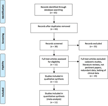
After running the search string in PubMed, an initial check was performed considering the inclusion and exclusion criteria, full-text retrieval and manual cross-checking of references, and a total of 65 titles were obtained. Among these, 5 literature reviews, 1 animal study and 23 non-English language titles were excluded. By reading the abstracts, one cadaveric study and five articles clearly unrelated to the topic of the analysis were also discarded. Among the remaining 30 articles, 1 cadaveric study, 1 review of the literature, 9 articles unrelated to the topic of this research, 1 article repeating the same results found in another article and 7 articles lacking clinical data were excluded at this stage. Finally, 11 of 65 articles were considered eligible and chosen for the analysis (Figure 1).
Preferred Reporting Items for Systematic Reviews and Meta-Analyses (PRISMA) guidelines flow diagram identifying and selecting the articles used in this review.

The studies were published between 1948 and 2018. A total of 2323 women were considered in this review. Overall, 1805 women (77.7 per cent) were considered ‘pregnant’, and 518 women (22.3 per cent) were considered ‘non-pregnant’.
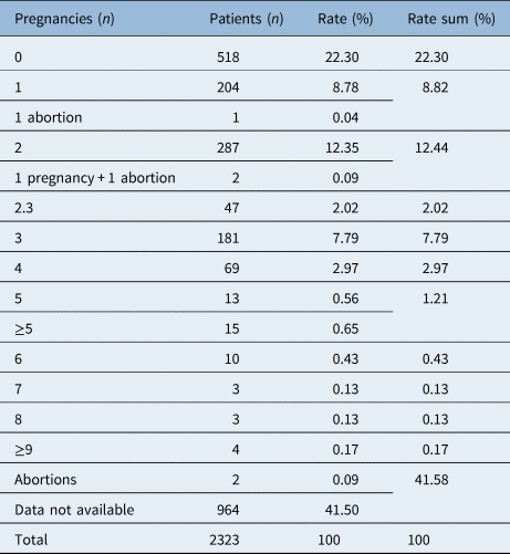
Regarding the number of pregnancies, data were not available for 966 cases, 2 of which reported ‘abortions’ without further details. The exact number of pregnancies was reported for 770 women (Table 1).
Number of pregnancies among patients affected by otosclerosis

Two patients experienced one pregnancy and one abortion. Forty-seven women were described altogether as having 2.3 pregnancies as an average. Two patients were described as having abortions (not quantified)
In 7 articles, hearing assessment was performed with audiometric evaluations; air and bone conduction, air–bone gap and median pure tone audiometry were analysed. The analysed frequencies were 0.5–4 kHz in 3 of 7 studies, 0.125–8 kHz in 1 of 7 studies, while the range was not specified in 3 of 7 studies. Assessment was not specified in four articles.
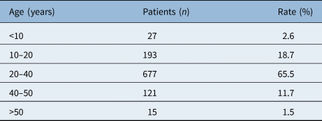
Age of onset of otosclerosis and subsequent diagnosis was reported for 1108 of 2323 women (47.7 per cent). Age distribution is shown in Table 2. The age of fertility was considered between 10 and 50 years,Reference Bordini and Rosenfeld19,Reference Gold, Crawford, Avis, Crandall, Matthews and Waetjen20 so overall there were 43 patients for whom a diagnosis of otosclerosis was given when non-fertile and 1065 when fertile.
Summary of age groups regarding onset of otosclerosis

Ages were obtained for 1108 of 2323 women
Regarding the course of otosclerosis, onset during pregnancy occurred in 14 patients, hearing worsening during pregnancy occurred in 213 and hearing worsening after pregnancy occurred in 26. By adding these numbers to those regarding patients who were not further characterised, hearing change with pregnancy occurred in 737 patients. By stratifying how many women had onset or not during pregnancy, hearing worsening during pregnancy, hearing worsening after pregnancy and hearing change with pregnancy, the prevalence of the disease could be calculated as 1 per cent for onset during pregnancy, 21 per cent for hearing worsening during pregnancy, 4 per cent for hearing worsening after pregnancy and 44 per cent for hearing change with pregnancy.
Statistical results
Two groups could be obtained among pregnant women by considering those who had onset during pregnancy and those whose disease arose separately from pregnancy. Among non-pregnant women, subdivision could be made similarly by considering women who were diagnosed within their fertile period and in their non-fertile period. According to the only available data regarding age of onset, 4 groups were obtained: women who had pregnancy and onset of disease at the same time (14), women who had pregnancy and onset of disease in two unrelated times (1259), non-pregnant women who had a diagnosis during their fertile period (316) and non-pregnant women who were diagnosed during their non-fertile period (10). According to the chi-square test and Fisher's exact test, the onset of otosclerosis was significantly related to pregnancy (p < 0.0001). By considering women who had pregnancy and disease onset during their non-fertile period (33) instead of those who had pregnancy and disease onset at two unrelated moments only, the previous analysis again returned a statistically significant correlation (p < 0.0001).
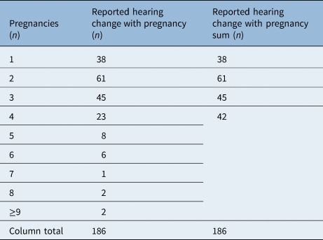
Other evaluations were carried out considering the correlation between the number of pregnancies and the above-mentioned parameters. Series presented as a whole group were not taken into account because the exact characterisation of every single patient was not possible. The same assumption was made for averages. The distribution of hearing change with pregnancy cases in relation to the number of pregnancies is shown in Table 3. Statistical analysis was performed with the chi-square test considering women who had 4 or more pregnancies altogether and comparing them to those who had 1, 2 or 3 pregnancies. Correlation between the number of pregnancies and hearing change with pregnancy was statistically significant (p = 0.003).
Stratification of cases of hearing change with pregnancy in relation to the number of pregnancies

Data across the studies showed 186 of 737 patients who had hearing change with pregnancy. Regarding the remaining 551 of 737, data on the number of pregnancies were lacking. Patients with four or more pregnancies have been grouped for statistical purposes.
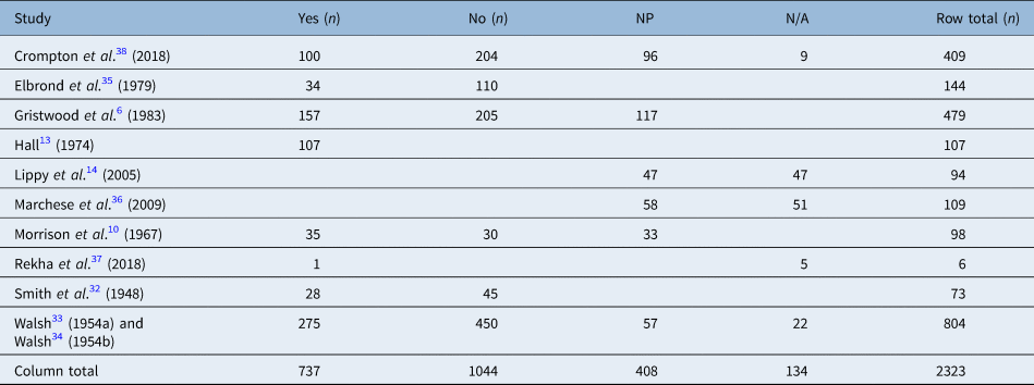
All results from across the studies are summarised in Table 4.
Summary of all studies included in the review

Yes = number of women who had hearing worsening related to pregnancy. No = number of women who did not experience hearing worsening related to pregnancy. Cases from two studies by Walsh (1954a and 1954b) were considered together because although these papers presented the same cases, Walsh 1954b reported two additional commentaries. NP = number of women who had no pregnancies. N/A = number of cases where data were not available.
Discussion
The aetiology of otosclerosis has been described as the sum of multiple factors. Several theories have been suggested regarding possible causes, such as genetic background, viral infections, autoimmune disorders and endocrine factors.Reference Markou and Goudakos17,Reference McKenna, Mills, Galey and Linthicum21–Reference Grayeli, Sterkers, Roulleau, Elbaz, Ferrary and Silve30 It has also been frequently reported that clinical otosclerosis is more prevalent in women than in men, and this has led to the aetiological hypothesis of sex hormones.Reference Podoshin, Gertner, Fradis, Feiglin, Eibschitz and Sharf5,Reference Gristwood and Venables6,Reference Lippy, Berenholz, Schuring and Burkey14,Reference Vessey and Painter31 Indeed, it has been reported that the female to male ratio is 2:1, even though from a histological point of view this difference has not been confirmed.Reference Markou and Goudakos17 Furthermore, the age of onset of this disease is similar to the typical age range for pregnancy, and therefore a correlation between the two has been suggested.Reference Gristwood and Venables6
As reported, the diagnosis of otosclerosis is achieved with audiometric evaluation, stapedius reflex analysis and CT-scan.Reference Molteni, Fabbris, Molinari, Alicandri-Ciufelli, Presutti and Paltrinieri3 It should be noted that the selected studies in the present review were published at very different times including some that were published prior to the introduction of CT imaging. Therefore, the criteria followed for the diagnosis were not the same for all patients, and in some cases, there may be a possibility of misdiagnosis. On the other hand, for analysis purposes, we could not make any difference among the selected women, and we considered them as all affected by the disease.
Among the selected articles,Reference Gristwood and Venables6,Reference Morrison10,Reference Hall13,Reference Lippy, Berenholz, Schuring and Burkey14,Reference Smith32–Reference Crompton, Cadge, Ziff, Mowat, Nash and Lavy38 the most recent was the study by Crompton et al.Reference Crompton, Cadge, Ziff, Mowat, Nash and Lavy38 in which a British cohort with otosclerosis was studied retrospectively from 2011 to 2017. Consistent with the literature, women comprised 65 per cent of the cohort so that the female to male ratio was 1.9:1.
As reported in the Results, there was a statistically significant correlation between pregnancy and disease onset. However, we are aware that there was a large disparity between group sizes when performing the chi-square test (pregnancy and otosclerosis onset at the same time (14), pregnancy and onset at two unrelated times (1259), non-pregnant women who had a diagnosis during their fertile period (316) and non-pregnant women who had a diagnosis during their non-fertile period (10). In order to distinguish between ‘fertile’ and ‘non-fertile’, we performed another analysis by considering women who had been pregnant and who had disease onset during their non-fertile age (33 cases). Once again there was a statistically significant difference (p < 0.0001), but the size of the groups was still heterogeneous.
By considering all these aspects, no robust conclusions can be obtained to answer the main question of this paper. However, we could calculate that prevalence (which can be interpreted as the probability of disease) is 1 per cent for onset during pregnancy, 21 per cent for hearing worsening during pregnancy and 4 per cent for hearing worsening after pregnancy. These rate differences may be because audiological evaluation is not routinely performed in pregnant women, and the data collected are from retrospective studies, often obtained by subjective evaluation. This leads to a probable under- or overestimation of the real occurrence of the disease. Assessing the course of an already diagnosed disease is easier than giving a new diagnosis, and sometimes this new diagnosis results from a complex evaluation process; this may explain why onset during pregnancy is 1 per cent and disease deterioration is higher during pregnancy. On the other hand, a correct evaluation of hearing worsening after pregnancy would be given by comparing audiometric evaluations before and after pregnancy; these data are not generally collected, so this may explain why the prevalence of hearing worsening after pregnancy is only 4 per cent. However, a further hypothesis is that pregnancy does have an influence on the disease, leading to a higher percentage for disease worsening compared with the rates of onset or subsequent deterioration of the disease.
By considering any case of hearing change with pregnancy, our study shows a prevalence of 44 per cent. Accordingly, we can state that among women who have finally been diagnosed with otosclerosis, almost 1 in 2 had an influence given by pregnancy. A comparison with the general female population, and also knowing who did not suffer from otosclerosis, was not achievable with the results of this study, since this was not the main purpose.
The number of women who had otosclerosis onset during pregnancy, hearing worsening during pregnancy or hearing worsening after pregnancy were very few compared with women who did not. Because of these large discrepancies, a statistical analysis could not be performed. The results were more comparable regarding hearing change with pregnancy. In this case, a significant result with similar cohorts could only be obtained by considering women who had four or more pregnancies altogether. The chi-square test returned a p-value of 0.003, which means that as the number of pregnancies increased, so did the cases with hearing impairment. This seems to be the only particularly significant result, leading us to consider the negative influence of pregnancy on otosclerosis.
Regarding the originality of the present study, (see Materials and Methods), six reviews were obtained with the searched terms.Reference Schrauwen15–Reference Markou and Goudakos17,Reference Booth39–Reference Panossian and Wikman41 The topic of pregnancy has been addressed in some cases, but no definitive answers could be given and sometimes descriptions of the previously published articles were made. According to our search, the most recent review of the literature was published by Schrauwen.Reference Schrauwen15 That article referred to Lippy et al.,Reference Lippy, Berenholz, Schuring and Burkey14 who stated that hormonal factors typical of pregnancy do not have an influence on the disease, even if symptoms of otosclerosis often arise during or shortly after childbearing. As explained above, this is one of the differing and controversial opinions regarding this topic. After the last review, four articles were published: two of them have been analysed in this studyReference Rekha, Ramalingam and Parani37,Reference Crompton, Cadge, Ziff, Mowat, Nash and Lavy38 , one was discarded because of non-adherence to the purpose of this researchReference Liao, Lu, Li and Chen42 and one moreReference Shiny Sherlie and Varghese43 was discarded because no patient characterisation was present, so no data were available for this study. We compared the results from across the studies obtaining estimations of probability (prevalence) and obtained one statistically significant result on the influence of pregnancy on otosclerosis, which was not reported in any of the analysed articles.
Limitations of the present study
The selected articles have been published over a long period of time, during which medical knowledge and diagnostic techniques have improved. Therefore, data collected from the most recent articles were very different from those obtained from the oldest. The reported results were obtained with all of the considerations reported in the Results and Discussion. Because of the high heterogeneity of the data, the use of a funnel plot and the test for small-effect size were not appropriate,Reference Terrin, Schmid, Lau and Olkin44,Reference Ioannidis, Rothstein, Sutton and Borenstein45 so risk assessment for bias was not performed in this study.
Conclusion
The present study examined data on changes in the onset or course of otosclerosis among women who had experienced pregnancy. According to our review, pregnancy seems to have an influence on the disease. It particularly seems to influence a worsening in women who already had a diagnosis: this influence is represented by a prevalence of 21 per cent. Furthermore, by considering any change in hearing that may occur with pregnancy, this prevalence rises to 44 per cent. With the same assumption, a relationship emerged between the number of pregnancies and hearing change.
A correlation emerged with regard to onset of the disease, but it derived from a comparison between pregnant and non-pregnant women; the latter were, of necessity, divided by different criteria than pregnant women because of lack of the triggering factor (i.e. pregnancy). Moreover, this correlation emerged from analysis of numerically heterogeneous groups.
Otosclerosis has a multifactorial aetiology, and evaluation of the role of a single cause is quite difficult, so it is not always clear whether hearing deterioration is because of a specific factor or whether it is a result of the natural course of the disease. Because of all these considerations, we cannot express firm statements, but our conclusion is that pregnancy seems to have a negative influence on the disease.
Competing interests
None declared