Introduction
Since the introduction of the carbon dioxide (CO2) laser for laryngeal surgery in the 1970s, transoral endoscopic laser surgery has become a preferred modality for treatment of early stage glottic cancer.Reference Kerr, Mark Taylor, Rigby, Myers, Osborn and Lambert1–Reference Lee, Chun, Kim, Kim, Oh and Hong4 Radiation therapy is the alternative treatment and demonstrates equivalent voice and oncological outcomes relative to surgery. However, endoscopic laser surgery has the benefits of decreased treatment cost and superior rates of laryngeal preservation.Reference Kerr, Mark Taylor, Rigby, Myers, Osborn and Lambert1–Reference Lee, Chun, Kim, Kim, Oh and Hong4 Additionally, surgery provides a laryngeal salvage option in the setting of cancer recurrence after radiation.Reference Ganly, Patel, Matsuo, Singh, Kraus and Boyle5
Carbon dioxide laser excision of early glottic cancer results in a wound characterised by tissue loss and healing by secondary intention. The healing process is heterogenous between patients and ranges from rapidly healing wounds to prolonged healing with or without granulation development.Reference Rioja, Blanch, Borés, Bernal-Sprekelsen and Vilaseca6–Reference Jeong, Kim, Ahn, Sung, Kim and Ahn8 Granulation can be a normal component of the benign healing process; however, this proliferative tissue may also conceal a persistent or recurrent cancer. This creates a diagnostic challenge during surveillance endoscopy in discriminating between benign granulation tissue and cancer recurrence.
Despite this complexity, there is a paucity of evidence to guide surgeons as to when biopsy should be pursued to evaluate for cancer following endoscopic CO2 laser surgery. A premature biopsy before completion of the healing process subjects a patient to unnecessary procedural risks. Conversely, a delay in surgical intervention could delay a recurrent cancer diagnosis and definitive treatment. Given this, we conducted a retrospective study of patients with early glottic cancer who underwent transoral endoscopic CO2 laser surgery in order to describe the healing timeline and correlate with patient characteristics and surgical factors. The purpose was to report the healing timing after CO2 laser surgery for early glottic cancer and to understand the clinical significance of granulation in order to inform post-operative clinical surveillance.
Materials and methods
The Institutional Review Board of Boston University School of Medicine approved this protocol (H-38447). We retrospectively reviewed patients with early glottic cancer (cT1 and cT2) who underwent endoscopic transoral CO2 laser excision from 2008 to 2019. Patients without recorded videos of endoscopic examinations or who had inadequate follow up were excluded from assessment. Adequate follow up was defined as regular post-operative endoscopic examinations until the surgical wound healed or biopsy was performed. The decision to pursue biopsy versus continued wound observation was made based on clinical characteristics and endoscopic examination findings. Patient demographic data and medical co-morbidities were obtained. Surgical and pathological characteristics including tumour depth, extent of vocal fold involvement and anterior commissure involvement were also collected. Evaluation of the pre-operative laryngoscopy examinations was utilised to determine extent of vocal fold involvement, which was categorised as more than 75 per cent or less than 75 per cent.
All previously recorded videos of post-operative, in-office endoscopic exams were independently reviewed by three otolaryngologists with experience in treating early glottic cancer. Reviewers evaluated for the presence of granulation, time to completion of healing or time to additional surgical intervention. All reviewers were blinded to patient characteristics, surgical characteristics and post-operative follow-up interval. Surgical sites were rated as having: (1) granulation, (2) leukoplakia, (3) wounds or (4) healed.
For assessment of inter-rater reliability between each pair of raters across the four categories for agreement, the Fleiss kappa statistical measure was used. Three reliability coefficients were produced: rater 1 versus rater 2, rater 1 versus rater 3 and rater 2 versus rater 3. Kappa statistics are classically interpreted as: more than 0.81, near perfect agreement; 0.61 to 0.81, substantial agreement; 0.41 to 0.60, moderate agreement; 0.21 to 0.40, fair agreement; 0.01 to 0.20, no to slight agreement; and less than 0, no agreement.Reference Stemler9 Spearman's rho correlation was computed as a correlation matrix amongst the three raters to evaluate inter-rater reliability for consistencies.Reference Ferguson10 Values between 0.2 and 0.39 are considered weak consistency, 0.4 and 0.59 as moderate consistency, 0.6 and 0.79 as strong consistency, and 0.8 and above as very strong consistency.Reference Stemler9
Patient characteristics were summarised using descriptive statistics. Comparisons were made using the Fisher's exact test. The Kaplan–Meier method was used to estimate time to events. P-values less than 0.05 were considered to be statistically significant.
Results
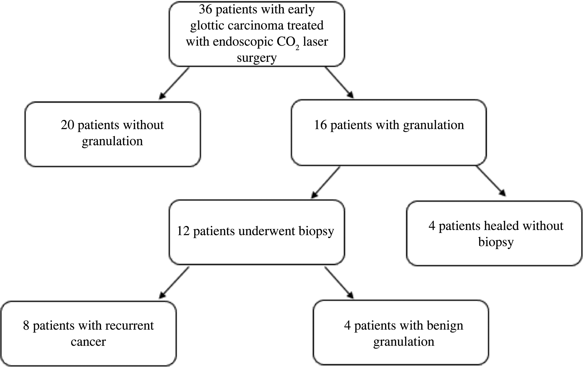
We identified 36 patients who underwent transoral CO2 laser surgery for early glottic cancer (Figure 1). Thirty-two patients (89 per cent) were male and 14 (39 per cent) were more than 65 years old at the time of surgery. History of smoking and co-morbid diabetes were present in 33 patients (92 per cent) and 7 patients (19 per cent), respectively. Clinical tumour stages included T1a (n = 16, 44 per cent), T1b (n = 14, 39 per cent) and T2 (n = 6, 17 per cent). Cancer involved the anterior commissure in 13 of 35 patients (37 per cent), and 23 patients (64 per cent) had cancer encompassing more than 75 per cent of the vocal fold length. Resection depth was available for 21 of 36 patients (58 per cent) and included subepithelial lesions or ligament (n = 13, 62 per cent), and muscle (n = 8, 38 per cent). Four patients (11 per cent) had a history of radiation therapy.
Patient flow chart. CO2 = carbon dioxide

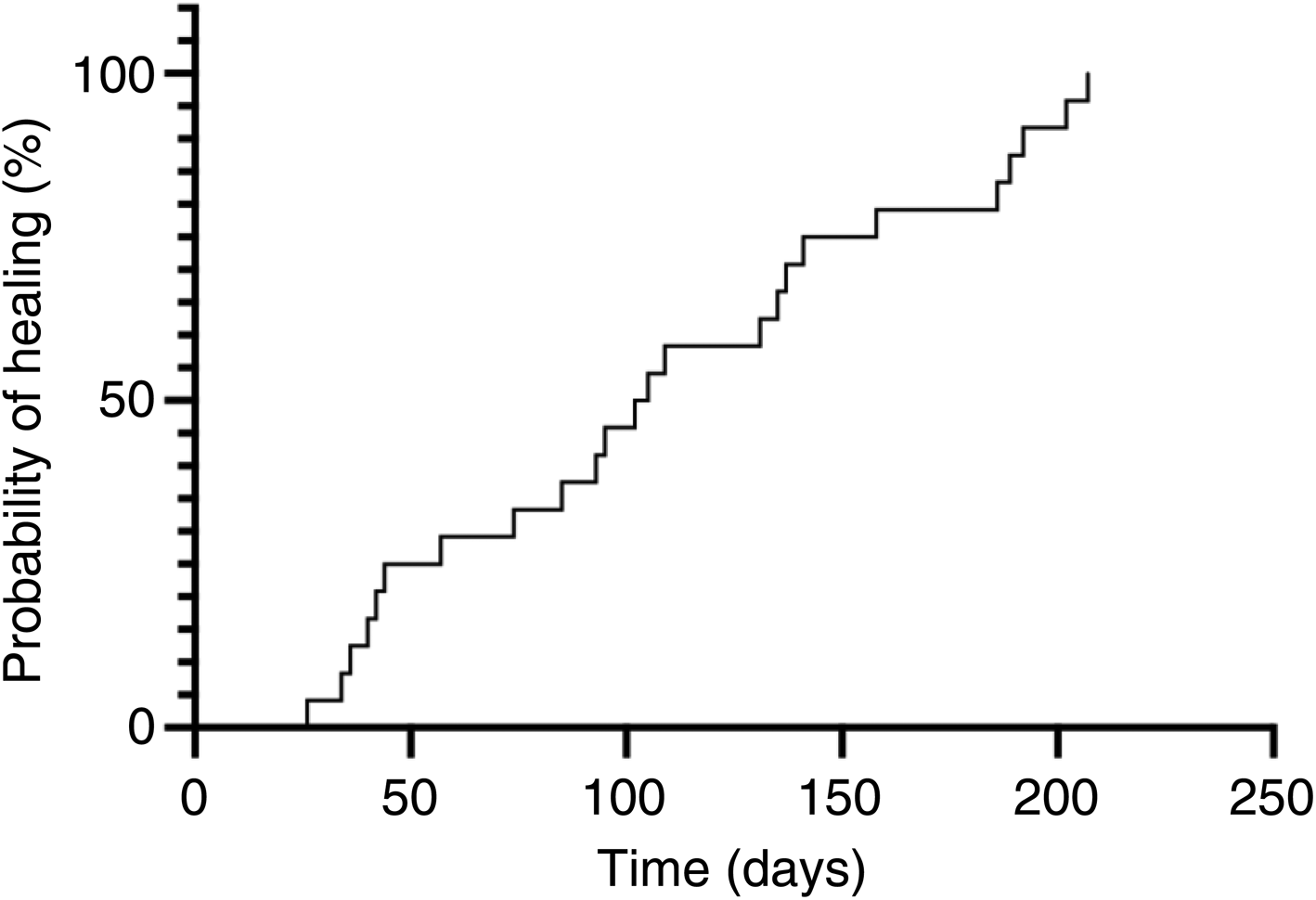
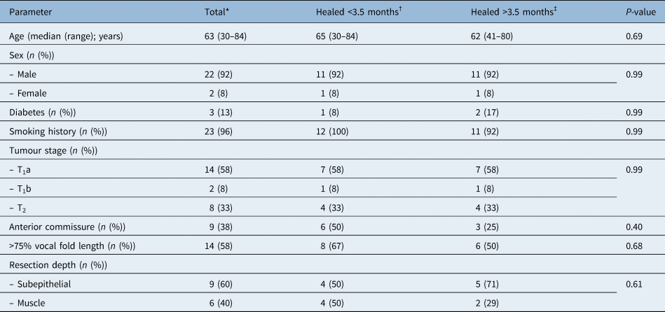
Twenty-four out of 36 patients (67 per cent) healed without the need for additional surgical intervention. The median time to heal for these 24 patients was 107 days (range, 34–207 days), with 4 patients (27 per cent) taking more than 6 months to heal (Figure 2). There was no observed difference in the studied variables between patients who healed more than 107 days or less than 107 days after surgery (p > 0.05 for all comparisons; Table 1) including for age, smoking history and tumour stage.
Probability of wound healing over time. The median time to heal was 107 days for patients without biopsy.

Patient and surgical characteristics associated with healing relative to 3.5 months

*n = 24; †n = 12; ‡n = 12. A total of 24 patients healed without the need for further surgical intervention. Data on diabetes and smoking history was available for 3 and 23 patients, respectively. Anterior commissure involvement, vocal fold length and resection depth were available for 9, 14 and 15 patients, respectively.
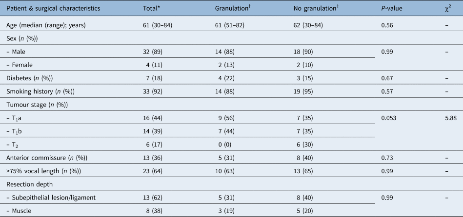
Sixteen patients (44 per cent) developed granulation following CO2 laser surgery. All patients who developed granulation tissue had a T1a or T1b tumour; however, this finding was not statistically significant (p = 0.053). Other studied variables were not associated with granulation development (p > 0.05 for all variables; Table 2). Three of the 4 patients (75 per cent) with a history of radiation developed granulation tissue.
Patient and surgical characteristics associated with presence of granulation

*n = 36; †n = 16; ‡n = 20. A total of 36 patients were evaluated for the presence of granulation tissue after endoscopic carbon dioxide laser surgery. Diabetes and smoking history were available for 7 and 33 patients, respectively. Anterior commissure involvement, vocal fold length and resection depth were available for 13, 23, and 21 patients, respectively.
Among the 16 patients with granulation tissue, 4 (25 per cent) healed without additional surgical intervention. The median time to heal among the 4 patients who developed granulation tissue and did not require repeat biopsy was 160 days versus 104 days for those who healed without granulation. Four of 24 patients (17 per cent) healed after more than 6 months, with 2 with granulation and 2 without. Initial post-operative follow up ranged from 7 to 72 days with a median of 15 days.
A total of 12 of 16 patients (75 per cent) underwent biopsy of vocal fold wounds; of these, 8 of 12 (67 per cent) had biopsy with cancer recurrence. Biopsies performed equal to or less than 3.5 months after the initial CO2 laser surgery demonstrated cancer in 1 of 4 specimens (25 per cent). For biopsies performed more than 3.5 months after the initial CO2 laser surgery, cancer was found in 7 of 8 specimens (87.5 per cent). Biopsy of granulation more than 3.5 months after initial surgery was associated with 28-fold increased odds of glottic cancer compared with equal to or less than 3.5 months (odds ratio, 28.00; 95 per cent confidence interval, 1.088–373.3). Two of the four patients with history of radiation underwent granulation biopsy, and cancer recurrence was identified in one patient.
For the 36 patients, 103 endoscopic examinations were reviewed. Inter-rater reliability was moderate in agreement between all 3 raters (kappa, 0.435, 0.546, 0.556). Spearman's rho was moderate to strong in consistency (ρ = 0.576, 0.634, 0.725). All reliability coefficients were statistically significant (p < 0.0001).
Discussion
In this study, we sought to describe the healing timeline and significance of granulation following transoral CO2 laser surgery for patients with early glottic cancer. We found that vocal fold wounds heal at a median of 3.5 months after surgery. Furthermore, we identified that granulation that persists beyond this timeframe has an increased risk of harbouring cancer.
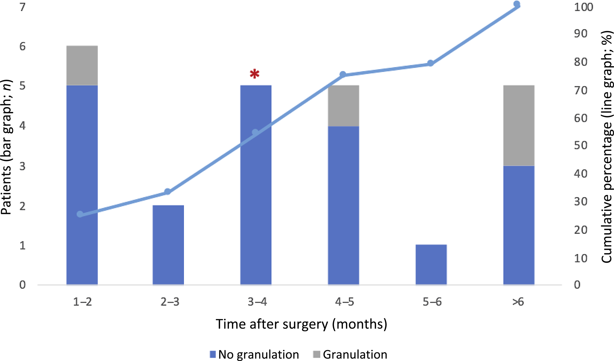
Nearly 70 per cent of patients in this cohort healed following surgery without undergoing biopsy (Figure 3). The median interval to healing was 3.5 months; however, six patients took more than six months to heal and were clear outliers. In a previous study, Shen et al.Reference Shen, Xu, Wang and Fan7 reported the mean time to heal after surgery in 29 patients with T1 glottic cancer was 88.1 days (± 19 days). The longer interval to healing observed in this study may be attributed in part to the inclusion of T2 cancer.
Healing timeline for patients who healed without biopsy with and without granulation tissue. The bars depict the number of patients who healed within that timeframe. The line graph depicts cumulative percentage healed. The median time to heal was 107 days indicated by the asterisk.

We did not identify patients or surgical characteristics that were associated with delayed time to heal. Co-morbid diabetes and tobacco abuse were two factors hypothesised to contribute to delayed healing. Diabetes results in dysregulation of all stages of wound healing, which is attributable in part to poor vascularity and immune system function.Reference Okonkwo and DiPietro11 It is possible that the smaller wounds resulting from CO2 laser surgery are not substantially affected by a poorer healing substrate. Although diabetes is known to negatively impact laryngotracheal healing after subglottic and tracheal injuries, its impact on healing of the vocal folds proper is not as well studied.Reference Nicolli, Carey, Farquhar, Haft, Alfonso and Mirza12 Smoking tobacco has also been shown to negatively impact many aspects of wound healing; however, in this study, smoking was not associated with delayed healing of glottic wounds.Reference McDaniel and Browning13 The reported history of smoking, in contrast to active smoking during wound healing, may contribute to this finding.
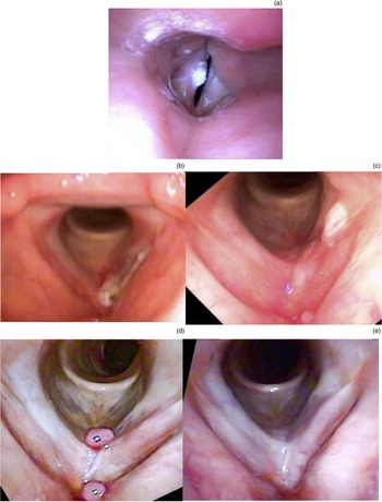
Nearly half of the patients in our cohort developed granulation tissue, which corroborates previously reported incidence of granulation in 53.8 per cent of wounds from Tis to T3 cancer by Rioja et al.Reference Rioja, Blanch, Borés, Bernal-Sprekelsen and Vilaseca6 This finding contrasts the lower incidence (35 per cent) of granulation tissue after potassium titanyl phosphate (KTP) laser surgery for early glottic cancer.Reference Tracy, Hron, Van Stan and Burns14 The higher incidence of granulation tissue after a CO2-laser induced injury in comparison with injury incurred by the KTP laser may be attributed to the deeper thermal spread of the CO2 laser as well as histopathological differences in wounds created by the laser.Reference Carew, Ward, LaBruna, Torzilli and Schley15,Reference Mallur, Branski and Amin16 Despite the frequently observed development of granulation, one quarter of those patients healed without undergoing biopsy. Figure 4 shows serial endoscopic examinations of a vocal fold wound that developed granulation tissue and subsequently healed. This patient's healing was delayed with respect to the median time to heal in this cohort (154 vs 107 days).
Images showing healing with granulation. Progression of healing of a patient with T1a cancer who developed granulation tissue after carbon dioxide laser surgery. (a) T1a cancer on posterior left vocal fold, (b) wound at 2 weeks post-operatively, (c) granulation tissue at 6 weeks post-operatively, (d) granulation tissue at 16 weeks post-operatively and (e) resolution of granulation tissue observed at 22 weeks.

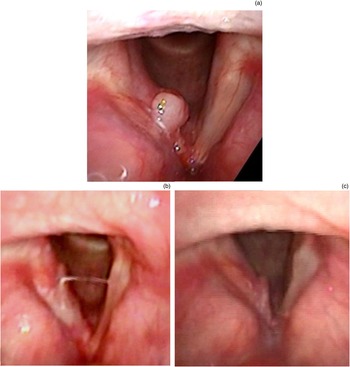
Glottic cancer recurrence is not easily distinguishable by clinical appearance. Endoscopic evaluation is substantially confounded in the setting of granulation which can mask recurrence. In this retrospective review, surgeons elected to pursue biopsy out of concern for cancer recurrence in two-thirds of patients with granulation. In addition to endoscopic findings, patients’ clinical history including surgical factors and medical co-morbidities are considered when determining the need for biopsy. We found that granulation persisting 3.5 months following surgery had a significantly higher odds of glottic cancer within the biopsy in comparison with biopsies performed prior to 3.5 months. This finding suggests that granulation tissue present after 3.5 months should be biopsied to evaluate for cancer recurrence. Our timeline for recommending biopsy is slightly earlier than that recommended by Shen et al.,Reference Shen, Xu, Wang and Fan7 who noted that wounds not healed after four months were associated with an increased risk of residual cancer. Jeong et al. evaluated 46 patients with T1 glottic cancer and found a 33 per cent likelihood of cancer recurrence in wounds not healed by 100 days (3 of 9 wounds). In our cohort, the likelihood of recurrence was higher after 107 days of a non-healing wound, which strengthens the recommendation for biopsy after this timepoint.Reference Jeong, Kim, Ahn, Sung, Kim and Ahn8 As a case example, Figure 5 shows serial endoscopic examinations of a T1a cancer that subsequently recurred after surgery.
Granulation with cancer recurrence. Cancer recurrence in a patient with T1a cancer after carbon dioxide laser surgery. (a) T1a cancer on right vocal fold, (b) wound with granulation at 6 days post-operatively and (c) wound with granulation at 2.5 months post-operatively that was positive for cancer on biopsy.

One unexpected finding of this review was that all patients who developed granulation had T1a or T1b cancer, and there was no granulation in wounds resulting from T2 cancer surgery. An increased incidence of granulation after treatment of T2 cancers was expected because of the larger extent and invasion, which therefore requires a greater resection with resultant increased thermal injury and greater inflammatory response.Reference Carew, Ward, LaBruna, Torzilli and Schley15 Other studies have correlated surgery for T2 cancers with an increased incidence of granulation development.Reference Rioja, Blanch, Borés, Bernal-Sprekelsen and Vilaseca6 Our findings could be explained by the small sample size of seven patients with T2 cancers in addition to the known variability of healing between patients. No other studied patient or surgical characteristics were associated with the formation of granulation tissue. Specifically, co-morbid diabetes and smoking history, which have classically been associated with the formation of granulation tissue were not significantly correlated with granulation.Reference Rioja, Blanch, Borés, Bernal-Sprekelsen and Vilaseca6,Reference Nerurkar and Shah17 The lack of association between diabetes and granulation is likely due in part to the limited number of patients with diabetes (n = 3). The lack of association between smoking history and granulation could be explained by the high incidence of patients who were previously smokers (92 per cent).
• Carbon dioxide laser surgery for early glottic carcinoma results in excellent oncological and voice outcomes
• Some wounds created by the laser form granulation tissue that can mask carcinoma recurrence
• There is paucity of evidence to guide surgeons as to when biopsy to evaluate for cancer should be pursued when granulation tissue persists
• Most patients heal after endoscopic carbon dioxide laser surgery without the need for further biopsy
• Granulation tissue that persists for more than 3.5 months warrants biopsy because of a 28 times increased risk of cancer
To our knowledge, no previous studies have evaluated wound healing in patients who underwent salvage CO2 laser surgery after radiation. Of the four patients in this study with history of radiation, three of four (75 per cent) developed granulation in comparison with 14 of 32 patients (44 per cent) without history of radiation. Although the number of patients with history of radiation is too small to draw conclusions, it is conceivable that increased incidence of granulation results from radiation changes to surrounding tissue. Higher rates of recurrence are also expected in this population given the challenges with navigating margins in the background of abnormal radiated tissue.Reference Haubner, Ohmann, Pohl, Strutz and Gassner18 Given this, recurrent cancer after radiation may require multiple laser surgical procedures to ultimately eradicate glottic cancer; however, salvage transoral CO2 laser for glottic cancer therapy has demonstrated good oncological outcomes and allows for laryngeal preservation.Reference Steiner, Vogt, Ambrosch and Kron19,Reference Puxeddu, Piazza, Mensi, Ledda, Argiolas and Peretti20
Previous studies examining wound healing after surgery for glottic cancer have small sample sizes, and the inclusion of this cohort adds to a growing body of literature evaluating factors that contribute to wound healing and formation of granulation. Limitations of this study include its retrospective design and comparably small sample size. Despite the recommendation for monthly follow up for oncological surveillance, variable follow up limits the accuracy of interval for wound healing; wounds may have healed prior to scheduled appointments and healed during the intervening timeframe. Given this, our study may overestimate the median time to heal after endoscopic CO2 laser surgery for glottic cancer. Future directions should include larger prospective studies with more frequent endoscopic examinations to ascertain a more precise timeline for healing. This could further narrow the timing for pursuing repeat biopsy of granulation and delayed healing wounds.
Conclusion
Development of granulation tissue after CO2 laser surgery for early glottic cancer is common and was not significantly impacted by patient co-morbidities or surgical factors. Most patients after endoscopic CO2 laser surgery heal without the need for biopsy, but granulation that persists for more than 3.5 months warrants a biopsy because of a 3.5-fold increased risk of cancer within the wound.
Competing interests
None declared