Introduction
Foreign body ingestion is a common presentation to paediatric otolaryngologists. While coins are the most frequently ingested item, the reported variety of materials consumed by the paediatric population is remarkable. Ingestion of pointed objects more typically occurs in psychiatric patients, and the diagnosis is unfortunately sometimes only made at autopsy.Reference Webb1, Reference Djokic, Atanasijevic, Savic and Nikolic2 While most foreign bodies will pass spontaneously, the risks associated with needles are only too well highlighted by the presented case report, in which cardiac tamponade occurred following ingestion of a straight needle.
Case history
A nine-month-old boy had originally presented to his paediatrician with a one-month history of a wheeze, dry cough and intermittent fever. A routine chest X-ray had been performed, identifying a linear, metallic foreign body. The patient was then referred to Children's Hospital Boston for further management.
On clinical examination, the child showed no evidence of respiratory distress and was tolerating his regular diet. Clinical examination was unremarkable, with normal heart and breath sounds.
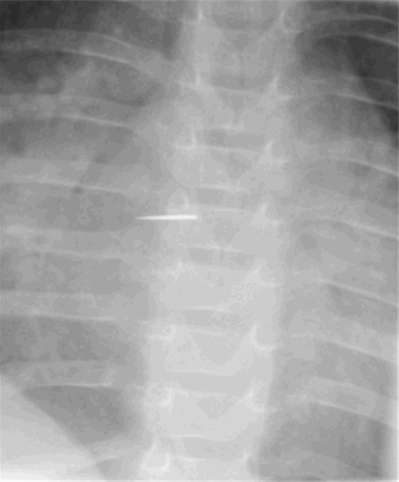
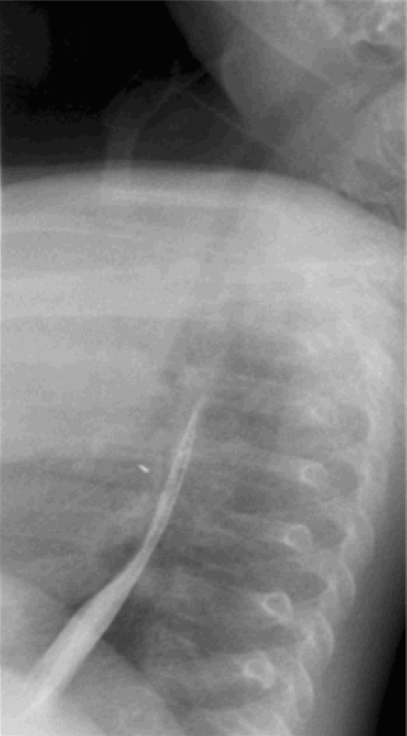
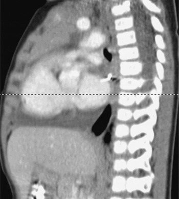
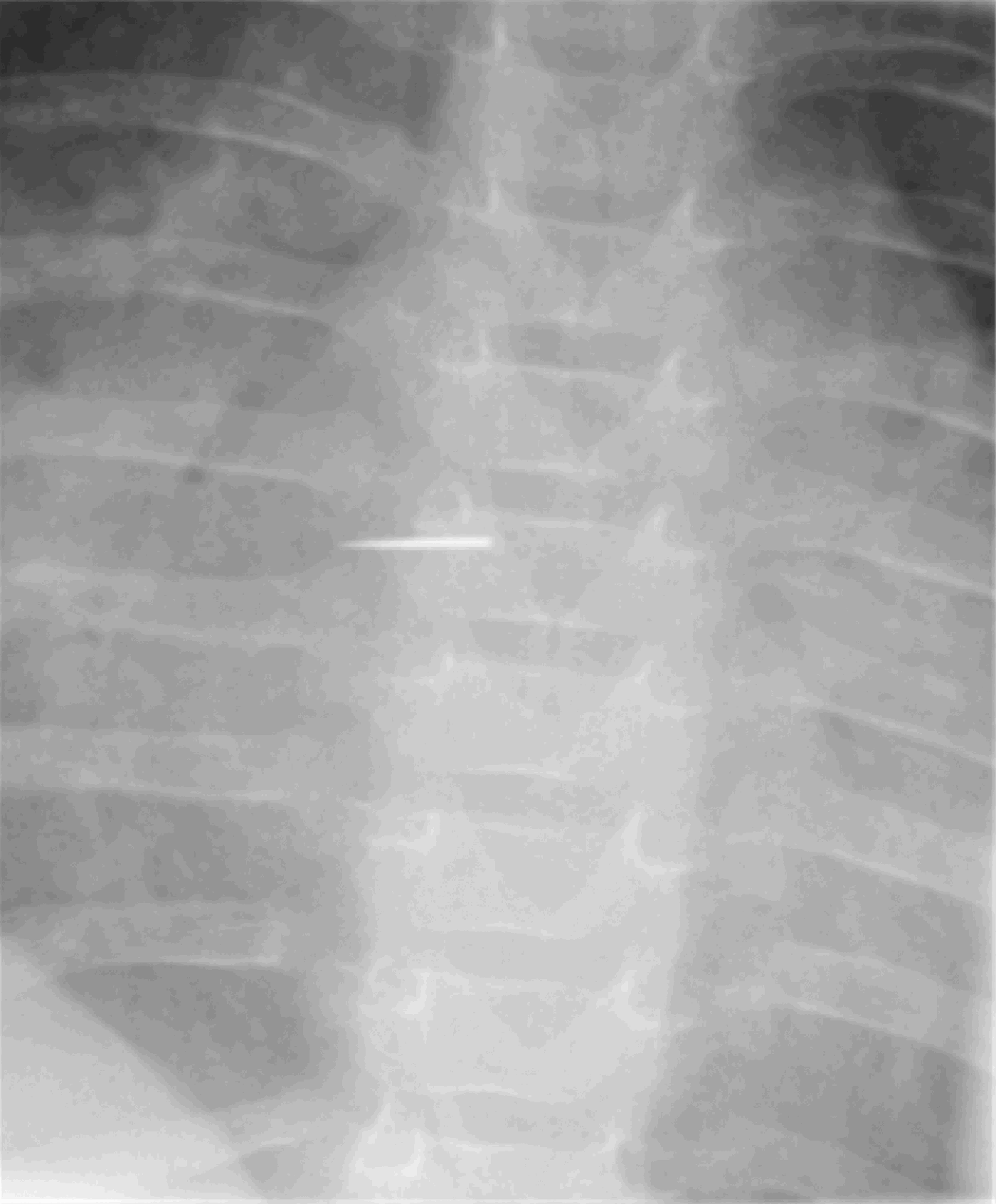
Antero-posterior and lateral plain radiographs were repeated, confirming the presence of a foreign body close to the level of the carina (Figure 1). A modified barium swallow was performed which demonstrated an intact oesophageal wall with no extravasation of contrast, with the foreign body located anterior to the oesophagus (Figure 2). A contrast-enhanced computed tomography (CT) scan of the patient's chest showed a 1.2 cm, metallic foreign body in the posterior mediastinum below the carina, lodged between the left atrium and the oesophagus, with one end abutting the left inferior pulmonary vein (Figure 3). In addition, a large, diffuse pericardial effusion was present (Figure 3). An echocardiogram confirmed the CT findings, but also identified early cardiac tamponade.
Antero-posterior chest X-ray illustrating the needle.
Modified barium swallow illustrating the needle lying anterior to the oesophagus.
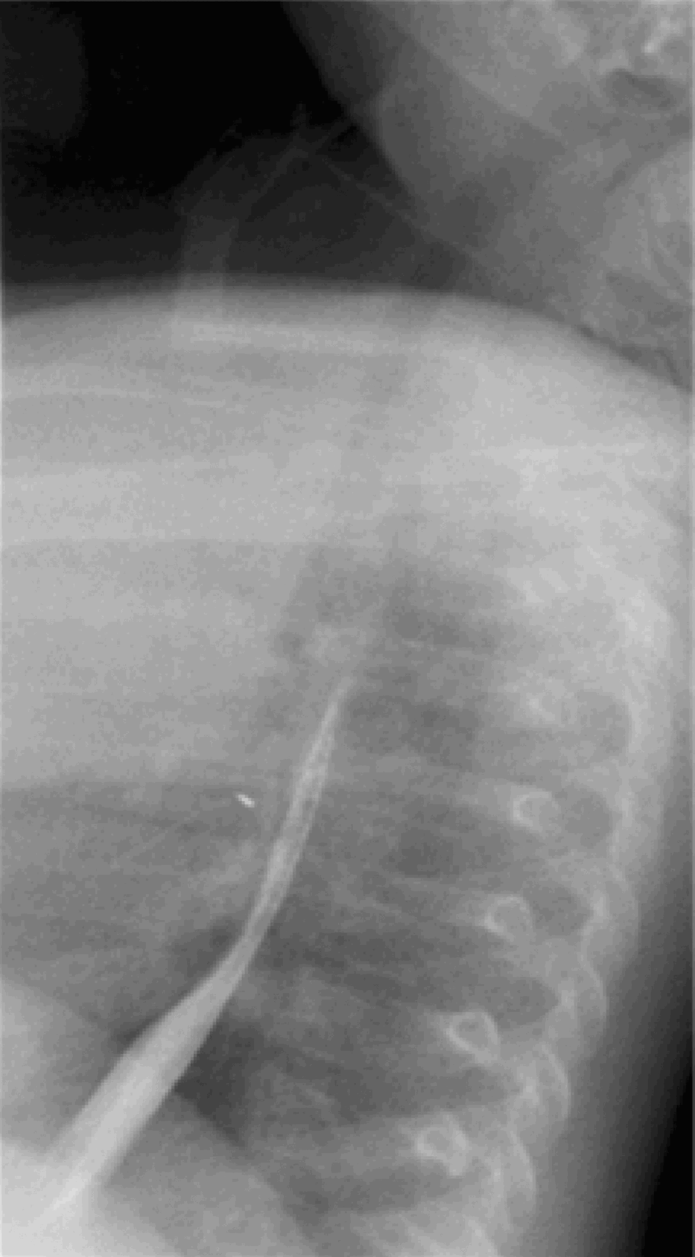
Contrast-enhanced, sagittal computed tomography scan illustrating the needle abutting the left inferior pulmonary vein, and also showing a pericardial effusion.

A cardiothoracic surgical opinion was obtained, and the patient was taken to the operating theatre for a rigid bronchoscopy and a right thoracotomy. The rigid bronchoscopy (to the right and left mainstem bronchi) was unremarkable. Following the right thoracotomy, the needle was extracted from inside the pericardium, and a large, serosanguinous pericardial effusion was drained. No breach of the cardiac wall was identified.
Post-operatively, the patient recovered without complications. Follow-up echocardiography failed to show reaccumulation of the effusion. At the time of writing, the infant continued to do well.
Discussion
The outer pericardium is composed of fibrous tissue and has a limited ability to stretch. Once fluid enters the pericardial space, its pressure increases. If this accumulation occurs slowly, the space may expand and contain the fluid before evidence of tamponade occurs is better. However, with ongoing accumulation, intrapericardial pressure increases, the systemic venous return is decreased and cardiac output drops, resulting in so-called ‘tamponade physiology’. Untreated, this can lead to shock and cardiac arrest.
Cardiac tamponade is a rare complication of needle ingestion which has previously been reported in adults.Reference Djokic, Atanasijevic, Savic and Nikolic2, Reference Cekirdekci, Avan, Ilkay and Yildirim3 More common causes of pericardial effusion include trauma, pericarditis, hypothyroidism and myocardial rupture. The classical presentation is known as Beck's triad: hypotension, jugular venous distension and muffled heart sounds. The relative absence of clinical findings in this child would suggest an interval of several weeks between ingestion and presentation.
• Paediatric foreign bodies may present with vague and nonspecific symptoms. It is important to have a high index of suspicion in managing such cases
• This paper describes the case of a nine-month-old infant who presented with a wheeze, cough and fever following ingestion of a needle
• This patient developed pericardial effusion and cardiac tamponade as a consequence of the needle ingestion, and required a thoracotomy for retrieval
To gain access to the pericardium and the heart, pointed objects have several viable routes available: embolisation, inhalation and direct penetration from the chest wall or stomach have all been reported.Reference Gyrtrup, Andreassen, Pedersen and Mortensen4–Reference Potek and Wright9 Most of the cases of needle ingestion reported are the result of self-injurious behaviour. Some have been missed clinically on presentation, with the diagnosis only made at post mortem. This highlights the importance of suspecting a pericardial effusion when there is clinical suspicion or radiographic evidence of an ingested sharp foreign body in the mediastinum (i.e. outside the oesophagus and trachea), particularly in a child (in whom many of the classical signs and symptoms may be lacking).
The precise sequence of events affecting our patient will never be known; however, it seems most plausible that the needle was swallowed, became lodged in the anterior wall of the oesophagus, and then migrated through it and into the pericardium. Intra-operatively, the needle was located anterior to the oesophagus, within the pericardium. No tract was identified from the oesophagus, but there was evidence of a localised inflammatory reaction in the area.
To our best knowledge, the current patient represents the youngest reported presentation of cardiac tamponade secondary to needle ingestion. The lack of clinical findings on presentation emphasises the importance of a high index of clinical suspicion for cardiac injury in such clinical and radiologically enhanced presentations.