Introduction
The plant microbiome, including bacteria and fungi, are fundamental partners throughout the developmental stages, including seed germination (Bulgarelli et al., Reference Bulgarelli, Schlaeppi, Spaepen, van Themaat and Schulze-Lefert2013; Shahzad et al., Reference Shahzad, Khan, Bilal, Asaf and Lee2018). Seeds play a crucial role in the life cycle of spermatophytes. Bacteria present inside disseminating organs can be important for the evolution of seedling-associated microbiota (Johnston-Monje and Raizada, Reference Johnston-Monje and Raizada2011; Truyens et al., Reference Truyens, Weyens, Cuypers and Vangronsveld2015). Although endophytic microbes have been identified in plants, seed-borne endophytic bacteria have not yet been fully explored. Seed-borne endophytes may play a key role in producing various phytohormones, enzymes, antimicrobial and antifungal compounds, secondary metabolites, in eliminating various stresses, promoting seed conservation and facilitating seed germination or possible biocontrol in the soil (Shahzad et al., Reference Shahzad, Khan, Bilal, Asaf and Lee2018). These dynamics are particularly crucial for symbiotic and parasitic plants, whose existence depends on their currently poorly understood complicated relationships with microorganisms, parasites and hosts. Seed endophytes may come from different plant organs, as a result of the transfer to seeds through vascular connections or gametes, causing colonization of the embryo and endosperm (Malfanova et al., Reference Malfanova, Lugtenberg and Berg2013). Next, endophytes could be transferred via the seed to benefit the next generation (Truyens et al., Reference Truyens, Weyens, Cuypers and Vangronsveld2015). Furthermore, importantly, the bacterial endophytic communities may be similar across the next consecutive seed generations (Sánchez-López et al., Reference Sánchez-López, Thijs, Beckers, González-Chávez, Weyens, Carrillo-González and Vangronsveld2018) and may contribute to adaptations to respond rapidly to environmental challenges and increase plant fitness (Truyens et al., Reference Truyens, Beckers, Thijs, Weyens, Cuypers and Vangronsveld2016). Thus far, 131 bacterial genera from four different phyla and 25 different plants have been reported as seed endophytes, belonging to the Proteobacteria (80 genera) and the γ-Proteobacteria (41 genera); the majority were similar to common soil strains (Truyens et al., Reference Truyens, Weyens, Cuypers and Vangronsveld2015).
Holoparasitic plants from the Orobanchaceae family parasitize on the roots of a variety of cultivated and wild plants, by creating an intrusive organ-haustorium and establishing a direct connection with the host vascular system. Apart from the exchange of various substances, this connection also allows for the exchange of endophytic microorganisms (Iasur Kruh et al., Reference Iasur Kruh, Lahav, Abu-Nassar, Achdari, Salami, Freilich and Aly2017). Parasitic plants are characterized by the production of a large number of some of the smallest seeds in the world's flora, usually less than 0.5 mm in length, frequently called ‘dust seeds’. The obligate root parasite Phelipanche ramosa (L.) Pomel (syn. Orobanche ramosa L.) is one of the most aggressive and economically important broomrape weeds and attacks various crops, especially tomato and tobacco (Solanaceae), as well as many other cultivated, ornamental and wild plants from the Brassicaceae, Cannabaceae, Fabaceae, Apiaceae and Asteraceae. P. ramosa occurs natively in Eurasia and North Africa, but its range is even wider due to introduction into Australia and North America (Piwowarczyk et al., Reference Piwowarczyk, Mielczarek and Guzikowski2018). P. ramosa has an extraordinary capacity for propagation because each individual plant may produce up to 500,000 extremely small seeds (usually between 200 and 400 μm) (Joel et al., Reference Joel, Bar, Mayer, Plakhine, Ziadne, Westwood and Welbaum2012), with reticulate testa with fibrillary ornamentation of the periclinal wall (Piwowarczyk, Reference Piwowarczyk2015). Resistance of the seeds to negative environmental conditions enables them to stay viable in the soil for more than 20 years (Linke et al., Reference Linke, Sauerborne and Saxena1992; Joel et al., Reference Joel, Bar, Mayer, Verdoucq, Westwood, Adkins and Ashmore2007). Furthermore, the ecological fitness of P. ramosa seeds is not limited to morphological and physiological features. It is suggested that the seed microbiota of P. ramosa may play a crucial role in geografic and host co-specialization. Genetic analysis of the composition of P. ramosa seed microbiota (Huet et al., Reference Huet, Pouvreau, Delage, Delgrange, Marais, Bahut, Delavault, Simier and Poulin2020) revealed the presence of many microorganisms present in the soil environment as well as in the host plants.
The relationship between the bacterial composition of the seed and the host plant can be explained by the transfer of the microbiome between the parasite and the host plant. Recent reports on the microbiome of parasitic plants indicate a relationship between the host and the parasitic plant, as exemplified by in vitro studies on Phelipanche aegyptiaca and tomato (Iasur Kruh et al., Reference Iasur Kruh, Lahav, Abu-Nassar, Achdari, Salami, Freilich and Aly2017), and in situ studies on Orobanche hederae and ivy (Fitzpatrick and Schneider, Reference Fitzpatrick and Schneider2020), indicating that both the plant parasite and its host may transfer bacteria between them. The seeds of parasitic plants only germinate in response to germination stimulants, like strigolactones, present in the rhizosphere of host plants (Zwanenburg et al., Reference Zwanenburg, Pospíšil and Ćavar Zeljković2016). It is suggested that plants have the ability to select a valuable microorganisms with a great potential for seed fitness, for example, a strain producing germination stimulants. Species have been described which promote the production of germination stimulants, for example, Sphingobacterium (Meulenbeld and Hartmans, Reference Meulenbeld and Hartmans2001) which supports the production of isothiocyanates which were also detected in seed core microbiota P. ramosa. The presence of Sphingobacterium sp. suggests a mutualistic relationship between bacteria and parasitic plant (Huet et al., Reference Huet, Pouvreau, Delage, Delgrange, Marais, Bahut, Delavault, Simier and Poulin2020).
The aim of our research was to isolate and characterize endophytic bacteria from P. ramosa seeds by using molecular methods. This paper describes, for the first time, seed endobiome isolates of P. ramosa, and presents the characterized potential of its dominance. Seed microbiome interaction studies can further develop our knowledge on the importance of the seed microbiota of parasitic plants and may lead to the development and viability of seeds and their germination.
Materials and methods
Species studied and seed collection
P. ramosa seeds were collected in September 2016 in Poland from severely infested tomato fields in one locality in Świętokrzyskie voivodeship, Sandomierz county, near Szewce (50°37′ N, 21°37′ E, 145 m a.s.l.). After harvesting, the material was dried under prevailing conditions. Voucher specimens were deposited in the Herbarium of the Jan Kochanowski University in Kielce (KTC).
Surface sterilization
Seed surfaces were sterilized using a previously described method (Wright and Wang, Reference Wright and Wang2014). Briefly, after being placed in glass tubes, seeds were surface sterilized using concentrated sulphuric acid for 5–8 min, and then the seeds were rinsed five times for 3 min with sterile, distilled water. To verify that the surface sterilization was properly performed, seeds were placed onto LB medium plates broth (10 g 1−1 tryptone, 5 g l−1 yeast extract, 5 g l−1 NaCl and 15 g l−1 agar, pH 7.2), Christensen (1 g l−1 peptone, 1 g l−1 glucose, 5 g l−1 NaCl, 2 g l−1 Na2HPO4, 0,012 g l−1 phenol red, 15 g l−1 agar, 20 g l−1 urea pH 6.8) and Sabouraud dextrose (5 g l−1 peptone, 40 g l−1 dextrose, 15 g l−1 agar), and then incubated at 30°C for 5 days.
Isolation of bacteria
To obtain endophytic bacteria, sterilized seeds were crushed under aseptic conditions using sterile and/or autoclaved tools, and then the crushed seeds were placed onto LB agar plates and incubated at 30°C for 5 days. Non-sterile seeds were placed onto LB medium plates and incubated at 30°C for 5 days for control condition.
DNA isolation
The two unique bacterial colonies from plates with surface-sterilized, homogenized seeds and the three most dominate types of colony obtained from plates contained bacteria from non-sterile seeds were selected and streaked on fresh LB agar plates to isolate a pure strain for DNA isolation. DNA was isolated using BIONEER kits (AccuPrep® Genomic DNA Extraction Kit), according to manufacturer's instructions.
16S rDNA gene fingerprinting
The amplification of the 16SrDNA gene fragment of the bacteria with universal 27 F primers (5′-AGA GTT TGA TCM TGG CTC AG-3′) and 1492R (5′-CGG TTA CCT TGT TAC GAC TT-3′) was performed on five bacterial isolates in accordance with Jiang et al. (Reference Jiang, Dong, Zhang, Yu, Chapman and Fields2006). The obtained product served as a sequencing template with primers: 27F-5′-AGAGTTTGATCMTGGCTCAG-3′; 515F-5′-GTGCCAGCMGCCGCGGTAA-3′; 800R-5′-TACCAGGGTATCTAATCC-3′; 1492R-5′-CGGTTACCTTGTTACGACTT-3′. Sequencing was carried out on an ABI3730xl apparatus using the BigDye Terminator v3.1 Cycle Sequencing Kit; Thermo Fisher Scientific. The resulting corrective sequences were used to assemble and obtain a consensus that was compared to the database of reference sequences, EzBioCloud.
BOX PCR
BOX amplification with the oligonucleotide sequence of the primer BOXA1R 5′-TACGGCAAGGCGACGCTGACG-3′ was performed on five bacterial isolates in accordance with Jarocki et al. (Reference Jarocki, Podleśny, Komoń-Janczara, Kucharska, Glibowska and Targoński2016). PCR was executed in the total volume of 50 μl of the reaction mixture containing 1 U (for BOXA1R-PCR) of Taq DNA polymerase, 200 μM of each deoxynucleoside triphosphate, 1 μM of primer, 50 ng of bacterial DNA and PCR buffer (GenoPlast Biochemicals). Amplification was conducted with template denaturation at 94°C for 4 min, followed by 35 cycles (denaturation for 1 min at 94°C, annealing for 1 min at 40°C). Amplified products were resolved in 1.5% (w/v) agarose gel in a Tris-acetate-EDTA buffer (TAE) and visualized under UV light (Syngene) after staining with ethidium bromide. For analysis of band patterns after electrophoresis, GelAnalyzer 2010a was used.
Phylogenetic trees
Phylogenetic trees were based on five first multifast sequences and created in phylogeny.fr (Dereeper et al., Reference Dereeper, Audic, Claverie and Blanc2010)
Results
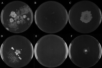
After 5 days of incubation at 30°C, plates with non-surface sterilized seeds revealed numerous bacterial colonies (Fig. 1A, B). Fungus was observed on Sabouraud dextrose plates (data not shown), and bacterial colonies were present on Christensen with urea agar plates (Fig. 1D). Christensen agar plates turned from light orange to pink within 24 h, suggesting that bacterial strains present on the seed surface are capable of urease production. Plates with sterilized seeds with no observed bacterial colonies demonstrated the effectiveness of the sterilization (Fig. 1A, E). On plates with homogenized sterile seeds, one large colony located between seeds fragments both in LB and Christensen was observed (Fig. 1C, F) but not on the Sabouraud plate (data not shown). The three most dominant types of colonies from plates with non-sterile seeds and the single colony from homogenized sterile seeds were streaked to obtain a pure culture for DNA extraction and further study. After streaking, two types of colonies obtained from the homogenized sterile seeds were observed.
Bacterial growth on plates. (A–C) LB medium plates, (D–F) Christensen medium plates. (A,D) Bacterial grown on plates with non-sterile seeds; (B,E) Plates with sterile seeds (control); (C,F) Bacterial grown on plates with homogenized sterile seeds.

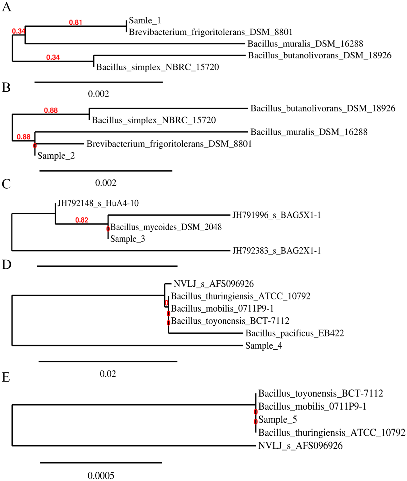
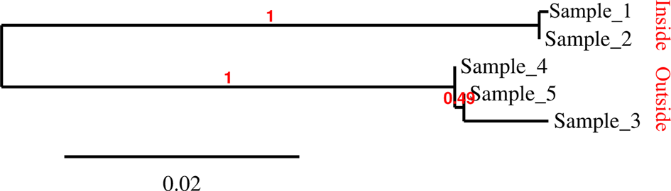
For taxonomic identification, 16S rDNA genes from five bacterial isolates were amplified and analysed. One of the bacterial isolates from the P. ramosa sterilized and homogenized seeds was classified as Brevibacterium frigoritolerans (100% compatibility with B. frigoritolerans DSM 8801) – Sample 1 (Fig. 2A). The second endophyte was 99.9 and 99.8% compatible with B. frigoritolerans DSM 8801 and Bacillus simplex NBRC 15720, respectively (Fig. 2B). All three bacteria strains isolated from the surface of non-sterile seeds are related to the Bacillus genus (Fig. 2C–E). One strain was classified as Bacillus mycoides (100% compatibility with B. mycoides DSM 2048) – Sample 3 and the other two strains showed 100% compatibility with B. mobilis, Bacillus toyonensis and B. thuringiensis – Samples 4 and 5 (Fig. 3).
Phylogram of seed isolates. (A,B) Endophytes, (C–E) Surface bacteria.

Phylogram of 16S rDNA sequence isolated bacteria.

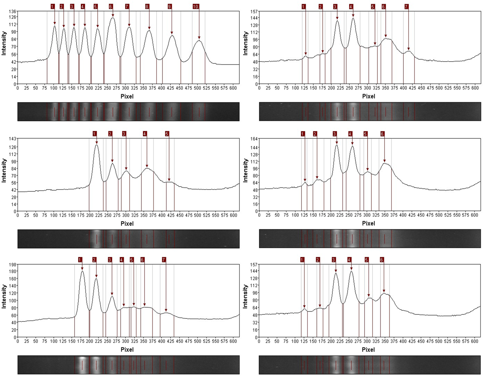
To determine the clonal differentiation of isolated endophytes, non-coding BOXA1-R sequences were used. Results of BOX PCR shows four different band patterns, suggesting that four out of five samples were genetically different (Fig. 4). Two endophytes differed in the band of approximately 700 pb and a band of approximately 350 pb for sample 2. Samples 3–5 had a very similar band pattern. Sample 3 differed from sample 4 and sample 5 by band approx. 200 pb. Samples obtained from the seed surface differed from endophytes samples by an additional band 900 pb.
Analysis of band patterns obtained by BOX amplification. (A) Marker, (B) sample 1, (C) sample 2, (D) sample 3, (E) sample 4 and (F) sample 5.

Discussion
P. ramosa is one of the most important root parasites in agriculture. Expansiveness of P. ramosa resulted from producing large quantities of long-living seeds (Joel et al., Reference Joel, Bar, Mayer, Verdoucq, Westwood, Adkins and Ashmore2007; Piwowarczyk et al., Reference Piwowarczyk, Mielczarek and Guzikowski2018). Endophytes may be one of the factors promoting seed viability and germination (Truyens et al., Reference Truyens, Weyens, Cuypers and Vangronsveld2015). This study's aim was to isolate and identify P. ramosa seed endophytes and compare the diversity of endophytes to seed surface bacteria. Accordingly, we obtained two types of colonies of Gram-positive rod-shaped endophytic bacteria. Analysis of 16S rDNA showed that strains are closely related to B. frigoritolerans. Preliminary information about seed endophytes of P. ramosa is based on metagenomic DNA and shows that the seed endobiome profile consists of 64.54% Proteobacteria, 29.64% Bacteroidetes, 3.37% Actinobacteria and 2.21% Firmicutes; the diversity of the endobiome depends on the host and field (Huet et al., Reference Huet, Pouvreau, Delage, Delgrange, Marais, Bahut, Delavault, Simier and Poulin2020). The Brevibacterium genus belongs to the Actinobacteria, indicating that isolated bacteria belong to one of the non-dominant groups of bacteria present in seeds of P. ramosa. Based on 16 sRNA sequences, the first isolated endophyte had 100% homology with B. frigoritolerans. Previous studies have shown that B. frigoritolerans may be a component of tomato microbiota contribute to a decrease of infections by Pseudomonas syringae, which suggests a mutualistic relation between B. frigoritolerans and tomato (Köhl et al., Reference Köhl, Medeiros, Lombaers-van der Plas, Groenenboom-de Haas and van den Bosch2020) and possibly a transfer of bacteria between parasite plant and host, as previously described (Iasur Kruh et al., Reference Iasur Kruh, Lahav, Abu-Nassar, Achdari, Salami, Freilich and Aly2017; Fitzpatrick and Schneider, Reference Fitzpatrick and Schneider2020). Representative strains of B. frigoritolerans were described as strictly aerobic, chemoorganotrophic members of the genus Brevibacterium. B. frigoritolerans is an environmental bacterium and was originally isolated by Delaporte and Sasson from the arid soils of Morocco (1967). A unique feature of B. frigoritolerans is its tolerance to low temperature (Selvakumar et al., Reference Selvakumar, Sushil, Stanley, Mohan, Deol, Rai, Ramkewal, Bhatt and Gupta2011).
Moreover, studies on the Brevibacterium genus suggest the presence of bioremediation activity and resistance to high concentrations of metals which can be used by plants colonizing new, difficult-to-survive areas. For example, the presence of the seed core microbiome of Crotalaria pumila (Sánchez-López et al., Reference Sánchez-López, Thijs, Beckers, González-Chávez, Weyens, Carrillo-González and Vangronsveld2018), and root endophytes of Prosopis laegivata (Román-Ponce et al., Reference Román-Ponce, Li, Vásquez-Murrieta, Sui, Chen, Estrada-de los Santos and Wang2015) grow on multi-metal contaminated mine residues. The occurrence of this bacterium may be related to the expansion of the parasitic plant Orobanche lutea in post-industrial areas with toxic metals (Zn, Cd, Pb) (Turnau et al., Reference Turnau, Jędrzejczyk, Domka, Anielska and Piwowarczyk2018), but this requires further research. It is worth noting that B. frigoritolerans isolated from Aloe vera rhizosphere has multiple plant growth promotion attributes, such as phosphate solubilization, IAA production and siderophore production, which are expressed differentially at sub-optimal temperatures (Meena et al., Reference Meena and Saharan2017). This is important as previously referenced data suggests that seed endophytes may promote seed germination. In addition, studies investigating the seed endophytes of rice show that endophyte isolates from salt-tolerant cultivars improved germination under salt stress conditions of a salt-sensitive cultivar (Walitang et al., Reference Walitang, Kim, Madhaiyan, Kim, Kang and Sa2017). Resistance to high concentrations of metals, tolerance to hot and dry or cold conditions and plant growth promotion features (Liu et al., Reference Liu, Guo, Salam, Li, Zhang, Han, Mohamad and Li2016) may indicate that B. frigoritolerans adapt to severe environmental conditions, influences seed survival and may affect seed viability and germination.
Studies on B. frigoritolerans and its influence on the growth and development of highly invasive Flaveria bidentis have shown that B. frigoritolerans enriches the soil with elements, such as N, P and K, thereby increasing plant biomass. In addition, B. frigoritolerans has a positive effect on root development by increasing the duration and absorption of nutrients. Furthermore, B. frigoritolerans can act as a mycorrhizal helper bacterium that increases the population of Rhizoglomus intraradices, influencing the competitive growth of the invasive plant (Du et al., Reference Du, Chen, Li, Chen, Xu and Zhang2020). On the other hand, data show that this bacteria occurs in insect gut and can be a novel entomopathogen of Anomala dimidiata and Holotrichia longipennis (Scarabaeidae: Coleoptera) (Selvakumar et al., Reference Selvakumar, Sushil, Stanley, Mohan, Deol, Rai, Ramkewal, Bhatt and Gupta2011). This is notable with respect to entomopathogenic potential because P. ramosa is attacked by the monophagous fly Phytomyza orobanchia (Diptera: Agromyzidae), whose larvae can reduce seed production by 30–90%, and this is the most effective biological control agent of broomrape (Klein and Kroschel, Reference Klein and Kroschel2002; Piwowarczyk et al., Reference Piwowarczyk, Mielczarek and Guzikowski2018). It is possible that this bacteria could be transferred by seeds of P. ramosa to P. orobanchia larvae or other insects during the feeding process. Many insect species are reported to feed on broomrape species, including Scarabaeidae representatives (Linke et al., Reference Linke, Sauerborne and Saxena1992), which may have a broader impact on insect microflora or co-evolutionary adaptations. The second strain isolated from P. ramosa seeds showed highly similar homology to both B. frigoritolerans and Bacillus muralis; the difference is 1 or 2 nucleotides, respectively. Differences between colony shape and two different BOX PCR patterns indicate that the two isolates are very similar but derived from different strains. B. muralis and B. frigoritolerans are closely related and are present in soil, for example, painted murals environments (Wang et al., Reference Wang, Liu, Liu, Chen, Pan, Zheng and Chen2016; Tomassetti et al., Reference Tomassetti, Cirigliano, Arrighi, Negri, Mura, Maneschi, Gentili, Stirpe, Mazzoni and Rinaldi2017). Furthermore, bacteria closely related to B. muralis have been described as plant growth-promoting rhizobacteria (PGPR) of avocado (Persea americana Mill.) (Méndez-Bravo et al., Reference Méndez-Bravo, Cortazar-Murillo, Guevara-Avendaño, Ceballos-Luna, Rodríguez-Haas, Kiel-Martínez, Hernández-Cristóbal, Guerrero-Analco and Reverchon2018). Interestingly, we isolated only two strains of bacteria, with an analysis of metagenomic DNA showing high diversity of the P. ramosa seed endobiome. Obtained results may be due to many factors, such as sterilization method or used culture medium. In addition, the number of seed endobiome isolated strains decreases with storage time (Truyens et al., Reference Truyens, Weyens, Cuypers and Vangronsveld2015) and studies were performed using 1-year stored seeds. Moreover, results based on metagenomic DNA give information about all bacteria which also were present in the seeds but do not show which bacteria were still alive. It is worth noting that isolated strains belonged to bacteria which are highly resistant to environmental conditions and adapted to survive in seeds inside. Moving forward, it is important to determine if the same strains of bacteria are isolated from the seeds of P. ramosa growing in different environmental conditions and hosts. The presence of B. frigoritolerans in arid soils suggests that endophytes of P. ramosa seeds may support seeds in acquiring resistance to solarization, which may be important in the future for the parasite's biocontrol.
In the present study, P. ramosa seed surface bacteria were also isolated. The three most numerous colony types were further identified. One surface isolate (sample 3) showed 100% homology to B. mycoides. The strains of samples 4 and 5 had 100% homology with B. toyonensis, B. thuringiensis and B. mobilis. Though different types of colonies, sample 4 and sample 5 had similar BOX PCR patterns which suggest that they may be the same strain. All isolated bacterial strains were closely related and belong to the Bacillus cereus group (Liu et al., Reference Liu, Du, Lai, Zeng, Ye, Xu and Shao2017; Yi et al., Reference Yi, de Jong, Frenzel and Kuipers2017). It is worth noticing that Bacillus bacteria are widespread in the soil environment and are often a component of the plant microbiome. The Bacillus genus is very common as an endophytic species in angiosperm groups such as rice (Pal et al., Reference Pal, Kumar, Verma, White, Verma, Kumar, James and White2019). These bacteria are known for their promotion of plant growth (PGPR) and their biocontrol properties. Curiously, bacteria such as B. mycoides or B. thuringiensis are described as PGPR. B. mycoides is omnipresent in the soil and the plant rhizosphere. Some isolates of B. mycoides are a group of beneficial, plant growth-promoting and biocontrol activity types for various crops, such as sugar beet or cucumber (Bargabus et al., Reference Bargabus, Zidack, Sherwood and Jacobsen2002; Neher et al., Reference Neher, Johnston, Zidack and Jacobsen2009). B. mycoides cells were described as bacteria capable of colonizing a plant's internal tissue. Endophyte strains are characterized by different biochemical and genetic features influencing competences of colonization (Yi et al., Reference Yi, de Jong, Frenzel and Kuipers2017). Interestingly, B. thuringiensis has been described as bacteria that can suppress the growth of Ralstonia solanacearum and the development of wilt symptoms in tomato plants (Hyakumachi et al., Reference Hyakumachi, Nishimura, Arakawa, Asano, Yoshida, Tsushima and Takahashi2013). Also, B. thuringiensis is used as a biopesticide against caterpillars (Raymond and Federici, Reference Raymond and Federici2017). Isolation of B. thuringiensis from plant seeds growing in a tomato field can be the subject of horizontal transmission between a host plant and their parasite. Given the properties of Bacillus bacteria to promote plant growth and their ability to form endospores, it seems useful to add Bacillus inoculates to soil. On the other hand, studies show that this type of supplementation changes the biodiversity of endophytes, which may affect plant development (Gadhave et al., Reference Gadhave, Devlin, Ebertz, Ross and Gange2018).
Based on genetic analysis, obtained isolates may be split into two closely related groups corresponding to endophytic and surface bacteria. Results showed that differences between the composition of endophytic and surface bacteria might not be accidental, particularly when the composition of the seed endobiome is associated with the plant and a habitat as described for P. ramosa (Huet et al., Reference Huet, Pouvreau, Delage, Delgrange, Marais, Bahut, Delavault, Simier and Poulin2020). Association of Bacillus bacteria with plant and especially seed endobiomes might be a result of endospore formation, adapting the bacteria to harsh environmental condition.
Studies on the relationships of bacterial populations between parasites and their hosts are important. Understanding the role of bacteria in supporting the growth of parasitic plants and host infections is essential because of potential ecological, evolutionary, biotechnological and economic consequences.
Conclusions
We presented the first bacteria isolated from P. ramosa seeds. Two isolated strains were closely related and described as highly resistant to environmental conditions and as plant growth-promoting species, which suggests that the bacteria may facilitate seed germination or seed and seedling viability. Obtained information might help in the biocontrol of the plant parasite in the future.
Financial support
This work was supported by the Jan Kochanowski University grant No. SMGR.RN.20.116.607.
Authors’ contributions
R.P. is the originator of the research topic. R.P. and W.K. initiated the studies. R.P. identified and collected plant material. K.D. Set up of experimental details. K.D. and P.Ż. performed the laboratory experiments. K.D. analysed the research results. K.D. and R.P. wrote a draft of the manuscript. R.P. and W.K. contributed critical comments on the draft. All authors approved the final version of the manuscript.