Introduction
Conservation and management of fisheries stocks require a fundamental understanding about the basic life history patterns, habitat and exploitation status of a particular species. This requires detailed information on the basic reproductive physiology, spawning seasons and grounds, size at maturity, fecundity, sex ratios, age and growth-based population structure, particularly for commercially important species. Studies focusing on such information are critical in the quantitative assessment of fisheries stocks. Data on the reproductive biology of a species are essential for an assessment of the threshold of sustainable exploitation and for the design of management plans (King, Reference King2003; Jakobsen et al., Reference Jakobsen, Fogarty, Megrey and Moksness2009), and for considering potential in aquaculture development. Fish stocks are renewable natural resources that can be replenished through reproduction in natural habitats (Olusegun, Reference Olusegun2011). The replenishment success of any particular species is dependent on its recruitment capability under challenging environmental conditions. Thus, efforts should be made to understand the processes that drive and regulate the population biology of commercially important fish species.
The Bay of Bengal is one of the world's Large Marine Ecosystems (LMEs) and has a high biological diversity. To date, very little is known about the basic life history of many fishes from the region. Moreover, intensifying fishing pressure and inadequate management actions are leading to a decline in marine yield every year (DoF, 2017). The silver pomfret, Pampus argenteus (Euphrasen), which belongs to the family Stromateidae (butterfishes), is one of the commercially important marine fish species in Bangladesh and some other Asian countries such as Kuwait, India, Korea, China, Malaysia, Thailand and Japan (FAO, 1995; Dadzie et al., Reference Dadzie, Seedo and Shallal2000b; Divya et al., Reference Divya, Mohitha, Rahul, Shanis, Basheer and Gopalakrishnan2017). Pomfret production from the Bay of Bengal was 11,899 metric tons in the year 2017–18 (DoF, 2017) which comprises 1.82%, placing it at the fifth position, of the total marine fish production in Bangladesh, and this species commands a high market price.
Despite its commercial importance in many countries, information on the life history traits of this species are limited, especially in the Bay of Bengal. Pati (Reference Pati1981, Reference Pati1982) reported the fecundity and maturation of silver pomfret from the Indian waters of the Bay of Bengal but there is no comprehensive information regarding the reproductive biology of this species in Bangladeshi waters. Previous studies have reported on the food and feeding habits (Dadzie et al., Reference Dadzie, Seedo and Qattan2000a), spawning frequency, fecundity, egg weight and spawning type (Almatar et al., Reference Almatar, Lone, Abu-Rezq and Yousef2004), and seasonality in biological parameters (Akhter et al., Reference Akhter, Islam, Islam and Zahangir2020) of P. argenteus from different parts of the world, including the East China Sea, Korean waters and Kuwait waters. Dadzie et al. (Reference Dadzie, Seedo and Shallal2000b), Almatar et al. (Reference Almatar, Lone, Abu-Rezq and Yousef2004), and Lone et al. (Reference Lone, Ablani and Almatar2008a, Reference Lone, Ablani and Almatar2008b) (from Kuwait waters) and Lee & Jin (Reference Lee and Jin1989) (from the East China Sea) reported the reproductive biology of P. argenteus, which showed differences in the reproductive timing and maturation pattern between habitats. Thus, it is necessary to clarify the reproductive seasonality and complete gonadal maturational cycle of P. argenteus for successful conservation and management for the Bay of Bengal, Bangladesh, and also for considering regional aquaculture potential. Therefore, in this study, we aimed to elucidate the cyclic gonadal development and spawning seasons of this commercially important species from the Bay of Bengal, Bangladesh.
Materials and methods
Sample collection and processing
Samples of silver pomfret, P. argenteus (Euphrasen, 1788) were caught from the ‘South patches’ fishing ground of the Bay of Bengal (latitude: 20°50–21°40′N) and collected monthly from the fish markets in Cox's Bazar (11–20 fish of both sexes) from February 2016 to January 2018. A total of 144 females and 229 males were collected. The sexes of individual fish were determined using aceto-carmine gonad squashing methods, as described by Guerrero & Shelton (Reference Guerrero and Shelton1974). The total length (TL, cm), standard length (SL, cm), and total weight (TW, g) of individual fish were measured. Gonads were collected from both males and females, weighed (GW, g), and fixed in Bouin's solution for histological study.
Gonadosomatic index and fecundity
The gonadosomatic index (GSI) was calculated (gonad weight/TW) (Vladykov, Reference Vladykov1956) and expressed as percentages. The gravimetric method was used to estimate the fecundity (Hunter et al., Reference Hunter, Macewicz and Kimbrell1989) from randomly collected gravid female fish samples (n = 58) from different months of the year (4–5 individuals were sub-sampled monthly). At first, 7–8 subsamples from different parts of the individual ovary were cut and weighed. Subsequently, the number of eggs was counted for each sub-sample and the fecundity was calculated using the following formula: (weight of ovary/weight of sub-sample) × number of eggs in each sub-sample; and relative fecundity was also calculated from the absolute fecundity by dividing it by the total weight of fish.
Oocyte development and histological classification
The oocyte diameter of the fresh eggs was measured using an ocular micrometer set on a microscope (LABOMED, LX400, USA). About 2–4 specimens for both males and females were sub-sampled monthly for the histological study. The standard histological procedure was carried out on the gonad samples following dehydration in a graded series of alcohol, embedded in molten paraffin, stained with haematoxylin and eosin, and mounted with dibutyl phthalate xylene (DPX). Tissue samples were sectioned at 5 μm thickness and photographs were taken at 10 × magnification with a microscope (Optica B-190/Optica Vision Lite 2.1 software) and oocyte diameter (n = 15–22 oocytes/individual) was measured under a microscope. Changes in the gonadal phases throughout the year were described according to their morphometric appearance and gamete stages (types, organization, egg sizes, wall structure, presence of atresia, spermatids, yolk vesicle, yolk granules and post-ovulatory follicles) following Wallace & Selman (Reference Wallace and Selman1981), Nunez & Duponchelle (Reference Núñez and Duponchelle2009), Ma et al. (Reference Ma, Xie, Huo, Yang and Chen2012), Gardner et al. (Reference Gardner, Frazer, Jacoby and Yanong2015) and Amparo et al. (Reference Amparo, Cárdenas, Guevara and Pérez2017).
Statistical analysis
The Pearson correlation coefficient (r) was used to determine the relationships between SL and fecundity, and GSI and oocyte diameter. All statistical analyses were performed using Microsoft Excel and Microcal Origin 6.0 (OriginLab Co., USA), and results were presented as means ± SD.
Results
Gonadosomatic index
Pampus argenteus individuals collected during the experimental period were found to vary in length and weight. In males, the TL, SL and total weight (TW) were 23.62 ± 2.47 cm (mean ± SD), 16.00 ± 1.77 cm and 181.73 ± 57.28 g, while in females, the TL, SL and TW were 24.26 ± 3.71 cm, 16.73 ± 2.96 cm and 223.64 ± 131.51 g, respectively.
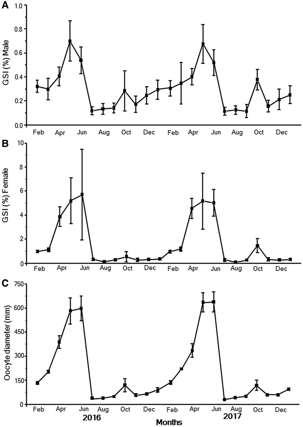
Gonadosomatic index was calculated monthly for both males and females using all the collected fish. The highest GSI value (4.17 ± 0.87 to 5.43 ± 2.89 in females and 0.41 ± 0.08 to 0.69 ± 0.17 in males) was observed from April–June. The second highest GSI values were observed in October (0.93 ± 0.68 in females and 0.34 ± 0.14 in males) in two years, which suggests that gonadal maturation may occur biannually in this species (Figure 1A, B).
Monthly changes in the (A) GSI (male, n = 229, 15–24 individuals each month), (B) GSI (female, n = 144, 8–15 individuals each month), and (C) oocyte diameter of Pampus argenteus collected from the Bay of Bengal. Values are presented as means ± SD.

Oocyte diameter
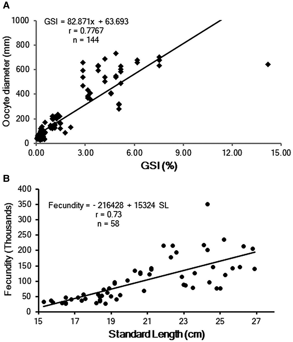
The annual changes in the oocyte diameter of P. argenteus throughout the year (15–22 oocytes from each fish, 4–8 fish in each month) were observed and compared with the changes in the GSI of females (Figure 1C). The oocyte diameter showed a similar pattern to that of the changes in GSI, and showed a linear relationship (y = 82.871x + 63.693, r = 0.78) with the GSI values (Figure 2A), which suggests the synchronized development of the gonads with increasing egg size. The highest oocyte diameter was observed in May (630.5 ± 96.7 μm, n = 16 individuals) when the fish are fully mature and the ovaries are at their largest, and the lowest in July (35.10 ± 7.13, n = 13 individuals), and when new eggs initiate development for the next spawning season. Size variation in oocytes was also observed in October with some large-sized oocytes (117.35 ± 32.34 μm, n = 14 individuals), which indicated ovarian maturity (Figure 1C).
The standard length–fecundity (A) and GSI–oocyte diameter (B) relationships of Pampus argenteus collected from the Bay of Bengal in different months from 2016–2018.

Fecundity
The randomly collected gravid females (n = 58), which ranged from 15.30–26.90 cm in SL, 275.01–617.60 g in TW, and with ovaries weighing from 18.46–46.50 g, were used for fecundity estimation. Fecundity was variable across the spawning season, from 26,109–350,371 (mean 102,019). The relative fecundity ranged from 102.52–673.37 eggs/g fish. The SL and fecundity were linearly correlated (fecundity = –216,428 + 15,324 SL, r = 0.73, Figure 2B).
Gonadal maturation stages in female
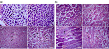
The gross ovarian stages and monthly ovarian development of P. argenteus were studied throughout the year and the results are summarized in Figure 3 (a–c). Each histological ovarian section was observed thoroughly and the different vitellogenic stages of ovarian maturation were described. The monthly changes of compositions of the oocyte stage were similar across the two years, and so were not discriminated for each year; overall results are shown in Figure 4A.
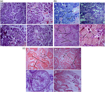
(a) Histological sections of gonads of female Pampus argenteus. Previtellogenic (A & B) and early primary vitellogenic stages (C & D). AT, atresia; OO, oogonia; CN, chromatin nuclear stage; PN, perinuclear oocyte; EO, empty ovary; GV, germinal vesicle; YV, yolk vesicle; YG, yolk granule; ZR, zona radiata. Scale bar = 50 μm. (b) Histological sections of female Pampus argenteus ovary. Advanced primary vitellogenic (B) and secondary vitellogenic stages (C & D). BV, blood vessel; GV, germinal vesicle; OC, ovarian cavity; PN, perinuclear oocyte; POF, post-ovulatory follicle; VO, vacuoles; YG, yolk granule; YV, yolk vesicle; ZR, zona radiata. Scale bar = 50 μm. (c) Histological sections of Pampus argenteus ovary. Ovary showing ripe stages (A & B), whereas (C) indicates fully ripe eggs with globules and oil droplets and (D) spent ovary. GV, germinal vesicle; YG, yolk granule; YV, yolk vesicle; ZR, zona radiata. Scale bar = 50 μm.

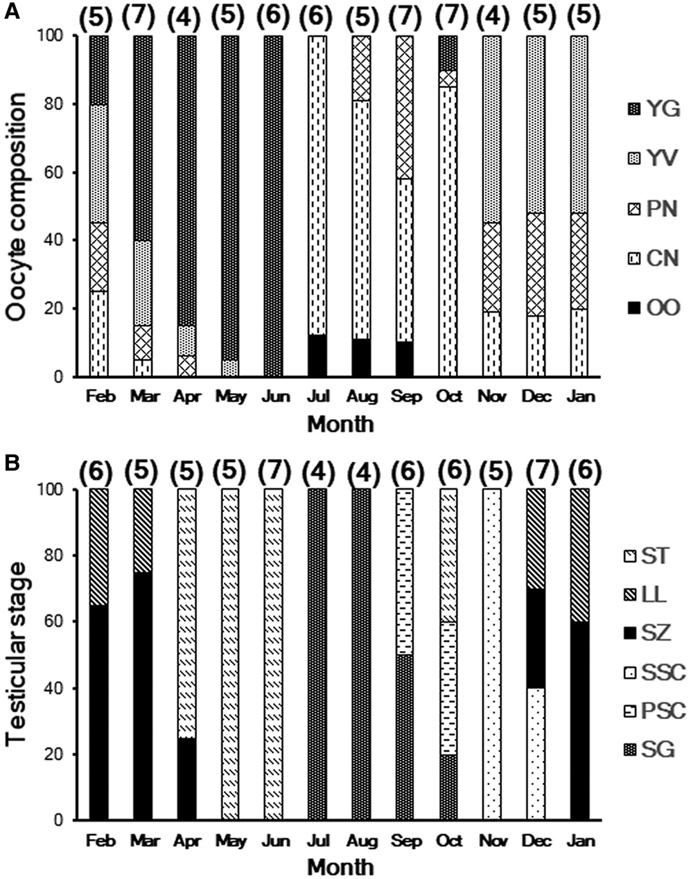
Oocyte (A) and testicular (B) composition of Pampus argenteus collected from the Bay of Bengal. Oocyte compositions are indicated as OO, oogonia; CN, chromatin nuclear stage; PN, perinuclear stage; YV, yolk vesicle stage; YG, yolk granule stage. Testicular stages are indicated as follows: SG, spermatogonia; PSC, primary spermatocyte; SSC, secondary spermatocyte; SZ, spermatozoa; LL, lobular lumen; ST, spermatid. Numbers in parentheses represent the number of fish analysed in each group.

Previtellogenic stage
This stage was characterized by a number of oogonia, chromatin nucleus, early perinucleus, and number of primary growth oocytes. The oocytes were very small in diameter with large and bright nuclei that were surrounded by basophilic cytoplasm, which was heavily stained with haematoxylin. This stage is often called the primary growth phase or immature stage of ovarian development, and the average oocyte diameter in this species was 42.5 ± 4.78 μm (n = 37). This stage was predominant from July to September (Figure 3a (A, B)).
Early primary vitellogenic stage
At this stage, some late perinucleus oocytes were found, but yolk vesicles appeared in the cytoplasm in the majority of oocytes. At the beginning of the vitellogenic stage, the oocyte diameter increased compared with that in the previous stage and became transparent and pinkish in colour. Blood capillaries were still not clear in this stage. The diameter of the ova was 57.5 ± 7.81 to 98.5 ± 28.7 μm (n = 38). This stage is sometimes called the early-maturing or secondary growth phase of the oocyte and is usually observed from November–January (Figure 3a (C, D), 3b (A)).
Advanced primary vitellogenic stage
The average size of the ova at this stage was 173.9 ± 10.12 μm (n = 19). The number and size of the yolk vesicle were increased compared with those in the former stages. The zona radiata became visible in the periphery at the end of this stage. Some advanced oocytes reached the primary globular stage. Oocytes appeared more rounded with small vacuoles. Ova were visible to the naked eye and the nuclei were partly hidden by the yolks. This is the development stage of the ovary which was primarily observed in February and March (Figure 3b (B)).
Secondary vitellogenic stage
The secondary vitellogenic stage was accompanied by the accumulation of eosinophilic yolk globules in the inner cortex. Yolk globules, sometimes with large oil droplets, mostly covered the cytoplasm. The nuclei contained some peripheral nucleoli. The zona radiata increased its thickness compared with that in the previous stage. The ova in the secondary vitellogenic stage were spherical, yellowish in colour, and could be counted using the naked eye. Blood vessels appeared on the surface of the ovary. The ova were filled with yolk and the average diameter was 286.5 ± 34.1 μm (n = 18). This stage is often known as the early yolk granule stage or early-maturing stage of ovarian development and was observed from March–April (Figure 3b (C, D)).
Ripe stage
The size of the oocyte increased rapidly by absorbing water to become fully hydrated. Post-ovulatory follicles (Figure 3b (D)) often co-exist with the yolk granules at this stage. The nuclei moved toward the animal pole and oil droplets merged together and formed a single oil droplet. The ovary was fully grown in this stage, light yellow in colour, and occupied the entire body cavity. Blood vessels were conspicuous in this stage. Large ova were often expelled from the vent with the application of slight pressure to the belly. For P. argenteus, this stage was more prominent in May and June (Figure 3c (A, B)). Some mature eggs with increased GSI values were also noticed in some females in October (Figure 3a (D)).
Spent or regressing stage
The weight of the ovary drastically decreased because of the release of eggs. Ovaries were shrunken, hollow, sac-like structures; small numbers of immature eggs were sometimes observed with denatured ova. Some post-ovulatory follicles were also seen at this stage. The spent fish were found in July. At this stage, some mature eggs also persisted in certain fish, indicating the possibility of a second spawning.
Monthly changes in the oocyte stages
Monthly changes in the oocyte compositions were found to vary depending on the stage of maturity and to have similar compositions across the two-year study period (Figure 4A), therefore, two-year samples were not discriminated during further observation. The chromatin nuclear (CN) and perinuclear (PN) stages were found almost throughout the year; however, their percentages gradually decreased upon development. The yolk vesicle (YV) stage was predominant from November–February, while after February, the yolk granule (YG) stage appeared. About 85% of the ovary was filled with yolk granules; this percentage increased to almost 100% by May. Oogonia (OO), along with the CN stage, were predominant from July.
Maturity stages in male
For histological observations, the degree of spermatogenesis and the annual testicular activity of P. argenteus were divided into the following developmental stages and the monthly testicular stages are presented in Figure 4B.
Immature stage
The testes in this stage were small, transparent, and pale yellow in colour. They occupied a very small part of the body cavity. The seminal lobules of the testes contained high numbers of spermatogonia and low numbers of spermatocytes. Spermatogonia were spherical in shape and stained with haematoxylin. Cells forming cysts were usually surrounded by Sertoli cells. The GSI value of the immature testes was 0.12 ± 0.04 (n = 31). This stage was mainly seen in July and August (Figure 5a (A)).
(a) Histological sections of testes of Pampus argenteus. Immature (A) and developing (B, C & D) testes. GE, germinal epithelium; LL, lobular lumen; PSC, primary spermatocyte; SG, spermatogonia; SSC, secondary spermatocyte; RT, regressing testes; SZ, spermatozoa. Scale bar = 50 μm. (b) Histological sections of testes of Pampus argenteus. Pre-spawning (A), ripe and spawning (B & C), and spent (D) stages of testes of Pampus argenteus. LL, lobular lumen; RS, residual spermatids; SZ, spermatozoa; ST, spermatids; TL, testicular lamellae. Scale bar = 50 μm.

Developing stage
The testes were slightly larger than those in the immature stage, and flat and translucent in colour. The germinal epithelium was seen throughout the testes in this stage. Spermatocyte formation was initiated. Secondary spermatogonia, primary spermatocytes and secondary spermatocytes were also observed. Spermatogonia had noticeable basophilic nucleoli and very faint stains; some became degenerated. No spermatozoa were present in the lumen. This stage was observed from September–December and the GSI values during this stage ranged from 0.17 ± 0.06 to 0.24 ± 0.07 (n = 74) (Figure 5B–D).
Pre-spawning stage
Testes were in the late development stages with spermatozoa in the lumina of the sperm ducts. All stages of spermatogenesis, such as spermatogonia, spermatocytes and spermatids, were usually seen in this stage; among them, spermatocytes were predominant in the sperm tissue. Many cysts containing spermatocytes were seen throughout the seminal lobule. Testes were creamy white, soft, and occupied 70% of the body cavity. The pre-spawning stage was usually observed from January to March and the GSI values ranged from 0.27 ± 0.08 to 0.33 ± 0.15 (n = 62) (Figure 5b (A)).
Ripe stage
Increases in the number of spermatids in the seminal lobules and spermatozoa in the lobular lumina and sperm ducts were observed. The number of spermatogonia and spermatocytes decreased with an increase in the number of spermatids (Figure 5b (B, C)). The testes of P. argenteus became ripe in April and were ready for spawning; at this time, sperm were not expelled from the vent by gentle pressure on the belly, which can help to distinguish the ripe male.
Ripe males were observed from May–June. Testes in this period were translucent and white, with some blood spots. At this time, sperm was expelled from the vent by gentle pressure on the belly. The testes were large in size and dominated by mature spermatozoa in the peripheral and central sperm ducts and throughout the lumina. An increase in the amount of sperm was found in October. At this time, mature spermatozoa were also observed in some males, which indicates the possibility of biannual spawning.
Spent stage
During this stage, the testes were flabby and shrank considerably because of the release of milt and a reduced chance of milt replenishment. The lobular walls became thicker. Blood vessels appeared dilated and blood cells were scattered. Some residual spermatozoa were sometimes seen in the lumina and sperm ducts, while spermatocytes and spermatids were not observed. Again, spermatogonia proliferation and germinal epithelium regeneration were initiated in the periphery of the testes (Figure 5b (D)). The GSI values were drastically reduced and the lowest values in the reproductive cycle were observed.
Monthly changes in testicular stages
The major testicular stages of P. argenteus were found to vary between months; particular stages were dominant in specific periods (Figure 4B). Similar compositions across the two-year study period were observed, therefore, the two-year samples were not discriminated. Spermatogonia (SG) were completely dominant from July–September, while in October, primary spermatocytes (PSCs) along with mature spermatids (~40%) were observed in the testes of P. argenteus. Spermatozoa (SZs) began to develop from December and continued their development until March. Mature spermatids (ST) abruptly appeared in April (~75% of the testes) and remained full (100%) until June.
Discussion
The present study observed gonadal maturation patterns of P. argenteus in the Bay of Bengal, Bangladesh and showed its reproductive seasonality, implying two spawning peaks: one in May–June and another in October. Further, the absolute and relative fecundity were estimated for this species which could be useful to estimate the potential stocks from the Bay of Bengal, Bangladesh.
In this study, the oocyte diameter and GSI value in both sexes showed a clear gonadal development pattern in P. argenteus. Histological analysis is the most accurate method for analysing gonadal maturity without error (West, Reference West1990), and our observations indicated that the gonadal maturity of P. argenteus increased from April to June in the Bay of Bengal. Ovarian development was similar to that described previously in some other Stromateidae species (Lone et al., Reference Lone, Ablani and Almatar2008a, Reference Lone, Ablani and Almatar2008b; Amparo et al., Reference Amparo, Cárdenas, Guevara and Pérez2017). Large proportions of the ovary were filled with yolk granules and the testes were filled with mature spermatids from May to June (Figure 4). The composition of oocytes at any particular stage contributes to changes in the development of the reproductive stage (Hinckley, Reference Hinckley1987). In July, a number of atretic follicles and empty spaces in the ovary were observed by histological analyses. Small numbers of oogonia in females and spermatogonia in males also began to develop in July, which indicated the initiation of gonadal development. CN and perinuclear oocytes appeared in September and continued their development until February, when YGs appeared. In May, multiple oil droplets were also fused together to form single droplets and mature eggs were expelled with slight pressure on the belly in May and June. These results suggest that the maturation of ovaries peaked in May and June. One of the most accurate methods of detecting fully mature fish is to observe the post-ovulatory follicles (POFs) (Arocha & Barrios, Reference Arocha and Barrios2009). A number of POFs were observed in the ovaries at the end of April. Some yolk granule-stage eggs were also observed in October in some fish, as well as those in the CN or perinuclear stages. POFs were also seen in the ovaries, which strongly suggests the biannual spawning potential of this species. In males, mature spermatids were found from April–June, which indicates that males reach maturity earlier than females. This study confirmed synchronized gonadal maturity in males and females, and the possibility of biannual spawning for the stock in the Bay of Bengal, with spawning in May–June and October.
The results of this study, which show two spawning peaks, are in accordance with several previous studies of P. argenteus in some areas. In the same region as this study, the Bay of Bengal, off the Orissa coast, India, Pati (Reference Pati1982) reported the maturation and spawning of P. argenteus by analysing the fecundity, and reported two spawning periods: one extended from February to April with a peak in April and the other extended from June to August with a peak in August. In the present study, in addition to fecundity, the oocyte diameter and histological examinations of gonads were conducted throughout the year and two spawning peaks were observed in the Bay of Bengal population of P. argenteus. In Kuwait waters, the minimum GSI for females (0.69 ± 0.11) was reported in January and the maximum was reported in June (7.90 ± 0.04), with a prolonged spawning period from June to October, with peak spawning in June (Lone et al., Reference Lone, Ablani and Almatar2008a). In another study in Kuwait waters, two spawning peaks (April and August) were also reported by Abu-Hakima et al. (Reference Abu-Hakima, Al-Abdul-Elah, El-Zahr and Akatsu1983) for this species. Almatar et al. (Reference Almatar, Lone, Abu-Rezq and Yousef2004) reported the extended spawning of silver pomfret from mid-May to early October in Kuwait waters. Though it is not clear whether they have two spawning peaks or an extended one in Kuwait waters, the slight difference in the spawning seasons between these studies may be because of the geographic position of the spawning ground, the environment, and the conditions of fish. Dadzie et al. (Reference Dadzie, Seedo and Shallal2000b) reported spawning peaks in May and August in both males and females in the Arabian Gulf. The spawning of P. argenteus in the East China Sea was reported to be from May–July (Lee & Jin, Reference Lee and Jin1989). Therefore, two spawning seasons might be common in this species, which is in accordance with the results of this study.
In this study, the significant dominance of the YG stage in females and mature spermatids in males was observed from May to June. Moreover, histological analyses showed the presence of yolk granules and spermatids in October, which indicated the probability of a second spawning. Several authors have reported the prolonged spawning seasons of other butterfishes: Pampus chinensis from September–November in Orissa, India (Pati, Reference Pati1979); Parastromateus niger from June–September and from January–February in India (Bapat et al., Reference Bapat, Deshmukh, Krishnamoorthi, Muthiah, Kagwade, Ramamirtham, Mathew, Krishna and Mukundan1982) and Pampus echinogaster from March–July in Korea (Oh et al., Reference Oh, Na and Kim2009). Spawning seasons are variable and species-specific and could be related to variations in the water temperature, photoperiod, spawning grounds and water currents. Water temperature may play a role in determining the breeding seasons in butterfishes, including those of P. argenteus. Jaswal et al. (Reference Jaswal, Singh and Bhambak2012) reported the monthly sea surface temperature (SST) over 40 years in the Bay of Bengal; their data have clear fluctuation patterns, with a peak in the water temperature in May–June and a sharp decrease after October. This matches the breeding pattern of P. argenteus in the Bay of Bengal, showing peaks from May–June and in October. Thus, increasing water temperature may be a trigger for the spawning of this species. In this study, a clear peak in the GSI in October was observed, along with the YG stage and mature spermatids, which confirms the two spawning peaks.
Fecundity is important to assess the reproductive potential of fish stocks (Islam et al., Reference Islam, Begum, Pal and Alam2008; Begum et al., Reference Begum, Islam, Pal and Alam2010). The fecundity estimated from randomly collected gravid fish varied from 26,109–350,371 in the Bay of Bengal, Bangladesh. Pati (Reference Pati1981) reported that the fecundity of P. argenteus in the Bay of Bengal ranged from 40,610–90,460 in fish (19.1–23.8 cm SL) from India. Dadzie et al. (Reference Dadzie, Seedo and Shallal2000b) also found that the fecundity of P. argenteus ranged from 28,965–455,661 (mean 164,558) in Kuwait waters, which is similar to the results of this study. Dadzie et al. (Reference Dadzie, Seedo and Shallal2000b) also reported that there were no strong correlations between the SL and fecundity; however, in this study, a positive correlation between the SL and fecundity was observed (Figure 2B). Variations in fecundity might be because of the differences in stocks, size of fish, and nutritional status of the respective habitats.
Conclusion
A clear confirmation of the development of gonadal maturation and gamete production is crucial for egg production, and hence is vital for fisheries management, stock assessment, artificial breeding and aquaculture. The results of this study provide the detailed maturation cycle and spawning seasons of P. argenteus in the Bay of Bengal, which are important for artificial breeding programmes and fishery management strategies. This study also provides a basis for further research on reproductive parameters, embryological and larval studies, and could help in the successful conservation of this species. Further investigations of spawning in controlled conditions, determination of gonadotropin levels throughout the year, and direct observations of the spawning behaviour will confirm the maturation profile of this species.
Acknowledgements
The authors wish to express their gratitude to the UGC-CVASU of Bangladesh for funding this research. The authors also express sincere thanks to the fishers who helped to collect samples. This study is a contribution from the Coastal Biodiversity, Marine Fisheries and Wildlife Research Centre of Chattogram Veterinary and Animal Sciences University. We also express our cordial thanks to T. Kitahashi (Niigata University) for his invaluable comments on the manuscript and all of the laboratory personnel of the Fish Biology and Biotechnology Department, CVASU, for their kind support.
Financial support
This work was financially supported by the University Grants Commission (UGC) of Bangladesh and Chattogram Veterinary and Animal Sciences University, Chattogram, Bangladesh.
Conflict of interest
The authors declare no conflict of interest.