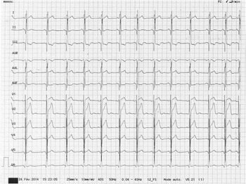
An asymptomatic 19-year-old man, without any familial or past personal cardiovascular history, was referred for a routine check-up before enrolling in a career in the army. The patient’s electrocardiogram (Fig 1) showed left posterior hemiblock, Q waves in inferior territories, and highly peaked T waves. Echocardiography showed a normally connected heart with thin left ventricular walls in the inferior and infero-lateral regions and associated hypokinesia, as well as structures described as “pillars”. No other anomaly was observed. Left ventricular ejection fraction was normal, calculated at around 50%. At this stage, the diagnosis remained unclear.
Left posterior hemiblock, Q waves in inferior territories, and highly peaked T waves.

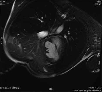
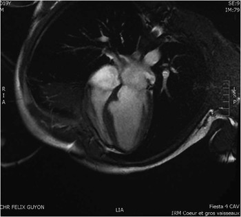
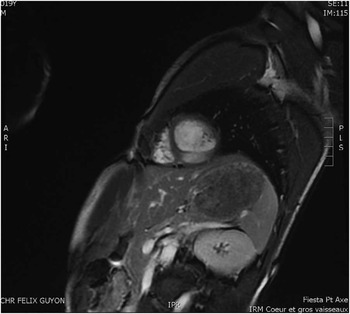
Thus, a cardiac MRI was performed that indeed confirmed the dysmorphic infero-septal muscular wall, without segmental kinetic change and normal left ventricular ejection fraction. No abnormal delayed enhancement was observed, neither endocardial nor myocardial. No trabeculations or fibrosis were observed, and no perfusion defects or evidence of thrombus formation were detected (Figs 2–4). On the basis of the MRI data, therefore, possible differential diagnoses such as non-compaction and aneurysm were ruled out. A congenital left ventricular diverticulum was the most likely diagnosis. Blood-thinning medication, so far, has not been deemed necessary owing to the diverticulum’s low profile, normal contractile function, and therefore low risk of thrombosis. This patient continues to be followed up with regular clinical assessment, electrocardiograms, and echocardiographic studies.
Four-chamber view. Pseudoaneurysm shape in the septum, without enhancement. No delayed enhancement and thus no fibrosis. Muscular diverticulum.

Four-chamber view. Left ventricular septal muscular diverticulum. Its small size does not harm right ventricular function.

Small-axis view. Isolated pseudo-trabeculation of the muscular diverticulum. No enhancement.

The aetiology of cardiac diverticula, their incidence, and natural history remain unclear. Most left ventricular diverticula are found at the left ventricular apex.1 Left-sided diverticula can be isolated and idiopathic, or be part of a syndrome such as pentalogy of Cantrell. They may also be associated with a congenital heart malformation such as tetralogy of Fallot, pulmonary atresia, or a ventricular septal defect, and in such cases they can be diagnosed prenatally or in infancy. A stand-alone diverticulum is considered benign, and its main complication is thrombosis. Depending on its size and location, a third-chamber effect or rupture may occur. Cases associated with ventricular arrhythmia have also been reported.
Acknowlegdements
We would like to thank Van Hung Chuong, MD, Sophie Morestin, MD, and Jean-Pierre Laissy, MD, PhD, for their role in the care of patient and counselling.
Financial Support
None.
Conflicts of Interest
None.