Introduction
The genus Belondira Thorne, 1939, with nearly 40 valid species, is a widespread dorylaimid taxon that has been recorded in many countries and territories, mostly in tropical areas. The study of its diversity has not received much attention during past decades. No monographic contribution has been published since the excellent revision by Ferris et al. (Reference Ferris, Ferris and Goseco1983), although regional studies of new and known Belondira species were provided by Shaheen & Ahmad (Reference Shaheen and Ahmad2005) for Costa Rica and by Naz & Ahmad (Reference Naz and Ahmad2009) for India, with an updated key to species identification. Interestingly, and more recently, Wu et al. (Reference Wu, Huang, Xie, Wang and Xu2016) have described a new species from China, including molecular data.
The study of nematode fauna from Iran has undergone very relevant progress thanks to the work of several nematological teams, who are discovering an incredible and unsuspected diversity of both free-living and plant-parasitic forms. In the course of several nematode surveys conducted in West Azerbayjan Province (north-western Iran), a population of Belondira was recovered from soil samples. Its detailed study has revealed that it belongs to a non-described species, which is characterized and described below. Besides, it represents the second species of this genus for which any DNA sequence data are available.
Materials and methods
Sampling, extraction, mounting and drawing
Nematodes were collected from different locations in 2014, extracted from soil samples by an improved Baermann funnel method (Whitehead & Hemming, Reference Whitehead and Hemming1965), heat-killed by adding boiling 4% formalin solution, and transferred to anhydrous glycerin, according to De Grisse (Reference De Grisse1969). Permanent slides were made and examined using an Olympus BH2 light microscope (Olympus, Tokyo, Japan). Pictures were taken using a Nikon Eclipse 80i light microscope provided with differential interference contrast (DIC) optics a DS-5M camera connected to a Nikon DS-U1 control box (Nikon, Tokyo, Japan) and a drawing tube. Images were edited using Adobe® Photoshop® CS. Drawings were prepared using the drawing tube (camera lucida) attached to the Nikon microscope.
DNA extraction, PCR and sequencing
Nematode DNA was extracted from single individuals and polymerase chain reaction (PCR) assays were conducted as described by Heydari et al. (Reference Heydari, Tanha Maafi and Decraemer2014). Single live nematode specimens from Belondira coomansi n. sp. were selected, examined in a temporary slide and transferred to a small drop of AE buffer (10 mm Tris-Cl, 0.5 mm EDTA; pH 9.0; Qiagen, Venlo, The Netherlands) on a clean slide and cut into small pieces using a sterilized razor blade. The suspension was collected by adding 20 μl AE buffer and 2 μl proteinase K (600 μg/ml) (Promega, Leiden, The Netherlands). The tubes were incubated at 65°C (1 h), then at 95°C (15 min) and finally at −80°C (15 min). One microlitre of extracted DNA was transferred to an Eppendorf tube containing: 2.5 μl 10× NH4 reaction buffer, 0.75 μl MgCl2 (50 mm), 0.25 μl deoxynucleoside triphosphate mixture (dNTPs; 10 mm each), 0.75 μl of each primer (10 mm), 0.2 μl BIOTAQ DNA Polymerase (Bioline, London, UK) and double-distilled water to a final volume of 25 μl. Primers for 28S rRNA gene (D2–D3 domain) amplification were: D2A (forward; 5′-ACAAGTACCGTGAGGGAAAGTTG-3′) and D3B (reverse; 5′-TCGGAAGGAACCAGCTACTA-3′) (De Ley et al., Reference De Ley, Félix, Frisse, Nadler, Sternberg and Thomas1999). The thermal cycling program was as follows: denaturation at 95°C for 2 min; followed by 35 cycles of denaturation at 94°C for 30 s, annealing at 55°C for 40 s and extension at 72°C for 80 s. A final extension step was performed at 72°C for 10 min. The PCR product was sequenced using the same primers with an ABI 3730XL sequencer (Bioneer Corporation, South Korea). The sequence was deposited in the GenBank database under accession number MF363124 (D2–D3 region of the 28S rRNA gene).
Phylogenetic analyses
The newly obtained sequence was aligned with other D2–D3 sequences available from GenBank using Dialing-TX alignment (Subramanian et al., Reference Subramanian, Kaufmann and Morgenstern2008). Outgroup taxa were chosen according to the results of previously published data. Sequence alignments were edited manually using BioEdit 7.2. (Hall, Reference Hall1999). The sequence dataset was analysed with Bayesian inference (BI) using MrBayes 3.2.6 (Huelsenbeck & Ronquist, Reference Huelsenbeck and Ronquist2001; Ronquist & Huelsenbeck, Reference Ronquist and Huelsenbeck2003). The analysis under the GTR model was initiated with a random starting tree and run with the Markov chain Monte Carlo (MCMC) method for 106 generations. For phylogenetic relationships, analyses were based on 28S rDNA. Coomansus gerlachei (accession number KM092524) was used as the outgroup. The tree was visualized and saved using the program FigTree v1.4.3 (http://tree.bio.ed.ac.uk/software/figtree/).
Results and discussion
Belondira coomansi n. sp.
Description
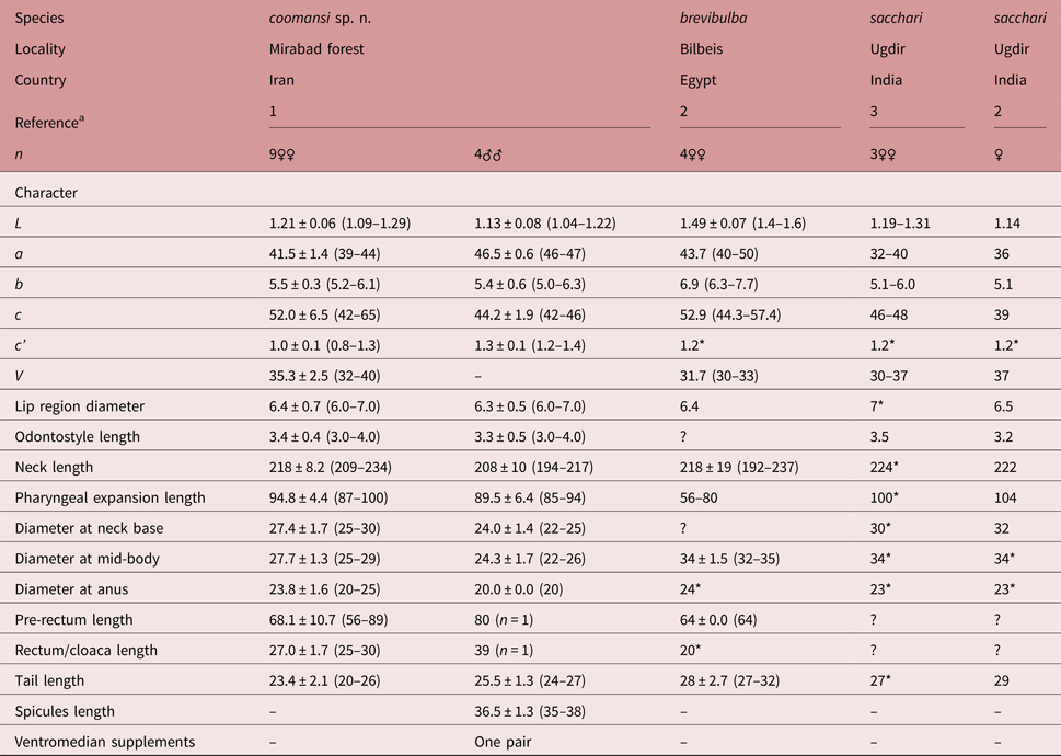
Measurements. See table 1.
Table 1.Morphometric data of Belondira coomansi n. sp. and similar species. Measurements in μm (except L, in mm), and in the form: mean ± standard deviation (range).

a Reference: 1, present paper; 2, Ferris et al. (Reference Ferris, Ferris and Goseco1983); 3, Suryawanshi (Reference Suryawanshi1972).
n, number of specimens observed; L, body length; a = L/maximum width; b = L/pharyngeal length; c = L/tail length; c' = tail length/body diameter at anus; V = distance of vulva from anterior end × 100/L.
* Morphometrics calculated from original illustrations and/or other original morphometrics.
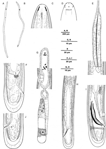
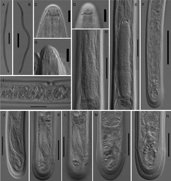
Adult. Slender to very slender (a = 39–47) nematodes (figs 1 and 2) of medium size, 1.04–1.29 mm long. Body cylindrical, visibly tapering towards the anterior end, less so towards the posterior end as the tail is broadly rounded. Upon fixation, body nearly straight to somewhat sigmoid. Cuticle two-layered, 1.0 μm thick at anterior region, 1.0–1.5 μm in mid-body and 7.0–9.0 μm on tail; outer layer thin, bearing fine but perceptible (even with light microscopy) transverse striation, and distinctly expanded at caudal region (see below); inner layer also thin throughout the body and thickened at caudal region. Lateral chord 5.5–7.5 μm wide, occupying one-quarter (23–25%) of mid-body diameter, and lacking any differentiation. Body pores, if present, obscure. Lip region narrow, rounded and totally continuous with the adjacent body; it is 1.8–2.1 times wider than high, and one-fifth to one-third (26–33%) of the body diameter at neck base; lips completely amalgamate, with inconspicuous papillae; labial and post-labial sclerotizations present, forming a perceptible cephalic framework. Amphid cup-shaped, its aperture 4.0–4.5 μm long, occupying up to three-quarters (53–60%) of lip region diameter. Odontostyle very short, thin, with narrow lumen and small aperture; it is about seven times as long as wide, less than one-half (0.44–0.47 times) of the lip region diameter long, and 0.27–0.33% of body length. Guiding ring apparently simple, at c. 6.0 μm from the anterior end. Odontophore difficult to observe and to measure with accuracy in the specimens examined. Pharynx typical of the genus, i.e. consisting of a slender and weakly muscular anterior region enlarging gradually; and the basal expansion, which is 6.6–8.0 times as long as wide, 3.1–3.3 times longer than the body diameter at neck base, occupies 42–45% of the total neck length, and appears to be surrounded by a spiral muscular sheath with nearly straight (slightly dextral) muscular bands. Cardia rounded, conoid, 9.5–11.5 × 5.5–7.0 μm. Pre-rectum 3.7–4.0 anal body diameters long. Tail rounded, even slightly clavate; outer cuticle layer distinctly expanded at the end, forming a hyaline space 5.0 μm thick in females and 3.5 μm thick in males, with less perceptible radial striation; inner cuticle layer also visibly thickened at tail tip.
Fig. 1.Line drawings of Belondira coomansi n. sp.: (A) female entire; (B) male, entire; (C) anterior region in median, lateral view; (D) lip region in surface, lateral view; (E) pharyngeal expansion; (F, J) female, caudal region; (G) female, genital system; (H) female, posterior body region; (I) male, caudal region and spicule.
Fig. 2.Light micrographs of Belondira coomansi n. sp.: (A) female, entire; (B) male, entire; (C, G) anterior region in median, lateral view; (D) lip region in surface, lateral view; (E) pharyngeal expansion; (F) female, posterior body region; (H) spiral muscular sheath of the pharyngeal expansion; (I) vagina and anterior uterine sac; (J–L) male, caudal region and spicules; (M, N) female, caudal region. Scale bars: (A, B) 200 μm; (C, D, G) 5 μm; (E, H–N) 20 μm; (F) 50 μm.
Female. Genital system mono-opisthodelphic. Anterior genital branch reduced to a uterine sac 47–70 μm long or 1.5–2.7 times the body diameter. Posterior branch well developed, 151–250 μm long or 12–20% of total length: oviduct 109–110 μm or 3.5–3.7 body diameters long; uterus very short, 15–16 μm or c. one-half of body diameter long. Vagina extending inwards 13–14 μm, occupying hardly less than one-half (43–45%) of body diameter: pars proximalis 8.5 × 13.0 μm, nearly hemispheroid; pars distalis 3.0–4.5 μm. Vulva apparently an oval transverse orifice. Rectum 22–30 μm or 1.0–1.3 times the anal body diameter long.
Male. Genital system diorchic, with opposite testes. Cloaca 39 μm or 2.0 body diameters long. In addition to the ad-cloacal pair, situated at 4.0 μm from the cloacal aperture, there is one pair of subventral supplements at 11.5–13.0 μm from the ad-cloacal pair, within the range of the spicules. Spicules slender, 8.0–8.8 times as long as wide, and 1.6–1.7 times the body diameter at level of the cloacal aperture; head 3.5–4.0 × 2.0–3.0 μm, occupying c. one-tenth (11%) of total length, and with its sides of similar length; median piece very thin; both hump and hollow hardly perceptible; posterior end 1.5–2.0 μm broad; curvature 150°. Lateral guiding pieces inconspicuous.


Taxonomic summary
Etymology. The species is named after Professor August Coomans (Ghent University, Belgium), the leading expert in the study of the morphology and taxonomy of dorylaims.
Type locality and habitat. The new species was collected in Mirabad forest, near Sardasht city, West Azarbayjan Province, north-western Iran (36°29′51.34″N, 45°19′2.43″E; 1284 m above sea level) from soil around the rhizosphere of unidentified grasses.
Type material. Female holotype (slide BBC001) and six (4 female and 2 male, slides BBC001 and BBC002) paratypes deposited in the Nematode Collection of the Department of Plant Protection, College of Agriculture and Natural Resources, University of Tehran, Karaj, Iran. Two female and two male paratypes at the Nematode Collection, University of Jaén, Spain. Two female paratypes deposited in the National Nematode Collection of the Department of Nematology, Iranian Research Institute of Plant Protection, Tehran, Iran.
Molecular characterization
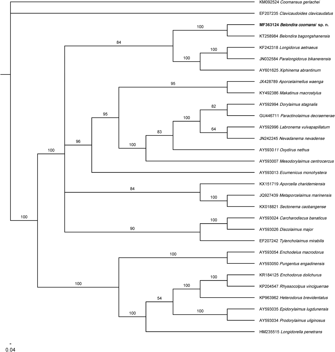
A 727 bp long sequence of the D2–D3 region of 28S rDNA has been obtained for the new species. The corresponding BLAST search shows a similarity value of 82% with Belondira bagongshanensis Wu, Huang, Xie, Wang & Xu, 2016 (accession number KT258984), the only Belondira sequences hitherto available from GenBank, differing from it in 136 nucleotides (fig. 3).
Bayesian tree derived from the molecular analysis of D2–D3 of 28S rDNA.

Diagnosis and relationships
Belondira coomansi n. sp. is characterized by its body length (1.04–1.31 mm); lip region continuous and 7.5–8.0 μm broad; presence of labial and post-labial sclerotization; odontostyle 3.0–4.0 μm long, with lumen and aperture inconspicuous; neck 194–234 μm long; pharyngeal expansion 85–100 μm long and occupying 42–45% of the total neck length; female genital system mono-opisthodelphic, anterior branch reduced to a uterine sac 45–70 μm long or 1.5–1.8 times the body diameter, posterior uterus 15–16 µm long or one-half of body diameter, V = 33–39; tail short and rounded in both sexes (20–26 μm, c = 42–57, c′ = 0.8–1.3) with the cuticle 6.5–9.0 μm thick at its tip and its hyaline layer 3.5–5.0 μm thick; spicules 32–38 μm long and only one ventromedian supplement pair at 11.5–13.0 μm from the ad-cloacal pair.
From a morphological perspective, and in having a small and slender (with no distinct lumen and aperture) odontostyle, the new species resembles Belondira brevibulba Ferris, Ferris & Goseco, 1983, Belondira sacchari Suryawanshi, 1972, Belondira tenuidens Thorne, 1964 and Belondira thornei Suryawanshi, 1972. It differs from B. brevibulba, from Egypt, in its smaller general size (body length 1.04–1.31 vs. 1.4–1.6 mm), longer pharyngeal expansion (85–100 μm or 42–45% of the total neck length vs. 56–80 μm or about 30% of the total neck length), larger female anterior genital branch (1.5–1.8 vs. less than 1.0 times the body diameter; 3.6–4.2 vs. 1.3–2.0% of total body length), and male as frequent as female (vs. male unknown). It differs from B. sacchari, which occurs in India (see also re-description by Ferris et al., Reference Ferris, Ferris and Goseco1983), in its shorter pharyngeal expansion (85–100 μm or 42–45% of the total neck length vs. 104 μm or 47% of total neck length), larger female anterior genital branch (1.5–1.8 vs. about 1.0 times the body diameter, 3.6–4.2 vs. 1.4% of total body length), shorter female tail (23–26 vs. 27–29 μm) with thickened outer layer not bearing (vs. bearing) distinct radial striation, and male known (vs. unknown, females without sperm cells).
The new species differs from B. tenuidens (see also re-description by Ferris et al., Reference Ferris, Ferris and Goseco1983) in its shorter odontostyle (3.0–4.0 vs. 5–6 μm), longer pharyngeal expansion in females (85–100 μm or 42–45% of the total neck length vs. 64–83 μm or about 33–39% of the total neck length), larger female anterior genital branch (1.5–1.8 vs. about 1.0 times the body diameter, 3.6–4.2 vs. 1.8–3.0% of total body length), caudal region bearing (vs. lacking) a thickened outer layer at its tip, and male ventromedian supplement located a short distance in front of the ad-cloacal pair, within the range of the spicules (vs. an appreciable distance in front of the ad-cloacal pair, outside the range of the spicules). The new species is also distinguishable from B. thornei (see also description by Ferris et al., Reference Ferris, Ferris and Goseco1983), a very similar species, in its shorter odontostyle (3.0–4.0 vs. 5–6 μm), larger pharyngeal expansion (85–100 μm or 42–45% of the total neck length vs. 67–83 μm or 38–41% of total neck length), and caudal region with thickened outer layer not bearing (vs. bearing) distinct radial striation.
The phylogenetic relationships of the new species, as derived from the molecular analysis, are presented in fig. 3. It forms a highly supported clade with B. bagongshanensis, as mentioned above, the only species of the genus for which the D2–D3 segment was available. The relationships of Belondira species with other dorylaimid taxa are not elucidated as they form a less-supported clade with members of the family Longidoridae, all of them included in a large group with deficiently resolved phylogeny.
Remarks
The new species belongs to a group of very similar species, with intricate delimitation. Its very short odontostyle (3.0–4.0 μm) apparently separates it from B. thornei and B. tenuidens (odontostyle 5–6 μm long). Relative to B. brevibulba and B. sacchari, it might form a west–east cline with a gradual increasing of the pharyngeal expansion: 56–80 μm or about 30% of the total neck length in B. brevibulba (from Egypt), 85–100 μm or 42–45% in B. coomansi n. sp. (Iran) and 100–104 μm or 47% in B. sacchari (India).
Financial support
This study was supported financially by the University of Tehran.
Conflict of interest
None.