I. INTRODUCTION
The continuing demand for environmentally friendly alternative energy technologies has led to much increased research activity in the area of thermoelectric research. For high temperature waste heat conversion applications, low-dimensional layered oxides have been found to have relatively high efficiency. The efficiency and performance of thermoelectric energy conversion or cooling are related to the dimensionless figure of merit (ZT) of the thermoelectric (TE) materials, given by ZT = S 2 σT/κ, where T is the absolute temperature, S is the Seebeck coefficient or thermoelectric power, σ is the electrical conductivity, and k is the thermal conductivity (Nolas et al., Reference Nolas, Sharp and Goldsmid2001). Examples of these oxides include NaCoO x (Terasaki et al., Reference Terasaki, Sasago and Uchinokura1997), Ca2Co3O6 (Mikami et al., Reference Mikami, Funahashi, Yoshimura, Mori and Sasaki2003; Mikami and Funahashi, Reference Mikami and Funahashi2005), Ca3Co4O9 (Masset et al., Reference Masset, Michel, Maignan, Hervieu, Toulemonde, Studer and Raveau2000; Mikami et al., Reference Mikami, Itaka, Kawaji, Wang, Koinuma and Lippmaa2002; Grebille et al., Reference Grebille, Lambert, Bourée and Petricek2004; Hu et al., Reference Hu, Si, Sutter and Li2005; Wong-Ng et al., Reference Wong-Ng, Hu, Vaudin, He, Otani, Lowhorn and Li2007), and Bi2Sr2Co2O x (Wang et al., Reference Wang, Venimadhav, Guo, Chen, Li, Soukiassian, Schlom, Pan, Wong-Ng, Vaudin, Cahill and Xi2009). Among these materials, the most efficient material, Ca3Co4O9, is a misfit layered oxide that has two monoclinic subsystems with identical a, c, β, but different b (Masset et al., Reference Masset, Michel, Maignan, Hervieu, Toulemonde, Studer and Raveau2000). Consequently considerable research has been conducted on cobaltates for thermoelectric applications, including phase diagram studies of ternary oxide systems including CaO and Co3O4 as two of the end members (Wong-Ng et al., Reference Wong-Ng, Liu, Martin, Thomas, Lowhorn and Otani2010, Reference Wong-Ng, Luo, Tang, Xie, Kaduk, Huang, Yang, Tang and Tritt2011, Reference Wong-Ng, Laws and Yan2013, Reference Wong-Ng, Laws, Talley, Huang, Yan and Kaduk2014).
In order to have materials with high enough efficiency for large-scale industrial applications, ZT of 2 or higher is a requirement. The search for cobaltate compounds with improved thermoelectric properties continues worldwide. The Y-type (Ba2 M 2Fe12O22) and W-type (Ba x Sr1− x )Co2Fe16O27) hexagonal ferrites have been synthesized for the first time back in the 1960s by Vinnik (Reference Vinnik1965; Vinnik et al. Reference Vinnik, Agranovskaya and Erastova1966) during his study of the phase diagram and crystal chemistry of the BaO–CoO–Fe2O3 system. The structure of Ba2 M 2Fe12O22 (M = Co, Zn) was characterized by Townes and Fang (Reference Townes and Fang1970) and Collomb et al. (Reference Collomb, Hadj Farhat and Joubert1989a, Reference Collomb, Muller, Guitel and Desvignes1989b). The X-ray patterns for the Y-type Ba2Co2Fe12O22 ferrite have been reported by Shin and Kwon (Reference Shin and Kwon1993). Bashkirov et al. (Reference Bashkirov, Dudchik, But`ko, Kris`ko, Kunitskii, Petrov, Shershavina, Shichkova and Fedorova2001) further evaluated the Ba–Sr and Ni–Co solid-state interdiffusion coefficients of the cations in Ba2 M 2Fe12O22 and Sr2 M 2Fe12O22 (M = Ni, Co) by the Boltzmann–Matano method. The structure of the W-type hexagonal ferrites was first studied by Braun (Reference Braun1957). In 1986, Collomb et al. (Reference Collomb, Wolfers and Obradors1986a, Reference Collomb, LambertAndron, Boucherle and Samaras1986b, Reference Collomb, Abdelkader, Wolfers, Guitel and Samaras1986c) used neutron diffraction to study the structure and magnetic characteristics of a number of hexagonal ferrites, including the W-type BaMg2Fe16O27 and BaCo2Fe16O27.
The Y-type and W-type hexagonal ferrites have found various applications in areas, such as microwave instrumentation as permanent magnets and elements of magnetic memory (Sugimoto, Reference Sugimoto1982). Extensive research work has been performed using these ferrites as model systems. For example, the BaCo2− x Zn x Fe16O27 system was used to study the effects of magnetic crystallographic anisotropy upon the process of magnetic ordering (Naiden et al., Reference Naiden, Maltsev and Ryabtsev1990).
There are three goals for this paper. Firstly (Ba x Sr1− x )2Co2Fe12O22 and (Ba x Sr1− x )Co2Fe16O27 compounds are investigated for the effect of size of Sr substitution on the Ba site on their structure. Since X-ray diffraction (XRD) is a non-destructive technique for phase identification, XRD patterns are especially important for phase characterization; therefore the second goal of this investigation was to determine the experimental patterns for (Ba x Sr1− x )2Co2Fe12O22 (x = 0.2, 0.4, 0.6, 0.8), and (Ba x Sr1− x )Co2Fe16O27 (x = 0.2, 0.4, 0.6, 0.8), and to make them widely available through submission to the Powder Diffraction File (PDF). The third goal of this study is to estimate the thermoelectric properties for selected members of (Ba x Sr1− x )2Co2Fe12O22 and (Ba x Sr1− x )Co2Fe16O27.
II. EXPERIMENTAL
A. Sample preparation
Both series of samples were prepared from stoichiometric amounts of SrCO3, BaCO3, Co3O4, and Fe2O3 using solid state high-temperature techniques. The starting samples were mixed, pelletized, and heat treated in air at 750 °C for 1 day and subsequently annealed at 950 °C for 15 h, 980 °C for 80 h with intermediate grindings, and 1250 °C for 60 h with intermediate grindings. During each heat treatment, the samples were furnace cooled. The heat treatment process was repeated until no further changes were detected in the powder XRD patterns.
B. X-ray Rietveld refinements and powder reference patterns
The (Ba x Sr1− x )2Co2Fe12O22 (x = 0.2, 0.4, 0.6, 0.8) compounds were measured as sintered disks, which were mounted in a deep cell using modeling clay. The (Ba x Sr1− x )Co2Fe16O27 (x = 0.2, 0.4, 0.6, 0.8) series were mounted as ethanol slurries on a zero-background cell. The X-ray powder patterns of the former samples were measured on a Bruker D2 Phaser diffractometer (30 kV, 10 mA, 5°–130° 2θ in 0.024° steps, 1 s step−1) equipped with a LynxEye position-sensitive detector (the purpose of identifying the equipment in this paper is to specify the experimental procedure. Such identification does not imply recommendation or endorsement by the National Institute of Standards and Technology). For all samples, the lower window of the detector electronics was increased from its default value of 0.11–0.19 V to minimize the effects of fluorescence. All patterns (especially those from the sintered disks) show evidence of granularity.
The Rietveld refinement technique (Rietveld, Reference Rietveld1969) with software suite GSAS (Larson and von Dreele, Reference Larson and von Dreele2004) was used to determine the structure of (Ba x Sr1− x )2Co2Fe12O22 and (Ba x Sr1− x )Co2Fe16O27. The structures of Ba2Co2 2+Fe12 3+O22 (Collomb et al., Reference Collomb, Hadj Farhat and Joubert1989a, Reference Collomb, Muller, Guitel and Desvignes1989b) and BaCo2 2+Fe16 3+O27 (Vinnik et al., Reference Vinnik, Agranovskaya and Erastova1966; Collomb et al., Reference Collomb, Wolfers and Obradors1986a, Reference Collomb, LambertAndron, Boucherle and Samaras1986b, Reference Collomb, Abdelkader, Wolfers, Guitel and Samaras1986c) reported previously were used as the starting models for the refinements, respectively. The site occupancies of Co/Fe reported by Vinnik et al. (Reference Vinnik, Agranovskaya and Erastova1966) was used in (Ba x Sr1− x ) Co2Fe16O2 7. The pseudovoight function (profile function #2) was used for the refinement of both series of compounds (Howard, Reference Howard1982; Thompson et al., Reference Thompson, Cox and Hastings1987)
Reference patterns were obtained with a Rietveld pattern decomposition technique. Using this technique, the reported peak intensities were derived from the extracted integrated intensities, and positions calculated from the lattice parameters. When peaks are not resolved at the resolution function, the intensities are summed, and an intensity-weighted d-spacing is reported.
The Seebeck coefficients of the materials were screened using a custom-made apparatus capable of performing temperature-dependent measurement of the Seebeck coefficient and electrical resistivity from 300 to 800 K. The details of the instrumentation are reported by Yan et al. (Reference Yan, Martin, Wong-Ng, Green and Tang2013).
C. Bond valence sum (BVS) calculations
The BVS values, for Ba, Sr, Fe, and Co were calculated using the Brown–Altermatt empirical expression (Brown and Altermatt, Reference Brown and Altermatt1985; Brese and O'Keeffe, Reference Brese and O'Keeffe1991). The BVS of an atom i is defined as the sum of the bond valences v ij of all the bonds from atoms i to atoms j. The most commonly adopted empirical expression for the bond valence v ij as a function of the interatomic distance d ij is v ij = exp[(R 0 − d ij )/B]. The parameter, B, is commonly taken to be a “universal” constant equal to 0.37 Å. The values for the reference distance R 0 for Ba–O, Sr–O, Fe2+–O, Fe3+–O, Co2+–O, and Co3+–O are 2.29, 2.118, 1.734, 1.759, 1.692, and 1.70, respectively (Brown and Altermatt, Reference Brown and Altermatt1985; Brese and O'Keeffe, Reference Brese and O'Keeffe1991). If more than one atom types occupy the same site, the resulting BVS is the weighted sum of each site occupancy.
III. RESULTS AND DISCUSSION
The two solid solution series of (Ba x Sr1− x )2Co2Fe12O22 and (Ba x Sr1− x )Co2Fe16O27 were successfully prepared and analyzed. Results of Seebeck coefficient measurements of both series of samples using our custom built screening tool (Yan et al., Reference Yan, Martin, Wong-Ng, Green and Tang2013) indicated that they are essentially insulators as they are highly resistive. No Seebeck coefficient signal could be measured with certainty. Tables Ia and Ib give the Rietveld refinement results for (Ba x Sr1− x )2Co2Fe12O22 and (Ba x Sr1− x )Co2Fe16O27, respectively. The profile coefficients for Simpson's rule integration of pseudovoight function (Profile function #2 with 18 terms) (Howard, Reference Howard1982; Thompson et al., Reference Thompson, Cox and Hastings1987) are also reported in Tables Ia and Ib.
Rietveld refinement residuals for (Ba x Sr1− x )2Co2Fe12O22. The profile coefficients for Simpson's rule integration of pseudovoight function (Profile function #2 with 18 terms) (Howard, Reference Howard1982; Thompson et al., Reference Thompson, Cox and Hastings1987) are listed. The rest of the terms are “0.00”.

Rietveld refinement residuals for (Ba x Sr1− x )Co2Fe16O27 The profile coefficients for Simpson's rule integration of pseudovoight function (Profile function #2 with 18 terms) (Howard, Reference Howard1982; Thompson et al., Reference Thompson, Cox and Hastings1987) are listed. The rest of the terms are “0.00”.

A. Structure description
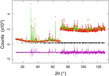
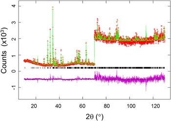
Figures 1 and 2 provide the Rietveld refinement results for (Ba0.8Sr0.2)2Co2Fe12O22 and (Ba0.6Sr0.4)Co2Fe16O27 as examples. The observed (crosses), calculated (solid line), and difference XRD patterns (bottom) are shown. The difference pattern is plotted at the same scale as the other patterns up to 70° 2θ. At higher angles, the scale has been magnified five times.
(Color online) Observed (crosses), calculated (solid line), and difference XRD pattern (bottom) for (Ba0.8Sr0.2)2Co2Fe12O22 by the Rietveld analysis technique. The difference pattern is plotted at the same scale as the other calculated peak positions up to 70° 2θ. At higher angles, the scale has been magnified five times.

(Color online) Observed (crosses), calculated (solid line), and difference XRD pattern (bottom) for (Ba0.6Sr0.4)Co2Fe16O27 by the Rietveld analysis technique. The difference pattern is plotted at the same scale as the other calculated peak positions up to 70° 2θ. At higher angles, the scale has been magnified five times.

Tables II provide the crystallographic data for (Ba
x
Sr1−
x
)2Co2Fe12O22 and (Ba
x
Sr1−
x
)Co2Fe16O27 which include cell parameters, Z values, and calculated density, respectively. The (Ba
x
Sr1−
x
)2Co2Fe12O22 compounds are rhombohedral with space group R
 $\bar 3$
m, Z = 3; while (Ba
x
Sr1−
x
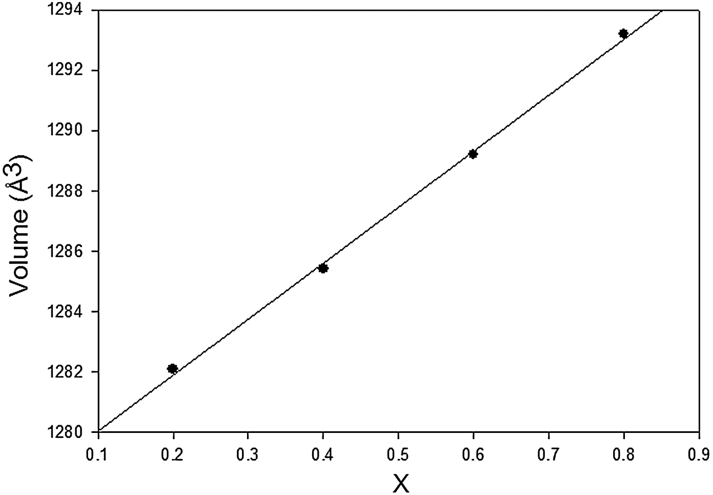
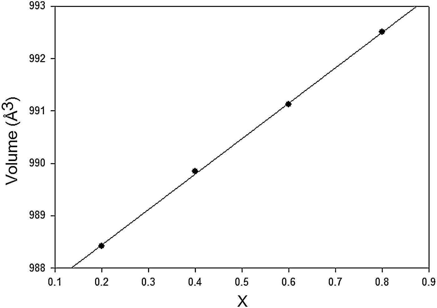
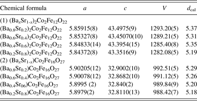
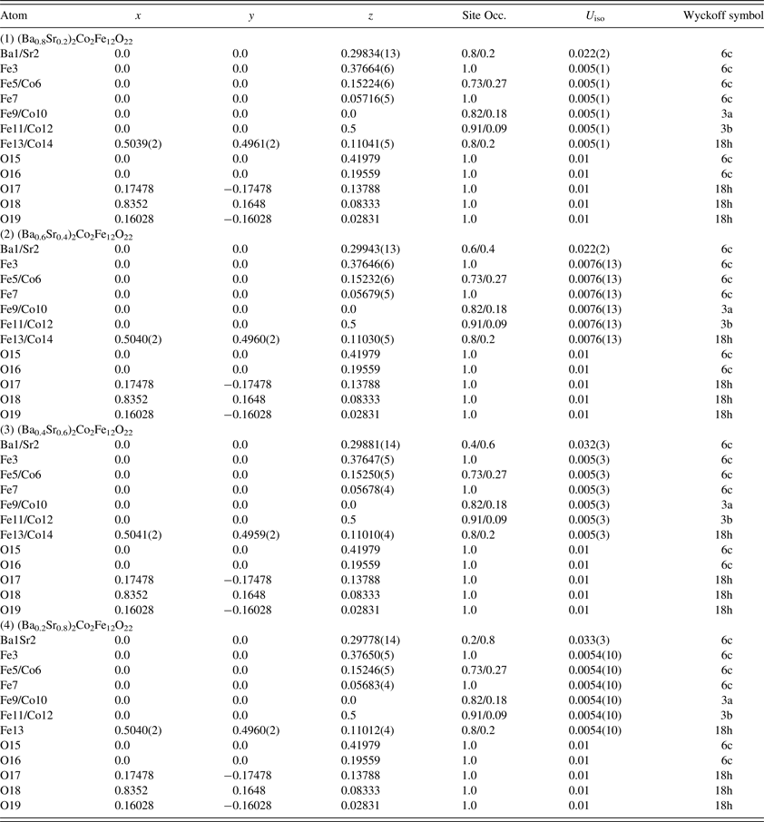
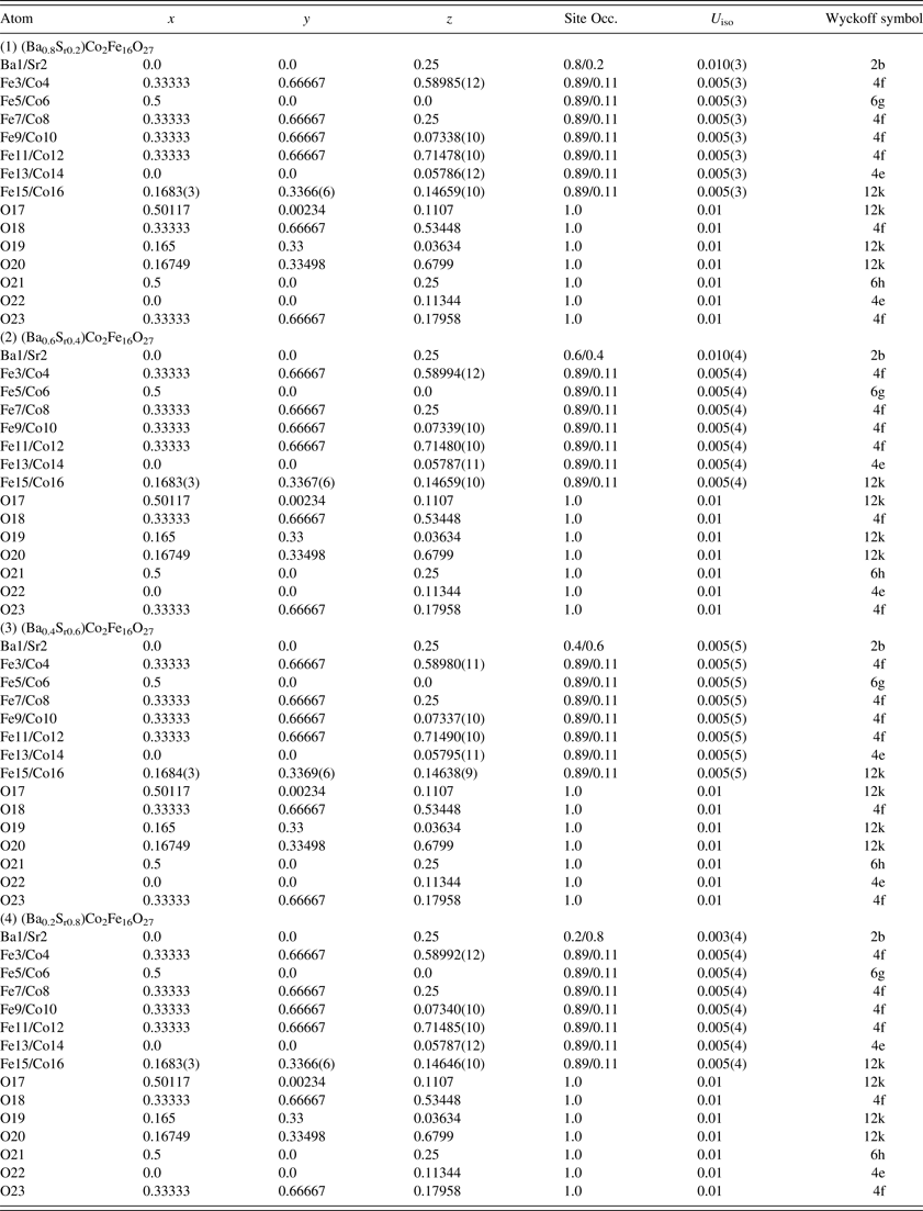
)Co2Fe16O27 are hexagonal with space group P63/mmc and Z = 2. As the ionic radius of Sr2+ is smaller than that of Ba2+ (Shannon, Reference Shannon1976), a monotonic increasing trend of unit-cell volume (as a function of x) is evidenced in Figures 3 and 4. The atomic coordinates and displacement parameters are listed in Tables IIIa and IIIb.
Plot of unit-cell volume, V, of (Ba x Sr1− x )2Co2Fe12O22 vs. x. A monotonic decrease of V is observed.

Plot of unit-cell volume, V, of (Ba x Sr1− x )Co2Fe16O27 vs. x. A monotonic decrease of V is observed.

Cell parameters for (Ba
x
Sr1-
x
)2Co2Fe12O22 (R
 ${\bar 3}$
m, Z = 3) and (Ba0.6Sr0.4) Co2Fe16O27 (P63/mmc, Z = 2).

Atomic coordinates and displacement factors for compounds for (Ba
x
Sr1-
x
)2Co2Fe12O22 [R
 $\bar 3$
m(No. 166), Z = 3].

Atomic coordinates and isotropic displacement factors for (Ba x Sr1- x )Co2Fe16O27 [P63/mmc (No. 194), Z = 2].

1. (Ba x Sr1− x )2Co2Fe12O22 (Y-type ferrite)
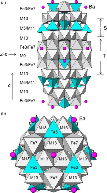
The (Ba x Sr1− x )2Co2Fe12O22 series is isostructural to the Y-type hexagonal ferrite, BaM 2+Fe6 3+O11, which belongs to a large family of hexagonal or trigonal ferromagnetic oxides (Collomb et al., Reference Collomb, Hadj Farhat and Joubert1989a, Reference Collomb, Muller, Guitel and Desvignes1989b). The crystal structure of (Ba x Sr1− x )2Co2Fe12O22 can be described as having a mixed hexagonal and cubic close-packed structure with 18 oxygen layers in the triple hexagonal cells. As shown in Figures 5(a) and 5(b), the structure can be considered as a stacking of S blocks (spinel block consisting of two oxygen layers) and T blocks (composed of two barium-containing oxygen layers sandwiched between two oxygen layers) along the hexagonal c-axis according to the sequence (TS)(TS)I(TS)II, whereas the symbols “I” and “II” refer to a 120° rotation of the three (TS) units with respect to each other around the c-axis, resulting in the rhombohedral symmetry. The structure is also sometimes referred to as (TS)3, meaning a repetition of three T and S blocks. The cation distribution of Co and Fe in the Y-type ferrite (Collomb et al., Reference Collomb, Hadj Farhat and Joubert1989a) is also reported in Table IIIa. Co atoms are mainly distributed unequally in the three octahedral sites (Fe9/Co10, Fe11/Co12, and Fe13/Co14) and in one tetrahedral site (Fe5/Co6). In summary, the Co atoms are mainly found in the Spinel layers, except a small amount was found in the octahedral z = 0 (Fe9/Co10) layer.
(Color online) Crystal structure of (Ba x Sr1− x )2Co2Fe12O22 based on X-ray powder data, showing the labeling of various atoms, and the two different coordination environments of the cation sites (M refers to the mixed Fe/Co sites): tetrahedral (blue) and octahedral (gray). (a) Structure viewed along the ab-axis, (b) structure viewed along the c-axis.

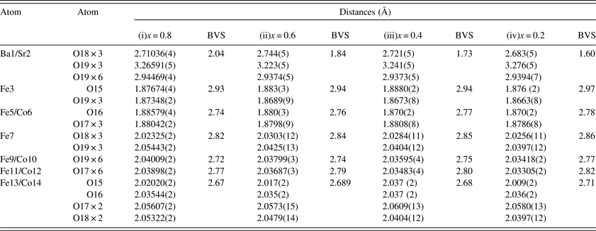
The bond distances of (Ba x Sr1− x )2Co2Fe12O22 are shown in Table IVa. The spread of the Fe/Co–O distance is rather narrow in these compounds [2.009(2)–2.0609(13) Å]. As the amount of the smaller size of Sr2+ that substituted into the Ba2+ sites (12-coordinated) increases, the BVS value decreases from 2.04 to 1.60 (as compared to their ideal values, see Table IVa), indicating an increase of tensile strain (or the cage is too large) for the increasing amount of Sr2+ ions. No Co-substitution was found in the Fe3 and Fe7 sites. In all Fe and Fe/Co sites, whether they are tetrahedral or octahedral sites, the BVS values are between 2 and 3, indicating a likely mixture of the Co2+ and Fe3+ valence. Also, these BVS values increase slightly for compositions from x = 0.8 to 0.2 across the table, indicating a slight decrease of tensile strain.
Bond distances and bond valence sum (BVS) values for (Ba x Sr1 − x )2Co2Fe12O22. The ideal BVS values are 2.0 (Ba1/Sr2 site), 3.0 (Fe3 site), 2.73 (Fe5/Co6 site), 3.0 (Fe7 site), 2.82 (Fe9/Co10 site), 2.91 (Fe11/Co12 site), and 2.80 (Fe13/Co14 site), respectively.

2. (Ba x Sr1− x )Co2Fe16O27 (W-type ferrite)
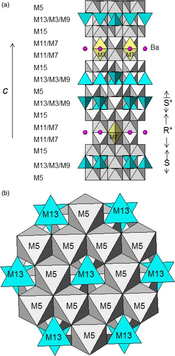
Crystal structure of the W-type ferrite, (Ba x Sr1− x )2Co2Fe16O27 (Collomb et al., Reference Collomb, Wolfers and Obradors1986a, Reference Collomb, LambertAndron, Boucherle and Samaras1986b, Reference Collomb, Abdelkader, Wolfers, Guitel and Samaras1986c), can be described as a stacking of the S(spinel) and R blocks along the c-axis. The layer containing the large cations and the two adjacent oxygen layers are usually referred to as R-blocks. The two S blocks are intercalated between the R-blocks, giving rise to a sequence
 $$ \cdots {\rm R}\, {\bar {\rm S}}\vert {\rm SR}\ast {\bar {\rm S}}\ast {\rm S}\ast {\rm R} {\bar {\rm S}}\vert {\rm SR}\ast \cdots \cdots$$
In this sequence, the | lines represent the boundary of the unit cell, the symbols*, and the overstrike ‘–’ mean the corresponding block is turned 180° around the hexagonal c-axis, and is deduced from the inversion operation, respectively [Figures 6(a) and 6(b)].
(Color online) Crystal structure of (Ba x Sr1− x )Co2Fe16O27 based on X-ray powder data, showing the unit cell outline, labeling of various atoms, and the three different coordination environment of the cation sites (M refers to the mixed Fe/Co sites): tetrahedral (blue), trigonal bipyramid (yellow), and octahedral (gray). (a) Structure viewed along the ab-axis and (b) structure viewed along the c-axis.

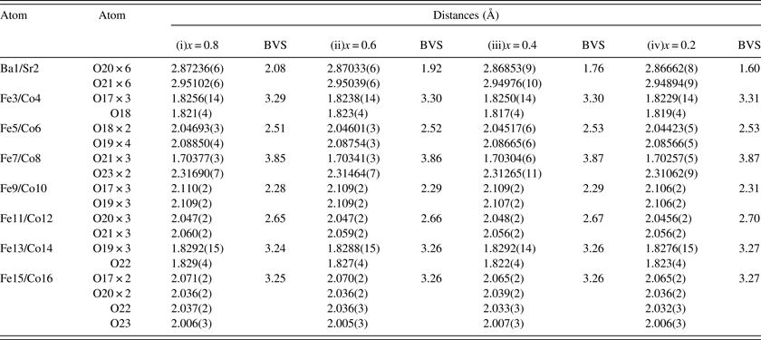
All Co-substituted Fe sites are disordered (Table IIIb). The configurations of these sites range from tetrahedral, trigonal bipyramidal, to octahedral (Table IVb). Based on the BVS calculations, among the four octahedral sites, three of them (Fe5/Co6, Fe9/Co10, and Fe11/Co12) are under tensile strain, only the Fe15/Co16 sites are under compressive strain. In the distorted fivefold coordinated trigonal bipyramid, the axial bonds are largely stretched [with an elongated Fe7/Co8-O23 distance of ≈2.31 Å) and the three equatorial bonds (Fe7/Co8)–O21 are relatively short (≈1.70 Å)]. The Fe7/Co8 site is under significantly compressive strain, as reflected in the large BVS values. The two tetrahedral sites (Fe3/Co4 and Fe13/Co14) are also somewhat under compressive strain.
Bond distances and bond valence sum (BVS) values for (Ba x Sr1 − x )Co2Fe16O27. The ideal BVS for Ba2+/Sr2+ is 2.0, for Fe3+/Co2+ is 2.89.

The Ba/Sr sites are all 12-fold coordinated (Table IVb). Similar to the (Ba x Sr1− x )2Co2Fe12O22 series, the BVS values for the Ba–Sr sites decreases from 2.08 to 1.60 as a function of the increasing amount of the smaller Sr2+ cation, indicating the change from a site without significant strain to tensile strain.
IV. REFERENCE XRD PATTERN
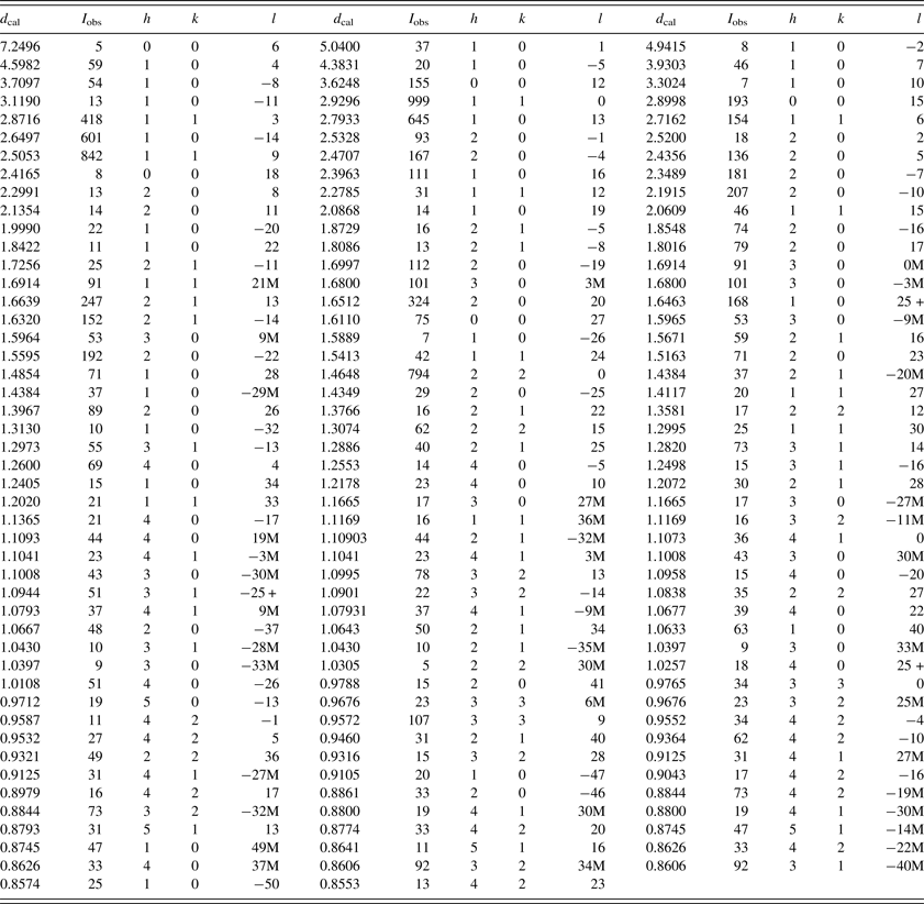
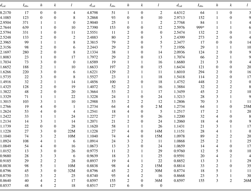
Examples of reference patterns, one from each of the Y-type and W-type hexagonal ferrite series, namely, (Ba0.8Sr0.2)2Co2Fe12O22 and (Ba0.6Sr0.4)Co2Fe16O27, are given in Tables V and VI. In these patterns, the symbols ‘M’ and ‘ + ’ refer to peaks containing contributions from two and more than two reflections, respectively. The particular peak that has the strongest intensity in the entire pattern is assigned an intensity of 999 and other lines are scaled relative to this value. In general, the d-spacing values are calculated values from refined lattice parameters. The intensity values reported are integrated intensities (rather than peak heights) based on the corresponding profile parameters as reported in Tables Ia and Ib. For resolved overlapped peaks, intensity-weighted calculated d-spacing, along with the observed integrated intensity and the hkl indices of both peaks (for ‘M’), or the hkl indices of the strongest peak (for ‘ + ’) are used. For peaks that are not resolved at the instrumental resolution, the intensity-weighted average d-spacing and the summed integrated intensity value are used. In the case of a cluster, unconstrained profile fits often reveal the presence of multiple peaks, even when they are closer than the instrumental resolution. In this situation, both d-spacing and intensity values are reported independently. All patterns have been submitted for inclusion in the Powder Diffraction File (PDF).
X-ray powder pattern for (Ba0.8Sr0.
2)2Co2Fe12O22 (R
 $\bar 3$
m, Z = 3), a = 5.85915(8) Å, c = 43.4975(9) Å, V = 1293.20(5) Å3, Z = 3, D
x
= 5.37 g cm−3. The symbols ‘M’ and ‘ + ’ refer to peaks containing contributions from two and more than two reflections, respectively. The particular peak that has the strongest intensity in the entire pattern is assigned an intensity of 999 and other lines are scaled relative to this value. The d-spacing values are calculated values from refined lattice parameters, and ‘I’ represents the integrated intensity values.

X-ray powder pattern for (Ba0.6Sr0.4)Co2Fe16O27 (P63/mmc, Z = 2), a = 5.90205(12) Å, c = 32.9002(10) Å, V = 992.51(5) Å3, Z = 3, D x = 5.29 g cm−3). The symbols ‘M’ and ‘ + ’ refer to peaks containing contributions from two and more than two reflections, respectively. The particular peak that has the strongest intensity in the entire pattern is assigned an intensity of 999 and other lines are scaled relative to this value. The d-spacing values are calculated values from refined lattice parameters, and ‘I’ represents integrated intensity values.

V. SUMMARY
Crystal structure and reference patterns of the (Ba x Sr1− x )2Co2Fe12O22 (x = 0, 0.2, 0.4, 0.6, 0.8) and (Ba x Sr1− x )Co2Fe16O27 (x = 0.2, 0.4, 0.6, 0.8) series of compounds have been determined. (Ba x Sr1− x )2Co2Fe12O22 belongs to the Y-type ferrite and (Ba x Sr1− x )Co2Fe16O27 belongs to the W-type ferrite structure. In both series of compounds, as the amount of the smaller size of Sr2+ that substituted into the Ba2+ sites increases, there is an increase of tensile strain (or the cage is too large) for the Ba/Sr sites. In the (Ba x Sr1− x )Co2Fe16O27 series of compounds, the fivefold coordinated trigonal bipyramids give rise to a significant compressive strain, as indicated by the relatively large BVS values. Results of Seebeck coefficient measurements of both samples indicated that they are essentially insulators as they are very resistive. Powder XRD patterns have been submitted for inclusion in the Powder Diffraction File (PDF).
ACKNOWLEDGEMENT
ICDD is acknowledged for the Grants-in-Aid assistance for the project