Case report
RK is a 5-year-old girl with double-inlet left ventricle, non-restrictive ventricular septal defect, and pulmonary stenosis who received a modified Blalock–Taussig shunt in infancy, a bidirectional Glenn shunt with main pulmonary artery ligation at 9 months, and a fenestrated extracardiac Fontan at 3 years of age without complication. At 5 years of age, a heart catheterisation showed a left ventricular end-diastolic pressure of 7 mmHg and a mean Fontan pressure of 10 mmHg, which did not increase with test occlusion of the fenestration that was subsequently closed with an Amplatzer® cribiform device (AGA Medical Corporation; Plymouth, Minnesota, United States of America) to remove this right-to-left shunt. A right internal mammary artery collateral was also successfully coiled at that time.
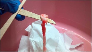
Over the subsequent 10 days, she began developing facial oedema, a progressive cough, and weight gain. Her home therapies were escalated over a period of months to include switching from enalapril to losartan, as well as several attempts to diurese with increasing doses of oral furosemide. These efforts failed to improve her persistent nocturnal hypoxia of 73%. Laboratory investigation showed hypoalbuminaemia and hyponatraemia. Shortly after admission, she expectorated a large mucus plug/cast (Fig 1). Culture of the plug was negative. Clarithromycin was started for its anti-inflammatory properties, and her fluid retention improved with albumin and furosemide boluses. An echocardiogram revealed a dampened velocity within the Fontan circulation, with increased flow reversal in the hepatic veins suggesting Fontan dysfunction. Normal left ventricular function and mild right-sided atrioventricular valve regurgitation were unchanged. Following diuresis of over 2 kg, she underwent cardiac catheterisation that found no obstruction of her Fontan anatomy, nor the presence of arterial or venous collaterals. Mean Fontan pressures were 18 mmHg with a left ventricular end-diastolic pressure of 13 mmHg. The cardiac index had decreased to 2.4 from 4.7 at the time of fenestration closure.
Expectorated bronchial cast.

On hospital day 5, sildenafil and spironolactone were added without improvement in her symptoms. Surgical therapies of last resort considered among providers included reopening the fenestration and less so thoracic duct ligation. Tissue plasminogen activator therapy was also considered, which prompted a review of the literature and subsequent consideration of a more affordable alternative, aerosolised heparin. On hospital day 7, administration of aerosolised heparin 5000 units every 4 hours produced a marked improvement in her oxygen saturations within 24 hours. Her saturations improved up to 94%, the cough became more productive and it was easier for her to expectorate secretions, and she even commented to the nursing staff that “she could breathe again”. She was discharged home with continued heart failure management and was continued on nebulised heparin 5000 units every 8 hours for over 5 months with stable saturations above 95%. Serial labs failed to show any increase in partial thromboplastin time when compared with pre-heparin levels during admission, and her mother reported an increase in respiratory symptoms on days she missed heparin treatments. However, by 12 months post-fenestration closure, she was successfully weaned off the aerosolised heparin. For her heart failure therapy, she was maintained on clarithromycin three times per week and daily aspirin, losartan, and spironolactone. On her most recent visit, she was asymptomatic with excellent somatic growth and fully saturated at 99%.
Discussion
Our patient presented with clinical findings and symptoms similar to others with Fontan physiology who have developed plastic bronchitis, which is the production of thick, rubbery casts within the tracheobronchial tree. As these casts increase in size and number, they pose a threat of airway obstruction and asphyxia. Our patient's haemodynamics suggested a minimal increase in transpulmonary gradient (from 3 to 5 mmHg), a decrease in cardiac output, and subsequent increase in left ventricular end-diastolic pressure (7 to 13 mmHg) with a 6-point increase in arterial saturation (88–94%) following our institutional preference of fenestration closure in stable Fontan patients. Despite attempts at optimising cardiac haemodynamics with diuresis, afterload reduction, and pulmonary vasodilators, we were unsuccessful in treating her respiratory symptoms. The application of aerosolised heparin produced a dramatic improvement in her symptoms shortly after initiation.
With the progressive development of plastic bronchitis, the single-ventricle patient will complain of a persistent cough and dyspnoea. Physical examination findings may include wheezing and decreased breath sounds. Expectoration or bronchoscopic removal of the rubbery, white casts is diagnostic. Chest radiographs may find atelectasis with small airway obstruction and hyperexpansion in the contralateral lung. Computed tomography will also demonstrate the obstruction of segmental bronchi with accompanying atelectasis, bronchiectasis, and consolidation.Reference Goo, Jhang and Kim 1 Literature on the pathophysiology has yet to provide a single aetiology and is believed to be more so multifactorial. The Fontan palliation is a derangement of pulmonary flow with a known sequelae of increased pulmonary pressures, decreased systemic ventricular preload, and decreased cardiac output. This combination is believed to incite inflammation, which can have a variable expression among Fontan patients.Reference Goldberg, Shaddy, Ravishankar and Rychik 2
Initially, as suggested by Seear et al,Reference Seear, Hui and Magee 3 classification of plastic bronchitis was based on the presence/absence of inflammatory cells into inflammatory type 1 casts and acellular type 2 casts. In contrast to the Seear's pathological classification, Brogan et alReference Brogan, Finn and Pyskaty 4 sought to predict outcomes by defining three subgroups of patient aetiologies – allergic/asthmatic, cardiac, and idiopathic – with cardiac malformations carrying a higher mortality (29%) and life-threatening event (41%) incidence. Plastic bronchitis classification has now evolved into a combination of the two systems in which patients are first categorised by disease association – structural heart disease, asthma/lymphatic disorders, and sickle cell disease – with further sub-categorisation by histology.Reference Madsen, Shah and Rubin 5
Plastic bronchitis therapy varies among institutions and is widely based on case reports and anecdotal experiences. Initial correction of anatomical or cardiac dysfunction can provide relief of elevated pulmonary venous pressures and symptoms if they are the sole perpetrator.Reference Barber, Burch, Tripple and Balaji 6 , Reference Yalcin, Özçelik and Celiker 7 Thoracic duct ligation has had limited success in eliminating elevated lymphatic pressures.Reference Shah, Drinkwater and Christian 8 Ventilator therapies have also shown some isolated benefit in the intensive care setting.Reference Zahorec, Kovacikova and Martanovic 9 Do et alReference Do, Chu and Berdjis 10 shared their experience of a single-ventricle, ventilator-dependent patient who failed to improve following left pulmonary artery stenting. A 24-hour course of tissue plasminogen activator produced marked reduction in ventilator settings, improved mobilisation of casts, and ultimately allowed extubation. Additional recent case reports continue to support the use of inhaled thrombolytics, including aerosolised heparin.Reference Kovesi and Gardin 11
For our application of aerosolised heparin, we incorporated the experience of our burn treatment centre in treating smoke inhalational injury. Parallels can be drawn from the pathophysiology of smoke inhalational injury in which following injury to the bronchial epithelium activation of the complement cascade results in leukocyte activation and release of free radicals. Pulmonary oedema ensues with plasma exudation of cellular debris, mucus, and fibrin, resulting in cast formation.
In the 1990s, aerosolised heparin found increasing favour in the treatment of smoke inhalational injury. Desai et alReference Desai, Mlcak, Richardson, Nichols and Herndon 12 retrospectively described their experience using pre-heparin era patients (n = 43) as controls against a treatment arm (n = 47) of children receiving 5000 units of aerosolised heparin every 4 hours for the 1st week following injury. Children receiving treatment benefited from decreased rates of atelectasis, reintubation, and mortality. These results were repeated in a similar review by Miller et alReference Miller, Rivero, Ziad, Smith and Elamin 13 identifying a number needed to treat of 2.7 and no report of adverse events. Over the past 20 years, aerosolised heparin has now become standard of care in the smoke inhalational injury population. The thrombolytic properties of heparin have been well documented with a mechanism of action that relies on its activation of antithrombin III and ultimately a reduction in fibrin clots within the common pathway. In addition, an in vitro study has described heparin's anti-inflammatory and anti-oxidative effects on the bronchial epithelium, which may contribute to the positive outcomes observed in the above studies.Reference Li, Lei and Zhou 14
The contribution of both fibrin and inflammation in the smoke inhalational injury and possibly plastic bronchitis patients with congenital heart disease does support a hypothesis that aerosolised heparin would be beneficial in our single-ventricle patients. The smoke inhalational injury population has demonstrated low complication rates, as did our patient with no increase in partial thromboplastin time over serial visits. Cost/benefit consideration would also support the use of aerosolised heparin. Our inpatient pharmacy reported an institutional expense of $417 for 1 month of heparin therapy versus $31,428 for 1 month of tissue plasminogen activator. The proven efficacy within the smoke inhalational injury population and a favourable cost/benefit ratio support the need for randomised clinical trials of aerosolised heparin in the treatment of Fontan patients with plastic bronchitis.