Introduction
Meningocoele of the fallopian canal has rarely been described. Only seven cases have previously been reported. We present an eighth case, which was unusual in that the patient presented with recurrent meningitis resulting in bilateral, profound, sensorineural hearing loss.
Case report
A nine-year-old child presented to the Birmingham Children's Cochlear Implant Centre with bilateral, profound, sensorineural hearing loss following recurrent pneumococcal meningitis. During routine investigation for cochlear implantation, abnormalities were noted on both the computed tomography (CT) and magnetic resonance imaging (MRI) scans.
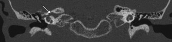
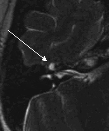
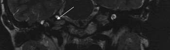
The right petrous apex was noted to be abnormal, with a widened fallopian canal commencing at the lateral end of the internal auditory meatus and extending to the region of the geniculate ganglion and proximal tympanic segment of the facial nerve. On CT, this manifested as a bony defect (Figures 1 and 2). The MRI appearances were consistent with a cerebrospinal fluid (CSF) communication along the proximal facial nerve, suggesting an extensive fallopian canal meningocoele. The signals from the right cochlea and lateral semicircular canal were diminished on the T2-weighted MRI images (Figures 3 and 4). On the enhanced T1-weighted images, there was some cochlear enhancement on the right, suggesting fibrosis within the inner ear.
Axial computed tomography scan demonstrating enlarged labyrinthine and geniculate segments of the right facial nerve (arrow).

Coronal computed tomography scan demonstrating enlarged labyrinthine and geniculate segments of the right facial nerve (arrow).

Axial, T2-weighted magnetic resonance imaging scan demonstrating high signal from the region of the geniculate ganglion (arrow), consistent with cerebrospinal fluid.

Coronal T2 weighted MR demonstrating high signal from the region of the right geniculate ganglion [shown by the arrow] and loss of high signal from within the cochlea.

The child was treated with bilateral cochlear implantation to enable auditory rehabilitation. There were no difficulties with implantation on either side, with no CSF leakage from the cochleostomy, and a full insertion was achieved despite the radiological changes seen on the right hand side. It was concluded that the recurrent meningitis had been secondary to the fallopian canal defect. To prevent further attacks, the right cochlear implantation was combined with a subtotal petrosectomy with blind pit closure of the ear canal. It was decided not to address the fallopian canal directly so as not to place the facial nerve at risk; thus, the medial wall of the middle ear was sealed with fascia and tisseel glue. The middle ear was then obliterated with abdominal fat and the eustachian tube occluded with soft tissue and bone wax.
Post-operative recovery was uneventful. The child gained an excellent result following implantation. Two years after the repair, there had been no further episodes of meningitis.
Discussion
Spontaneous CSF leakage through meningocoeles of the fallopian canal has been described in seven previously reported cases.Reference Harrington and Birck1–Reference Isaacson, Linder and Fisch6 This abnormality predisposes the individual to recurrent meningitis, due to contaminants from the middle ear reaching the CSF space.
Cerebrospinal fluid otorhinorrhoea is a potentially life-threatening condition that can be difficult to treat. It often occurs as a complication of skull base surgery, or due to trauma.Reference Gacek and Leipzig2,Reference Giddings and Brackmann7 Spontaneous leaks are more difficult to diagnose and are often not suspected at initial presentation, with resulting diagnostic delay and increased morbidity. Spontaneous CSF leaks into the temporal bone in adults are well described and occur through middle cranial fossa defects; spontaneous posterior fossa defects, although possible, have not been reported. In children, spontaneous CSF leaks and communications are typically seen in cases of defects of the lateral end of the internal auditory meatus, and can also be associated with defects in the oval and round windows.Reference Wetmore, Herrmann and Fisch8,Reference Ferguson, Wilkins, Hudson and Farmer9 In our patient, there was no history of CSF leakage, but the suspicion of a leak was raised in view of the history of recurrent meningitis.
In six of the reported cases of fallopian canal defects, there was no associated facial palsy. In the seventh case, reported by FishReference Isaacson, Linder and Fisch6, there was a coexistent facial motor weakness. This is thus rare, and was not a feature in our case.
The facial nerve and canal are embryonic derivatives of the second branchial arch (also known as the Reichert cartilage). The facial canal develops from a cartilaginous sulcus on the labyrinthine and tympanic portions of the otic capsule. Ossification of the facial canal begins relatively late in the development of the middle ear. Bone formation occurs in the 34th week of development, replacing the cartilaginous facial canal. In the late fetus and in the newborn, the facial nerve is normally enclosed in a true ossified canal. Incomplete ossification of the tympanic portion of the canal is a frequent occurrence, which results in dehiscence in the tympanic portion of the facial nerve in up to 30 per cent of temporal bones.
The facial nerve is normally sealed from the subarachnoid space by a tight sheath of dura extending laterally from the fundus along the facial canal. The seal is buttressed by the tight enclosure of the labyrinthine segment of the canal around the facial nerve. If this normally tight section is abnormally large, as seen in our patient, the subarachnoid space may extend out to fill the enlargement. Constant CSF pressure could cause a progressive stretching of facial nerve fibres without necessarily causing paralysis, as seen in our case. The constant pulsations may also cause erosion of surrounding bone, eventually leading to fistulisation.
Several localising studies are available for diagnosis, including radio-nucleotide scans, metrizamide-enhanced CT scans, high resolution CT scans with bone algorithm processing and MRI scans.Reference Nicklaus, Dutcher, Hengerer, Kido and Nelson10 In our patient, CT and MRI scans were vital in diagnosing the fallopian canal meningocoele.
Subtotal petrosectomy has been advocated by Fisch and Mattox to treat difficult CSF leaks.Reference Fisch, Mattox, Fisch and Mattox11 This procedure is used to prevent CSF leaks after skull base procedures, but can also control spontaneous adult and paediatric CSF fistulae.Reference Wetmore, Herrmann and Fisch8, Reference Bendet, Cerenko, Linder and Fisch12 It is employed for leaks into the middle ear through foramina such as the fallopian canal or cochlear aqueduct. Subtotal petrosectomy consists of complete exenteration of all pneumatic cell tracts of the temporal bone, with preservation of the otic capsule. In our patient, a middle-ear obliteration procedure was carried out. The isthmus of the eustachian tube was obliterated with bone wax and tisseel. The external auditory canal was closed in a double layer, blind sac fashion, and the middle-ear cavity was obliterated with abdominal fat.
• Meningocoeles of the fallopian canal are rare
• They can present with recurrent meningitis and, rarely, with facial weakness
• All children with recurrent meningitis should be investigated for skull base defects
• The diagnosis is based on high resolution computed tomography and magnetic resonance imaging
• Treatment aims to prevent further episodes of meningitis; this is best achieved by subtotal petrosectomy with obliteration of the middle ear and eustachian tube
• Appropriate auditory rehabilitation must be considered in all cases; in the presented patient, bilateral cochlear implantation was undertaken
However, tegmen defects are best approached by middle fossa craniotomy, in order to adequately construct a support for the temporal lobe. Meningocoele of the fallopian canal resulting in CSF communication into the middle ear and recurrent meningitis is a challenging entity, but thankfully rare. Detailed knowledge of the possible communication sites allows a more astute reading and interpretation of scans. We would recommend MRI and high resolution CT scans to establish the diagnosis.