Introduction
A biofilm is a community of micro-organisms encased within a self-produced, extracellular, polymeric substance which is attached to the surface of the micro-organisms. This is the natural state for 99 per cent of bacteria, with only 1 per cent existing in a planktonic, free-floating state. Biofilm and planktonic forms of the same bacterial species have been found to differ as regards gene transcription and phenotype expression.Reference Costerton, Veeh, Shirtliff, Pasmore, Post and Ehrlich1 Biofilms are by no means limited to bacteria; fungal pathogens are also capable of biofilm production.Reference Sanglard2
Biofilm biology
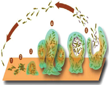
Biofilm formation involves attachment, growth and detachment (see Figure 1). Attachment is initially reversible and is based on electrostatic attraction. However, bacterial surface protein mediation and the formation of anchoring structures by some cells leads to irreversible adhesion.Reference Costerton, Montanaro and Arciola3 In pseudomonas bacteria, this stage is facilitated by type IV pili.Reference Davies, Parsek, Pearson, Iglewski, Costerton and Greenberg4 These are required for twitching activity and also to aid access to the nasal airway mucosa (which is covered in a moving, 200–250 µm thick mucous blanket).Reference Costerton, Veeh, Shirtliff, Pasmore, Post and Ehrlich1, Reference O'Toole and Kolter5
Biofilm formation. 1 = Individual cells populate the surface. 2 = Extracellular polymeric substance is produced and attachment becomes irreversible. 3 and 4 = Biofilm architecture matures and develops. 5 = Single cells are released from biofilm. Reprinted with permission.Reference Costerton, Montanaro and Arciola3

The processes of bacterial–substratum attachment and bacterial growth to a sufficient load result in co-ordinated chemical signalling between cells, known as quorum sensing.Reference Singh, Schaeffer, Parsek, Moninger, Welsh and Greenberg6 Gram-positive bacteria produce γ-butyrolactones and ‘auto-inducing peptides’; Gram-negative bacteria produce N-acylated homoserine lactones, quinolones and cyclic dipeptides.Reference Hastings and Greenberg7, Reference Dunn and Handelsman8 These compounds travel via diffusion and interact with the neighbouring bacterial surfaces and/or intracellular receptors. This communication results in gene induction, up-regulation of the proteome, and ultimately production of an extracellular, polymeric substance consisting of polysaccharides, nucleic acids and proteins.Reference Sutherland9, Reference Flemming, Wingender, Mayer, Korstgens, Borchard, Allison, Gilbert, Lappin-Scott and Wilson10 This substance forms a matrix, providing an ecosystem well suited to the needs of the biofilm colony, i.e. reduced nutrient and oxygen requirements; mediation for adhesion to surfaces; protection from unfavourable external conditions; a favourable anaerobic environment; and the formation of water channels allowing elimination of waste products and transportation of nutrients.Reference Costerton, Stewart and Greenberg11–Reference De Beer, Stoodley, Roe and Lewandowski13
Detachment of bacteria from a biofilm occurs due to external forces, or by active processes once a critical maturity level has been reached. Biofilm detachment may be facilitated by conduction of wave-like physical movement,Reference Stoodley, Lewandowski, Boyle and Lappin-Scott14 by enzymatic degradation of the extracellular matrix and by modulation of surface adhesions.Reference Lee, Li and Bowden15, Reference Baty, Eastburn, Diwu, Techkarnjanaruk, Goodman and Geesey16 Products of the detachment process stimulate further release of planktonic bacteria.Reference Baty, Eastburn, Diwu, Techkarnjanaruk, Goodman and Geesey17, Reference Cramton, Gerke, Schnell, Nicholas and Gotz18 Embolisation of bacteria to other areas may then occur, enabling the whole process to recommence.Reference Sanclement, Webster, Thomas and Ramadan19
Biofilm antibiotic resistance
Biofilms have increased antibiotic resistance. This occurs by several mechanisms. In biofilms, greater cell-to-cell contact between neighbouring micro-organisms facilitates easy plasmid exchange, enabling the evolution of antibiotic resistance.Reference Donlan20 In addition, biofilms may produce β-lactamases that deactivate β-lactam antibiotics;Reference Baggo, Heutzer, Anderson, Ciofu, Givskov and Holby21 the negatively charged matrix also repels positively charged aminoglycosides.Reference Walters, Roe, Bugnicourt, Franklin and Stewart22 Finally, the relatively slow bacterial growth within biofilms impairs antimicrobial effectiveness, as such drugs are more effective at killing rapidly growing cells.Reference Costerton, Stewart and Greenberg11
It was previously thought that the physical barrier presented by the biofilm reduced antibiotic penetration.Reference Stewart and Costerton23 However, more recent studies have found this not to be the case, with full antibiotic penetration of the biofilm demonstrated.Reference Walters, Roe, Bugnicourt, Franklin and Stewart22, Reference Konig, Schwank and Biaser24
The recalcitrant nature of biofilms may in part be due to specialised cells. When treated with antimicrobials, an initial 3- to 4-log drop in biofilm cell numbers is seen, followed by a plateau in which further increases in antimicrobial concentration have no additional killing effect.Reference Brooun, Tomashek and Lewis25 The ‘persister cells’ reform the biofilm when treatment is discontinued. Antimicrobials do not kill cells but rather cause damage that triggers programmed cell death. LewisReference Lewis26 has suggested that persister cells could represent slow-growing cells not susceptible to programmed cell death, due to mutations.
Biofilms in chronic rhinosinusitis
Chronic rhinosinusitis may become a prolonged, problematic condition which resists all attempts at antimicrobial and surgical treatment. In the US paediatric population, the estimated healthcare expenditure for chronic rhinosinusitis was $1.8 billion in 1996.Reference Chan, Abzug, Coffinet, Simoes, Cool and Liu27 Amongst US adults, chronic rhinosinusitis is one of the most common diseases, with 29.2 million adult cases diagnosed in 2002.28 The most common pathogens include the bacteria Streptococcus pneumoniae, Staphylococcus aureus, Haemophilus influenzae and Pseudomonas aeruginosa.Reference Benninger, Ferguson, Hadley, Hamilos, Jacobs and Kennedy29–Reference Finegold, Flynn, Rose, Jousimies-Somer, Jakielaszek and McTeague32
The role of biofilms as a major pathological aetiology in chronic rhinosinusitis would help explain the clinical manifestation of the disease. In chronic rhinosinusitis, mucosal changes result in favourable conditions for biofilm growth. Once a biofilm is established, its resistance to both host defences and external treatment leads to continual presentation of antigens (i.e. bacterial cell surfaces, fungal elements and exotoxins) and propagation of the chronic inflammatory process.Reference Harvey and Lund33
The objective of this review was to examine the current evidence regarding both biofilms and biofilm imaging techniques (in both chronic rhinosinusitis patients and healthy controls, investigated using mucosal biopsy samples). The review also aimed to discuss possible future research directions as regards biofilms and chronic rhinosinusitis.
Materials and methods
Search strategy
The search strategy was derived from the Cochrane Handbook for Systematic Reviews of Interventions.Reference Higgins and Green34 Medline (1966–2009), Embase (1988–2009), the Cochrane Library and Ovid (1966–2009) were searched for the terms ‘biofilm’, ‘rhinology’, ‘ENT’, ‘nose’, ‘otolaryngology’, ‘otorhinolaryngology’ and ‘chronic rhinosinusitis’. English language articles were retrieved and any relevant references cascaded to increase retrieval of relevant information.
Evaluation method
Two assessors (JK and LP) independently conducted a critical appraisal of studies identified by the literature search. Significant findings were incorporated into this review. The primary outcome assessed was the presence of biofilm in human mucosal biopsy samples taken from chronic rhinosinusitis patients and healthy controls. Data from the observational studies identified were analysed using guidance from the Meta-analysis of Observational Studies in Epidemiology group.Reference Stroup, Berlin, Morton, Olkin, Williamson and Rennie35
Results
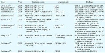
We identified 11 studiesReference Sanclement, Webster, Thomas and Ramadan19, Reference Cryer, Schipor, Perloff and Palmer36–Reference Psaltis, Weitzel, Ha and Wormald45 which examined biofilm formation in mucosal biopsy samples taken from patients with chronic rhinosinusitis (see Table I). Other identified studies assessed the ability of chronic rhinosinusitis isolates to form biofilms in vitro,Reference Ha, Psaltis, Butcher, Wormald and Tan46–Reference Bendouah, Barbeau, Harnard and Desrosiers48 documented evidence for biofilms in animal models of chronic rhinosinusitis,Reference Perloff and Palmer49–Reference Chiu, Antunes, Palmer and Cohen52 and demonstrated biofilms on silicone frontal sinus stents following functional endoscopic sinus surgery in adults.Reference Ramadan53
Evidence for biofilms in human mucosal biopsies: summary

Pt = patient; CRS = chronic rhinosinusitis; SEM = scanning electron microscope; EPS = Extracellular polymeric substance; TEM = transmission electron microscope; FISH = fluorescent in situ hybridisation; CSLM = confocal scanning laser microscopy; OSA = obstructive sleep apnoea
Discussion
The evidence for the presence of biofilms in mucosal biopsy samples from chronic rhinosinusitis patients is presented in Table I, and has been the subject of previous reviews.Reference Harvey and Lund33, Reference Hunasaker and Leid54 However, these studies used various different imaging modalities, introducing a potential bias in analysis.
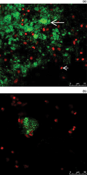
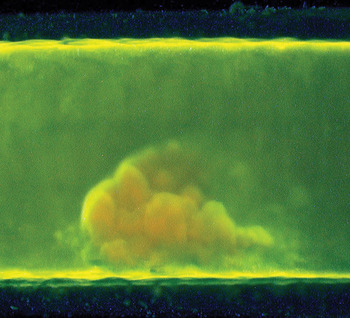
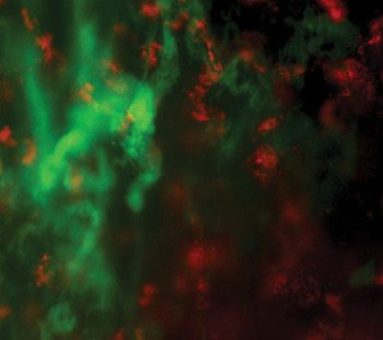
Previously described, relatively simple methods of biofilm imaging include transmission electron microscopy, scanning electron microscopy, confocal scanning laser microscopy, fluorescent in situ hybridisation species-specific probes (used with an epifluorescent microscope) and light microscopy (see Figures 2 to 6).
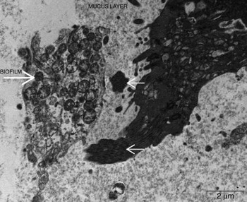
Transmission electron photomicrograph showing a mucous layer and cilia (small arrow) together with a cluster of bacteria surrounded by an extracellular polymeric substance matrix (large arrow). Published courtesy of Dr A J Psaltis and Professor P J Wormald, Department of Otorhinolaryngology, Head and Neck Surgery, The Queen Elizabeth Hospital, Adelaide, South Australia.

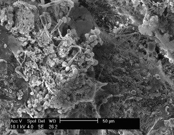
Scanning electron photomicrograph showing bacterial cocci with extracellular polymeric substance. Published courtesy of Dr A J Psaltis and Professor P J Wormald, Department of Otorhinolaryngology, Head and Neck Surgery, The Queen Elizabeth Hospital, Adelaide, South Australia.

Confocal scanning laser photomicrographs showing (a) intensely fluorescent bacteria (large arrow) surrounded by an extracellular polymeric substance haze and dead epithelial cells (small arrow) (×60), and (b) a high-power view of a biofilm (×180). Both figure parts published courtesy of Dr A J Psaltis and Professor P J Wormald, Department of Otorhinolaryngology, Head and Neck Surgery, The Queen Elizabeth Hospital, Adelaide, South Australia.

Flow chamber light photomicrograph showing a Pseudomonas aeuroginosa biofilm. Published courtesy of Professor R Chole, Washington University School of Medicine, Saint Louis, Missouri, USA.

Epifluorescent photomicrograph prepared with species-specific fluorescent in situ hybridisation probes, showing Haemophilus influenzae biofilms (red) with fungal elements (green). Published courtesy of Professor J Leid, Center for Microbial Genetics and Genomics, Flagstaff, Arizona, USA.

Studies of biofilm formation in human mucosal biopsy samples from chronic rhinosinusitis patients have demonstrated potentially significant differences in the sensitivity and specificity of imaging techniques used to detect biofilms. This could be related to inter-observer variation and/or the subjectivity inherent in some techniques (most notably scanning electron microscopy). Furthermore, variations in sample preparation techniques may result in biofilm shrinkage, damage or loss,Reference Wolf, Crespo and Reis55 and artefacts caused by dehydration and protein cross-linking may be confused for biofilms. In addition, although scanning electron microscopy is capable of demonstrating the three-dimensional structure of the biofilm matrix, it cannot clearly show bacteria within it. It may therefore be helpful to use scanning electron microscopy in conjunction with transmission electron microscopy, to enable demonstration of a cross-section and confirmation of the presence of bacteria.Reference Sanclement, Webster, Thomas and Ramadan19
Given the difficulties with these techniques, it is increasingly recognised that the current ‘gold standard’ for biofilm identification is confocal scanning laser microscopy used together with fluorescent in situ hybridisation probes.Reference Ha, Psaltis, Tan and Wormald50, Reference Hunasaker and Leid54
We observed marked heterogeneity in the patient demographics of the studies identified, and also in the subgroup classifications of chronic rhinosinusitis used (with some authors using terms such as eosinophilic mucin and allergic fungal chronic rhinosinusitis). The identified studies were conducted in tertiary referral centres, and therefore may have been biased in the spectrum of patients they recruited.
For the above reasons, we considered any meaningful meta-analysis problematic due to the potential biases involved. Therefore, we made no attempt to conduct statistical analysis. Rather, we present below a critical appraisal of the salient features of the recent literature, together with suggestions for future research directions.
Critical appraisal of evidence, and future research suggestions
Until recently, research concentrated on establishing the presence of bacterial biofilms. One studyReference Healy, Leid, Sanderson and Hunsaker43 has now demonstrated fungal elements in bacterial biofilms, using an epifluorescent microscope together with a pan-fungal fluorescent in situ hybridisation probe and species-specific fluorescent in situ hybridisation probes for the common bacterial pathogens (see Figure 6). This technique could not image a true fungal biofilm, but it was able to identify fungal elements. Fungal elements were demonstrated inside bacterial biofilms in patients with chronic rhinosinusitis; however, this study used only three controls, one of which also had fungal elements within bacterial biofilm.
The discovery of coexisting fungi within bacterial biofilms may imply that fungi contribute to the host inflammatory response. A mixed bacterial–fungal biofilm may involve a symbiotic relationship between the two microbe types, which enhances the metabolic needs and defences of the entire biofilm. We believe there is a need for a larger study than those previously undertaken, with more controls, in order to explore the presence of biofilm fungi in health, with the use of confocal scanning laser microscopy and species-specific fluorescent in situ hybridisation probes for common fungal and bacterial pathogens to enable identification of fungal and bacterial biofilm components.
Studies by both Healy et al. Reference Healy, Leid, Sanderson and Hunsaker43 and Sanderson et al. Reference Sanderson, Leid and Hunsaker39 have highlighted an interesting problem: the disparity between the bacterial species identified from mucosal biopsies, using fluorescent in situ hybridisation, and those grown from cultures of nasal swabs. In both studies, the biopsy process involved taking nasal swabs, using endoscopically guided transnasal culture techniques.
In Healy and colleagues' study,Reference Healy, Leid, Sanderson and Hunsaker43 imaging with species-specific fluorescent in situ hybridisation probes identified the predominant biofilm as H influenzae. This type of biofilm was found in 10 of 11 patients with chronic rhinosinusitis, generally in abundance. However, microbial culture results appeared to bear little relation to this finding, with no H influenzae cultured in any case. Conversely, some bacterial species (e.g. S aureus) were isolated in abundance on culture but were not identified in mucosal biopsies (using species-specific fluorescent in situ hybridisation probes).
Similarly, Sanderson et al. Reference Sanderson, Leid and Hunsaker39 consistently identified H influenzae biofilms in mucosal biopsies from chronic rhinosinusitis patients, using fluorescent in situ hybridisation probes, but did not identify this bacterial species in any nasal swab culture.
• Biofilms have been identified from mucosal biopsies taken from patients with chronic rhinosinusitis
• Further research is required on the presence of biofilms in health, and on their role in the pathophysiology of inflammation
• Universal adoption of a ‘gold standard’ biofilm imaging modality is needed; confocal scanning laser microscopy with fluorescent in situ hybridisation probes has been proposed by several authors
• This biofilm imaging modality should be combined with further investigation on the microbiology of chronic rhinosinusitis and the efficacy of traditional culture techniques used to identify pathogens
However, both Healy et al. Reference Healy, Leid, Sanderson and Hunsaker43 and Sanderson et al. Reference Sanderson, Leid and Hunsaker39 also found H influenzae biofilms (albeit in a less abundant form) in their control group, in one of three controls and in two of five controls, respectively. Healy et al. Reference Healy, Leid, Sanderson and Hunsaker43 proposed that swab culture techniques were inadequate to identify viable bacteria within biofilms, as such techniques depend on sections of the biofilm breaking off, releasing bacteria which then grow planktonically. Biofilms grow poorly when cultured on standard agar plates, with lower growth and metabolic rates.Reference Healy, Leid, Sanderson and Hunsaker43, Reference Bester, Woolfaardt, Joubert, Garny and Saftic56 This finding may be of significant importance for future research regarding both investigation of chronic rhinosinusitis pathophysiology and also guidance of chronic rhinosinusitis treatment via identification of bacterial pathogens from intranasal swabs. It may be necessary to rethink the accuracy of previous studiesReference Benninger, Ferguson, Hadley, Hamilos, Jacobs and Kennedy29–Reference Finegold, Flynn, Rose, Jousimies-Somer, Jakielaszek and McTeague32 which examined the microbiology of CRS and the traditional culture techniques used to identify pathogens.
We propose a larger study is warranted, which would compare the microbial species isolated from cultures of endoscopically guided intranasal swabs and biopsies versus those species identified by confocal scanning laser microscopy with species-specific fluorescent in situ hybridisation probes. Culture from biopsies would theoretically not rely on a swab breaking off sections of biofilm. Swabs and biopsies should be taken from the same area. The intranasal swab head should be small enough to be passed into the nasal cavity without contamination; it may even be necessary to insert a plastic sheath into the nasal cavity to protect the swab during its passage into the area of interest. The number of control patients should also be increased, in order to explore the role of H influenzae in health and to determine the statistical significance of biofilm presence.
Conclusion
Biofilms may play a significant role in chronic rhinosinusitis. However, great challenges lie ahead. Further research is required on the presence or absence of biofilms in health, and on the role they play in the pathophysiology of inflammation. It may be that biofilms are not present in every case of chronic rhinosinusitis; consequently, the significance of biofilm detection in some series needs to be considered carefully. This is particularly the case when different studies use a variety of biofilm imaging techniques.
There could be considerable benefit from universal adoption of a gold standard biofilm imaging modality. Several authors have already made strong arguments for the use of confocal scanning laser microscopy with fluorescent in situ hybridisation probes as the best technique for biofilm imaging. This imaging modality should be combined with further investigation of the microbiology of chronic rhinosinusitis and the efficacy of traditional culture techniques used to identify pathogens.