Marfan syndrome is an autosomal dominant disease caused by a mutation in chromosome 15, with a prevalence rate of 1/5000 individuals.Reference Dean 1 This syndrome affects males and females equally and is apparently evenly distributed among all ethnic groups. The phenotypic expression is rather diverse, and ~30% of affected individuals can exhibit new mutations, with >1200 mutations identified thus far, including missense and nonsense mutations, splice defects, or deletions.Reference Stheneur, Collod-Beroud and Faivre 2 Mutation in chromosome 15 leads to structural and qualitative abnormalities in the synthesis of fibrillin, a highly resistant glycoprotein, which is one of the main components of the extracellular matrix of connective tissue and is distributed throughout the entire body; however, its constitutional participation is greater in the cardiovascular, ocular, skeletal, and integumentary systems.Reference De Paepe, Devereux, Dietz, Hennekam and Pyeritz 3
Dissection and rupture of the proximal aorta are the main causes of death in untreated patients; however, clinical treatment and surgery have effectively reduced the occurrence of this type of fatality.Reference Braverman 4 Other frequent cardiac abnormalities in Marfan syndrome include mitral valve dysfunction, myocardial ischaemia, ventricular dysfunction,Reference Medeiros, Peres, Carvalho, Gun and De Luca 5 and electrocardiographic abnormalitiesReference Savolainen, Kupari, Toivonen, Kaitila and Viitasalo 6 , Reference Lawless and Best 7 such as non-sustained ventricular tachycardia (40%), premature ventricular contractions (35%), and ventricular tachycardia (8%).Reference Aydin, Adsay and Sheikhzadeh 8 These abnormalities may reflect cardiac disorders of both intrinsic and extrinsic origins.
The increase in heart rate at the onset of physical exercise is partially due to parasympathetic withdrawal and sympathetic activation that occur with increase in exercise intensity. The rapid reduction in heart rate after the completion of physical exercise is known as heart rate recovery.Reference Savin, Davidson and Haskell 9 , Reference Shetler, Marcus and Froelicher 10 A number of studies have investigated the potential of heart rate recovery as a predictor of autonomic dysfunction and death in healthy individualsReference Cole, Foody, Blackstone and Lauer 11 as well as patients with cardiovascular disease.Reference Shetler, Marcus and Froelicher 10 , Reference Nishime, Cole, Blackstone, Pashkow and Lauer 12 – Reference Morshedi-Meibodi, Larson, Levy, O’Donnell and Vasan 14 Researchers have concluded that heart rate recovery is a statistical criterion for a robust epidemiological association and is a strong, independent predictor of overall mortality in cardiac patients without a history of heart failure or coronary re-vascularisation.Reference Cole, Blackstone, Pashkow, Snader and Lauer 15 Moreover, studies have demonstrated strong correlations between an inadequate post-exercise heart rate recovery and both echocardiographicReference Guazzi, Myers and Ann Peberdy 16 , Reference Maeder, Duerring and Engel 17 and electrocardiographic abnormalities.Reference Negishi, Seicean, Negishi, Yingchoncharoen, Aljaroudi and Marwick 18 The utility of heart rate recovery analysis, especially in patients with heart disease, is justified by its consistent predictive capacity regarding death and its reflection of heart dysfunction of intrinsic and extrinsic origins. β-blocker therapy reduces adrenergic influence on the heart and is the current standard therapy to prevent aortic root enlargement; however, heart rate recovery can be changed by this therapy.Reference Racine, Blanchet and Ducharme 19
The aim of the present study was to evaluate heart rate recovery in patients with Marfan syndrome on and off β-blockers and compare them with a control group. To our knowledge, this is the first study of this type in Marfan patients.
Methods
The study population consisted of 12 patients diagnosed with Marfan syndrome and under β-blocker therapy for 3 months (group Marfan+βb) and 13 patients with Marfan syndrome without β-blocker therapy (group Marfan). All the patients were evaluated, diagnosed, and followed-up at the Cardiology and Genetics division of São Paulo Hospital (Federal University of São Paulo, Brazil). The eligibility criteria required patients to have either a normal aorta or minimal aortic dilatation of <37 mm, as determined by an echocardiogram, with no previous dissection, a recent echocardiogram (previous 6 months), the absence of signs and symptoms of cardiac ischaemia, neurological or motor disorders, and not having undergone heart surgery. Echocardiogram measurements were obtained and were carried out by a single examiner not involved in the study; 12 young university volunteers, paired for gender, age, and weight, with no cardiovascular, respiratory, metabolic diseases, and neurological or motor disorders, served as controls (group control). All the individuals of the three groups were sedentary and were not involved in sports or physical training over the past 2 years.
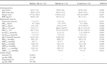
Clinical characteristics of the three groups are presented in Table 1. The Research Ethics Committee of Federal University of São Paulo approved the study and informed consent was obtained from all the participants.
Baseline demographics and submaximal exercise results of the three groups.

Δ=difference between rest and exercise peak; ANOVA=analysis of variance; BMI=body mass index; DBP=diastolic blood pressure; DR=Dosage range; HR=heart rate; L/T=lactate/total time; rec 1 min=first recovery minute; SBP=systolic blood pressure
Values are means±SD
* Marfan+βb versus Control
** Marfan versus Control
*** Marfan+βb versus Marfan
Exercise protocol
The individuals were submitted to a submaximal exercise test limited by 85–90% of maximal predicted heart rate. Maximum predicted heart rate (HRmax) was calculated using the formula of Tanaka et al,Reference Tanaka, Monahan and Seals 20 due to its greater accuracy in the general population and Brazilian population.Reference Camarda, Tebexreni and Pafaro 21
 $${\rm HR}_{{{\rm max}}} ({\rm pred})=208{\minus}(0.7{\times}{\rm age})$$
If the patient was taking β-blockers, the maximum predicted heart rate was adjusted by the following formula, in which “y” is the amount of medication (in milligrams) that the patient takes.Reference Godoi 22
 $$\,\%\,{\rm HR}_{{{\rm max}}} \:\,{\rm to}\,{\rm correct}={{({\rm y}{\plus}95.58)} \over {9.74}}$$
The exercise test was carried out on a stationary bike (Queens® – QH 1000, São Paulo, SP, Brasil), with warm up (unload) for 2 minutes, and progressive increments in intensity (range 10–20 W) every 2 minutes using the load control of the device. The participants were instructed to maintain a rate of 60 revolutions/minute. The exercise was interrupted when the participant reached the pre-calculated heart rate or manifested any signs or symptoms of exhaustion, dyspnoea, fatigue, myocardial ischaemia, and blood pressure equal to or above 160×100 mmHg.
Haemodynamic variables and blood lactate
Physiological variables, heart rate, and blood pressure were recorded after the patient remained at rest in a lying position for 5 minutes, when seated before beginning the exercise, at the time of interruption, and after 1 minute of recovery. Heart rate was obtained using a heart monitor (Polar® – S810, Polar Electro, Kempele, Finland), which was properly validated.Reference Nunan, Donovan, Jakovljevic, Hodges, Sandercock and Brodie 23 Blood lactate levels were determined from a blood sample obtained by puncture of the ring finger on the right hand, while at rest, in the sitting position, and at the time of interruption of exercise, using a lactometer (Accutrend® Lactate; Roche, Indianapolis, Indiana, United States of America).
Heart rate recovery
Absolute heart rate recovery (HRRabs) was determined by the difference between maximum heart rate (HRmax) and heart rate recovery after 1 minute of passive recovery.
Heart rate reserve in percentage values was calculated using the following formulaReference Lauer, Francis, Okin, Pashkow, Snader and Marwick 24 :
 $$\hskip-8pt {\rm HR}_{{{\rm reserve}}} (\,\%\,)=\left[ {{{{\rm HR}_{{{\rm max}}} {\minus}{\rm HR}_{{{\rm rest}}} } \over {{\rm HR}_{{{\rm max}}} \,({\rm pred}){\minus}{\rm HR}_{{{\rm rest}}} }}} \right]{\times}100$$
Heart rate recovery corrected by heart rate reserve in percentage (HRRcorr) was determined using the following formulaReference Lauer, Francis, Okin, Pashkow, Snader and Marwick 24 :
 $${\rm HRR}_{{{\rm corr}}} (\,\%\,)=\left( {{{{\rm HR}_{{{\rm max}}} {\minus}{\rm HRR}\,_{{{\rm 1}\:{\rm min}}} } \over {\left[ {{{{\rm HR}_{{{\rm max}}} {\minus}{\rm HR}_{{{\rm rest}}} } \over {{\rm HR}_{{{\rm max}}} \,({\rm pred}){\minus}{\rm HR}_{{{\rm rest}}} }}} \right]}}} \right){\times}100$$
Absolute heart rate recovery and heart rate recovery corrected by heart rate reserve were subsequently corrected by the lactate concentration measured at the end of physical exercise. These corrections were made due to the possible influence of age, heart rate reached, cardiac reserve, and metabolic stress on heart rate recovery.Reference Shetler, Marcus and Froelicher 10
Statistical analysis
After certification of data normality by the Kolmogorov–Smirnov test, two-way analysis of variance was used to determine differences between the groups, followed by Scheffe’s post hoc tests. Correlations between variables were calculated using Pearson’s correlations test. Data analysis was performed using IBM® SPSS® 20.0. A value of p<0.05 was considered to be statistically significant.
Results
Participant characteristics
As shown in Table 1, Marfan patients presented with significantly greater height than controls, despite similar body mass index. Echocardiogram showed aortic diameters of 37.4±5.9 versus 34.8±6.4 mm, left ventricle end-diastolic diameters of 50.2±5.5 versus 45.8±4.1 mm, and normal ejection fraction of 65.8±4.5 versus 67.2±9.1% in Marfan+βb and Marfan off therapy groups, respectively. No significant differences were observed between the groups.
Heart rate and chronotropic response
As expected, patients with Marfan syndrome and on β-blockers had lower rest heart rates (p=0.045) compared with the other two groups. Maximum heart rate was lower in both the groups of patients with Marfan syndrome in comparison with the control group, with lower maximum heart rate in the Marfan+βb group in comparison with the Marfan group (Table 1; analysis of variance=0.000). Similar findings occurred regarding heart rate recovery, which was lower in both groups of patients with Marfan syndrome (Table 1; analysis of variance=0.000). No significant differences were found among the three groups regarding systolic and diastolic blood pressures during rest, exercise, and recovery.
Metabolic response
Lactate concentration at rest was higher in patients with Marfan syndrome (Marfan+βb=4.0±1.1; Marfan=4.3±1.1; Control=3.2±0.9; analysis of variance=0.045). With physical exercise, a significant increase in lactate concentrations occurred in all the groups, even when corrected for total exercise time, with similar concentrations found in all three groups (Table 1).
Heart rate recovery
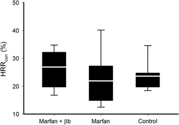
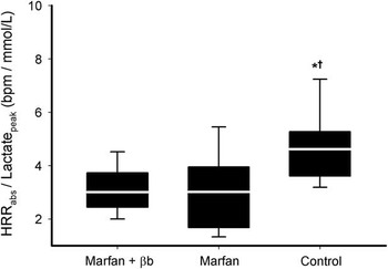
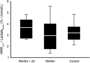
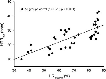
Absolute heart rate recovery was significantly slower in both groups with Marfan syndrome compared with controls (Marfan+βb: 22±6 versus Marfan: 22±8 versus Control: 34±9 bpm; analysis of variance=0.001) (Fig 1). A reduction of <34 bpm was found in all the patients with Marfan syndrome; however, no significant differences were found in heart rate recovery corrected by heart rate reserve (Marfan+βb=26±6 versus Marfan=22±9 versus Control=24±5 bpm; analysis of variance=0.248) (Fig 2). Absolute heart rate recovery corrected by peak lactate concentration was lower in both the groups with Marfan syndrome in comparison with the control group, with no significant differences between the Marfan groups (Marfan+βb: 3±1; Marfan: 3±1; Control: 5±1 bpm/mmol/L; analysis of variance=0.003) (Fig 3). We observed that the Marfan groups showed reduced heart rate recovery even when corrected for metabolic stress, suggesting that the intensity of the effort was not a determinant of recoverability. In addition, no differences were found between the Marfan groups and controls regarding heart rate recovery corrected by heart rate reserve and corrected by peak lactate concentrations (Marfan+βb: 4±1 versus Marfan: 3±1 versus Control: 3±1%/mmol/L; analysis of variance=0.283) (Fig 4), with no significant differences between the Marfan syndrome groups. There was absence of differences between the groups showing the influence of cardiac reserve in absolute heart rate recovery. Pearson’s coefficient correlation analysis for all individuals revealed a strong correlation (r=0.76; p=0.001) between absolute heart rate recovery and heart rate reserve as shown in Figure 5. The aortic dilatation did not correlate with cardiac reserve or heart rate recovery in absolute or corrected values (data not shown).
Absolute values of the heart rate recovery (HRRabs) in beats per minute (bpm). Values are expressed as mean and quartiles. *Marfan+βb versus Control. †Marfan versus Control.

Heart rate recovery corrected by cardiac reserve (HRRcorr). Values are expressed as mean and quartiles.

Absolute values of heart rate recovery corrected by peak lactate concentration (HRRabs/lactatepeak). Values are expressed as mean and quartiles. *Marfan+βb versus Control. †Marfan versus Control.

Heart rate recovery corrected by cardiac reserve and by peak lactate concentration (HRRcorr/lactatepeak).

Pearson’s correlation between heart rate reserve (HRreserve) expressed as percentage and absolute heart rate recovery (HRRabs) in beats per minute (bpm).

Discussion
The present investigation demonstrates, for the first time to our knowledge, that patients with Marfan syndrome have compromised post-exercise heart rate recovery. This behaviour seems to be independent of the use of β-blockers or the intensity of metabolic stress triggered by physical exercise; however, it was strongly correlated with the chronotropic response.
The high resting lactate concentrations in patients with Marfan syndrome was expected, especially among those who were under β-blocker therapy; two possible explanations acting either independently or simultaneously are offered: the important changes in ventricular performance seen in patients with Marfan syndrome, such as a reduced ejection fraction, an increase in end-systolic volume, and a reduction in systolic contraction velocity,Reference Medeiros, Peres, Carvalho, Gun and De Luca 5 , Reference De Backer, Devos and Segers 25 combined with vascular abnormalities and endothelial dysfunctionReference Nakamura, Itoh, Makita, Ohira, Arakawa and Hiramori 26 could result in low muscle perfusion and an increase in lactate concentrations during physical exercise; and the frequent muscle weakness, muscle fatigue, and myalgia in patients with Marfan syndrome,Reference Percheron, Fayet and Ningler 27 , Reference Giske, Stanghelle, Rand-Hendrikssen, Strom, Wilhelmsen and Roe 28 together with the high energy expenditure of skeletal muscles in Marfan patients, result in greater activity in oxidative and anaerobic energy pathways, leading to a higher production of lactate.Reference Crilley, Bendahan and Boehm 29
Heart rate recovery
Heart rate recovery has been used as a predictor of mortality and autonomic disorders in cardiovascular diseases.Reference Shetler, Marcus and Froelicher 10 , Reference Nishime, Cole, Blackstone, Pashkow and Lauer 12 – Reference Morshedi-Meibodi, Larson, Levy, O’Donnell and Vasan 14 Although this measurement does not have the same specificity as other evaluation methods of the autonomic nervous system, it strongly correlates with markers of parasympathetic activity in the early phases of recovery as well as markers of sympathetic activity in the late phases of recovery.Reference Imai, Sato and Hori 30 Therefore, alterations in heart rate recovery can suggest the possibility of autonomic dysfunction in patients with Marfan syndrome, which has not been previously mentioned.
Reductions >12 or 18 bpm in the first minute of recovery have been used as values of normality.Reference Shetler, Marcus and Froelicher 10 Nevertheless, these values are applied for maximum exertion tests, and therefore cannot be compared with those reported in the present study. Using a submaximum test (85% maximum heart rate) on a stationary bike, Danieli et alReference Danieli, Lusa, Potocnik, Meglic, Grad and Bajrovic 31 found a heart rate recovery of 52±11 and 33±9 bpm in trained individuals and controls, respectively. Similarly, Barack et alReference Barak, Ovcin, Jakovljevic, Lozanov-Crvenkovic, Brodie and Grujic 32 observed a heart rate recovery of ≅42 and ≅30 bpm in athletes and controls, respectively. Thus, we can observe the normal control group response and the impaired response of the Marfan group, as in present study the heart rate recovery of the control group (34±9 bpm) is in agreement with the values reported in the studies mentioned above, with significantly lower values found in patients with Marfan syndrome (Marfan+βb=22±6; Marfan=22±8).
As stated above, numerous mechanisms are involved in heart rate recovery, such as physical fitness, exercise intensity, rest, and maximum heart rate,Reference Shetler, Marcus and Froelicher 10 , Reference Lauer, Francis, Okin, Pashkow, Snader and Marwick 24 and it looks like the correction of absolute heart rate recovery by the chronotropic reserve is an adequate way, perhaps the most adequate of all. Considering this correction, no significant differences among groups were found (Fig 2), suggesting a strong influence of chronotropic reserve on heart rate recovery capacity (Figs 4 and 5). In other words, with an efficient chronotropic response, a fast heart rate recovery is expected. This behaviour is interpreted as a result of the interaction and integrity of the autonomic nervous system, as parasympathetic return is more efficient following sympathetic predominance,Reference Paton, Boscan, Pickering and Nalivaiko 33 such as during physical exercise. Using the same line of reasoning, we corrected heart rate recovery by peak lactate concentration with the aim of reducing the influence of metabolic stress, and, once again, deficient heart rate recovery in absolute values was found in the patients with Marfan syndrome, as well as an absence of differences regarding values corrected by cardiac reserve. These findings demonstrate that heart rate recovery is strongly determined by the chronotropic response, independent of the metabolic stress triggered by physical exercise. It should be pointed out that the chronotropic response also reflects the effect of sympathetic–parasympathetic modulation on the heart.Reference Colucci, Ribeiro and Rocco 34 An alteration in the aortic baroreceptors and peripheral chemoreceptors, as well as a compression in sympathetic branches due to aortic dilatation, could promote an autonomic dysfunction; however, we emphasise that, in this study, the observed dilatation was moderate and there was no significant correlation between the degree of aortic dilatation with the reserve chronotropic or heart rate recovery in absolute or corrected values.
β-blocker therapy
Owing to the impact of β-blocker therapy on the capacity to reach maximum heart rate, it is plausible to suppose that this therapy exerts an influence on heart rate recovery; however, this influence may be dependent on recovery time. The lower influence of sympathetic tone and the greater participation of parasympathetic activity in the first minute of heart rate recovery diminish the possibility of detecting a significant influence of β-blocker therapy. Desai et alReference Desai, De la Pena-Almaguer and Mannting 35 compared patients on β-blockers with those not on β-blockers and found no significant difference in heart rate recovery in the first minute, but the β-blocker group had lower heart rate recovery from the second to the 8th minute of recovery. On the other hand, Shetler et alReference Shetler, Marcus and Froelicher 10 found that the clinical administration of β-blockers had no significant impact on the predictive capacity of heart rate recovery.
Putative mechanisms of impaired heart rate recovery in Marfan syndrome
Previous studies have suggested a link between abnormal heart rate recovery and physical capacity;Reference Imai, Sato and Hori 30 however, this behaviour was not found when heart rate recovery was corrected by peak lactate concentration, total time, and Δlactate/total time (data not shown). Although patients with Marfan syndrome often exhibit reduced exercise capacity,Reference Medeiros, Peres, Carvalho, Gun and De Luca 5 , Reference Crilley, Bendahan and Boehm 29 this may not have been demonstrated due to the fact that a submaximal physical test was used. The buildup of muscle metabolitesReference Crilley, Bendahan and Boehm 29 and consequent increase in sympathetic activity may explain the reduced recovery capacity. Nevertheless, after correcting heart rate recovery by peak lactate concentration, smaller values were found in the groups with Marfan syndrome. Important changes in blood pressure may interfere with heart rate recovery reflexively through arterial baroreceptors; however, blood pressure values were similar in all groups. Indeed, it was demonstrated experimentally that, even considering marked baroreflex activity at the end of physical exercise, cardiac output and heart rate recovery adjust to this activity independently.Reference Walgenbach and Shepherd 36
The authors of the present study do not pretend to discard other mechanisms beyond autonomic dysfunction to account for abnormal heart rate recovery, as the present findings do not allow discarding such mechanisms; however, the correlation between heart rate recovery and cardiac reserve point towards this direction. In our opinion, heart rhythm control mechanisms of intrinsic and extrinsic origins are the main candidates. Patients with Marfan syndrome might have a high rate of arrhythmias, reduced atrioventricular conduction, and prolonged QT interval.Reference Savolainen, Kupari, Toivonen, Kaitila and Viitasalo 6 , Reference Aydin, Adsay and Sheikhzadeh 8 , Reference Chen, Fagan, Nouri and Donahoe 37 These abnormalities suggest a change in the electrical properties of the heart, as no associations were found between these findings and structural changes in the heart. Qualitative and structural changes in the synthesis of fibrillin could lead to changes in the matrix of the conduction tissue as well as the cardiac muscle tissue.Reference Maslen and Glanville 38 Changes in muscle structure and metabolismReference Percheron, Fayet and Ningler 27 – Reference Crilley, Bendahan and Boehm 29 , Reference Behan, Longman and Petty 39 as well as changes in the diastolic mechanical mechanisms of the myocardiumReference Savolainen, Nisula and Keto 40 have been demonstrated. Extrinsically, sympathetic hyperactivity and parasympathetic hypoactivity are the main candidates. Reduced ejection fraction, when present, combined with endothelial dysfunction may contribute to a decrease in output and inadequate perfusion. As a reflex, the aim of the increase in sympathetic activity is to improve the output; however, maintenance of low output contributes to dysfunction of the baroreflex, the central and peripheral chemoreflex, the ergoreflex, and the stretching reflex, consequently perpetuating excessive sympathetic activation.Reference Piepoli, Kaczmarek and Francis 41 This scenario is typically seen in patients with heart failure, but has not been reported in patients with Marfan syndrome; however, a reduced ejection fraction and both systolic and diastolic dysfunction are frequently seen in this group of patients,Reference De Backer, Devos and Segers 25 and diastolic dysfunction is a strong predictor of heart rate recovery.Reference Gharacholou, Scott and Borlaug 42 Thus, diverse cardiac dysfunctions of intrinsic and extrinsic origins may contribute to both the deficient chronotropic response and the reduced heart rate recovery.
Clinical implications
Heart rate recovery is a low-cost, easily obtained marker in different physical exercise situations – treadmill or stationary bike with maximum or submaximum intensity – and a strong predictor of mortality and autonomic dysfunction. With identified impaired heart rate recovery, the next step is to determine cut-off points and the identification of the mechanisms involved in this lower heart rate recovery, which are fundamental to the establishment of therapeutic strategies for exercise training or pharmacological therapeutics.
Study limitations
This study used a submaximal exercise test, on a bicycle ergometer, limiting the comparison of the results with studies using maximal tests on a treadmill. The lack of analysis of heart rate variability also prevented further analysis of the possible autonomical dysfunction in Marfan syndrome patients. The pathophysiology of an inadequate heart rate recovery has not been totally explained by our study, and due to lack of follow-up we could not confirm the predictive capacity for future cardiac events of heart rate recovery. Inhibitory signals from pulmonary stretch receptors to the cardiac vagal nerves must be considered when evaluating heart rate recovery. Marfan patients exhibit a reduced vital capacity,Reference Cipriano, Peres, Cipriano, Arena and Carvalho 43 which consequently could lead to a high respiratory rate for a given workload, interfering with heart rate recovery.
Conclusions
Patients with Marfan syndrome have slow post-exercise heart rate recovery and a diminished chronotropic response, which suggests possible autonomical dysfunction in this group of patients. The strong correlation between these variables suggests that chronotropic deficiency is a strong determinant of heart rate recovery. β-blocker therapy seems to have no negative effect on heart rate in the first minute of recovery in patients with Marfan syndrome.
Acknowledgements
The authors thank the participants of the present study and all the members of the Cardiology Department of Federal University of São Paulo who either directly or indirectly contributed to the present study.
Financial Support
P.A.P. has received support, in part, through an educational grant from the National Council for Scientific and Technological Development (CNPq), number 466/06/12.
Conflicts of Interest
None.
Ethical Standards
The authors assert that all the procedures contributing to this work comply with the ethical standards of the relevant national guidelines on human experimentation, of the National Commission for Ethics in Research (CONEP-Brasil), and with the Helsinki Declaration of 1975, as revised in 2008, and has been approved by the Institutional Committee of Federal University of São Paulo (UNIFESP), Brasil.