The construct of emotion regulation (ER) has received a great deal of attention in developmental psychology, developmental psychopathology, and neuroscience. From a developmental psychopathology perspective, microsocial, moment-to-moment interaction processes are likely critical for understanding how ER capacities emerge and develop and how they contribute to diverse pathways (e.g., Bronfenbrenner & Morris, Reference Bronfenbrenner, Morris and Lerner1998; Cole, Martin, & Dennis, Reference Cole, Martin and Dennis2004). It seems equally important to examine the biological mechanisms associated with effective versus ineffective ER capacities. Our main aim in the present study was to extend past research on ER by studying it in the context of parent–child interactions and in patterns of neurocognitive activity and relating the two levels of analysis. Specifically, we examined the brain processes associated with flexible or rigid parent–child interactions in children referred for behavior problems, because for them individual differences in ER capacities are critical predictors of developmental psychopathology outcomes (e.g., Bradley, Reference Bradley2003; Calkins, Reference Calkins1994; Calkins & Howse, Reference Calkins, Howse, Philippot and Feldman2004). Based on a dynamic systems (DS) approach and consistent with past research, the parent–child construct we used to capture ER was the dyadic flexibility of children's and parents’ emotional responses when engaged in conflictual situations (Granic, O'Hara, Pepler, & Lewis, Reference Granic, O'Hara, Pepler and Lewis2007; Hollenstein, Granic, Stoolmiller, & Snyder, Reference Hollenstein, Granic, Stoolmiller and Snyder2004; Lunkenheimer, Olson, Hollenstein, Sameroff, & Winter, Reference Lunkenheimer, Olson, Hollenstein, Sameroff and Winter2011). In our model, successful ER depends on the flexibility needed to shift emotional states, whereas poor ER is reflected by a rigid emotional state in which parent–child interactions become “stuck.” To date, parent–child emotional flexibility has been shown to correlate with and predict child psychopathology (for a review, see Granic & Patterson, Reference Granic and Patterson2006). This flexibility has been assumed to be related to children's capacity to regulate their own emotions, but no research has confirmed that these dyadic processes are linked to children's own ER capacities. Past research has also examined neurocognitive patterns underlying individual differences (e.g., McClure et al., Reference McClure, Monk, Nelson, Parrish, Adler and Blair2007; Monk et al., Reference Monk, Nelson, McClure, Mogg, Bradley and Leibenluft2006; Stadler et al., Reference Stadler, Sterzer, Schmeck, Krebs, Kleinschmidt and Poustka2007; Sterzer, Stadler, Krebs, Kleinschmidt, & Poustka, Reference Sterzer, Stadler, Krebs, Kleinschmidt and Poustka2005; Stieben et al., Reference Stieben, Lewis, Granic, Zelazo, Segalowitz and Pepler2007; van Goozen, Fairchild, Snoek, & Harold, Reference van Goozen, Fairchild, Snoek and Harold2007) and developmental (Lewis, Lamm, Segalowitz, Stieben, & Zelazo, Reference Lewis, Lamm, Segalowitz, Stieben and Zelazo2006) and treatment-related change in ER capacities (e.g., Lewis et al., Reference Lewis, Granic, Lamm, Zelazo, Stieben and Todd2008; Woltering, Granic, Lamm, & Lewis, Reference Woltering, Granic, Lamm and Lewis2011); however, these mechanisms have not been linked to parent–child processes. In the current study, we aimed to integrate parent–child and neurocognitive indices of ER and examine their associations. To determine what brain mechanisms are associated with “flexible” versus “rigid” styles of ER, we looked at the activation in two prefrontal areas responsible for appraisal, self-monitoring, and inhibitory control. Specifically, we examined the relation between dorsal prefrontal systems thought to mediate deliberate control and ventral prefrontal systems responsible for automatic or reactive control. Our hypotheses were based on the idea that ventrally mediated mechanisms support overlearned (rigid), ineffective regulatory processes, whereas dorsally mediated mechanisms allow for emotional flexibility and improved interpersonal functioning.
Emotional Flexibility in Parent–Child Interactions
Most clinicians and clinical researchers assume that the family interactions of children at risk for psychopathology are emotionally “negative” or “angry.” However, according to emotion theorists (e.g., Izard, Reference Izard1977; Magai & McFadden, Reference Magai and McFadden1995; Tomkins, Reference Tomkins1963), the expression of anger or any other negative emotion is not pathogenic; all emotions are adaptive and important to express in appropriate contexts. Researchers also hold that effective regulation of emotions is critical for healthy development (e.g., Cole et al., Reference Cole, Martin and Dennis2004; Southam-Gerow & Kendall, Reference Southam-Gerow and Kendall2002), and research supports the association between young children's poor ER and both externalizing (e.g., Eisenberg et al., Reference Eisenberg, Cumberland, Spinrad, Fabes, Shepard and Reiser2001; Rothbart, Ahadi, & Hershey, Reference Rothbart, Ahadi and Hershey1994; Zhou et al., Reference Zhou, Hofer, Eisenberg, Reiser, Spinrad and Fabes2007) and internalizing (Calkins, Reference Calkins1994; Calkins & Howse, Reference Calkins, Howse, Philippot and Feldman2004) outcomes. However, what is effective ER? Is it characterized by the inhibition of negative emotions, so that they never rear their ugly heads? In contrast, our research is based on the assumption that emotional flexibility is the hallmark of effective regulation (for a review, see also Kashdan & Rottenberg, Reference Kashdan and Rottenberg2010). When children and their parents can switch emotional responses more easily, especially during challenging interactions, they are essentially exploring alternative ways of interpreting and responding to the threatening and frustrating aspects of their interactions. Effective regulation involves the expression of negative emotions but also the working through of those emotions, leading to solutions to intractable problems, whereas the dampening of emotions (both negative and positive) indicates that one has already given up on novel possibilities for engagement and resolution. This conceptualization is similar to the control versus regulation distinction made by Cole, Michel, and Teti (Reference Cole, Michel and Teti1994), who suggest that ER often involves the adjustment and “dynamic ordering” of emotional behavior, not just dampening negative emotions. It is also consistent with Gross’ (Reference Gross1998a, Reference Gross1998b) distinction between reappraisal and suppression, where reappraisal implies context-specific adaptations and is considered the more effective strategy. Thus, parent–child interactions that predict more positive child outcomes are likely to be more emotionally flexible.
A number of previous studies support the link between emotional flexibility in parent–child interactions and reduced child behavior problems. One prospective study applied DS methods to analyze hundreds of parents and their prosocial and aggressive young children (Hollenstein et al., Reference Hollenstein, Granic, Stoolmiller and Snyder2004). Parent–child dyads were videotaped interacting with one another while they engaged in different types of activities (e.g., playing games, problem solving, cleaning up a mess). Results showed that rigid (inflexible) parent–child patterns measured in the fall of kindergarten predicted aggressive behavior 18 months later. Compared to normally developing children, those who developed problem behaviors expressed a smaller range of emotions and they became stuck with their parents for longer periods of time in one or very few emotional states. In another study, Granic and colleagues (Reference Granic, O'Hara, Pepler and Lewis2007) examined children referred for aggressive behavior problems and their parents before and after a 14-week treatment program combining parent management training and cognitive–behavioral therapy. Behavioral outcomes were assessed by reports from parents and clinicians, and home visits at pre- and posttreatment were videotaped while parents and children discussed both conflictual and positive topics. The results showed that significant improvements in children's externalizing behavior were associated with increases in parent–child emotional flexibility during the difficult problem-solving discussion. Dyads who improved still expressed negative emotions, but they acquired the skills to repair conflicts and shifted easily between negative interactions and mutually positive patterns.
A DS Method for Assessing Flexibility
In the current study, we examined differences in dyadic flexibility using a method based on DS principles. DS theorists use the concept of a state space to represent the variability (stochasticity) versus stability of the behavior of a given system. Behavior is conceptualized as moving along a real-time trajectory on this hypothetical landscape, sometimes remaining stuck in the vicinity of a specific attractor and sometimes shifting flexibly among a variety of weaker attractors (e.g., Lewis, Reference Lewis2000). Based on these abstract formalisms, Lewis, Lamey, and Douglas (Reference Lewis, Lamey and Douglas1999) developed state space grid (SSG) analysis, a graphical approach that quantifies observational data according to a map constructed from two ordinal variables. These variables code moment-to-moment changes in behavior or emotional expression, such that each cell on the grid represents a unique pair of values for the two codes. Granic and colleagues extended this methodology to represent dyadic behavior over one or more interaction episodes (e.g., Granic & Lamey, Reference Granic and Lamey2002; Granic, Hollenstein, Dishion, & Patterson, Reference Granic, Hollenstein, Dishion and Patterson2003; Hollenstein et al., Reference Hollenstein, Granic, Stoolmiller and Snyder2004).
SSGs are particularly well suited for looking at differences in dyadic flexibility. By coding the child's emotional states on one axis and the parent's states on the other axis, dyadic states can be represented by the sequence of concurrent codes (each represented by a specific cell) unfolding over time. Then, variables can be constructed to tap the number of transitions from cell to cell, the duration of behavior in each cell, and the spread or dispersion of behavior over the whole grid.
In the present study, we analyzed the emotional flexibility of parent–child interactions, videorecorded during a problem-solving episode, using variables derived from SSGs. To assess the emotional state, we used a standard affect coding system and arranged the codes into an ordinal series that could be represented along each axis (child and parent) of the grid. For each dyad in the study, a single grid was constructed, such that the variables derived from that grid showed a range of values from high flexibility (e.g., frequent movements from state to state, high dispersion over the grid) to low flexibility (e.g., few movements, low dispersion). These scores were then compared with the neural data.
Neurocognitive Approach
The neural correlates of ER are many and varied, but we have focused our research on two prefrontal systems presumed to mediate two distinct styles or modes of ER. It is useful to describe these modes of ER in terms of the distinction between reactive and effortful self-control proposed by Eisenberg et al. (Reference Eisenberg, Spinrad, Fabes, Reiser, Cumberland and Shepard2004). Reactive regulation includes implicit evaluations of objects or events as aversive or rewarding, eliciting immediate response tendencies (e.g., inhibition or approach) drawn from a limited range. In contrast, effortful regulation includes more deliberate or conscious cognitive manipulations that monitor, adjust, and select responses from a range of options (Gross, Reference Gross1998a, Reference Gross1998b; Rothbart & Bates, Reference Rothbart, Bates, Damon and Lerner1998). As cognitive processes responsible for effortful regulation emerge with development, higher-order regulation strategies are thought to supersede reactive regulation strategies, facilitating the successful pursuit of long-term or abstract goals (Diamond, Reference Diamond and Knight2002; Zelazo & Cunningham, Reference Zelazo, Cunningham and Gross2007).
We suggest that reactive ER is mediated by the ventral prefrontal cortex (vPFC), which includes the orbitofrontal cortex (OFC), ventromedial PFC (vmPFC), and ventral anterior cingulate cortex (vACC), regions that are adjacent anatomically and overlapping in function. The OFC is believed to mediate attention to the environment and facilitate further processing of perceptual information. The region allows us to anticipate whether a given stimulus will be rewarding or punishing and mediates gradual learning when the positive or negative aspects of a situation change. Moreover, the vPFC is activated when one inhibits or overrides automatic impulses for immediate gratification, delaying reward or avoiding punishment (Rolls, Reference Rolls2004). The vPFC has downward connections to the amygdala, hypothalamus, and brainstem nuclei as well as upward connections to the dorsal ACC (dACC). Its downward connections are integral to emotional states, and its activity has been frequently implicated in the regulation of emotion through inhibition of amygdala activation (Davidson, Jackson, & Kalin, Reference Davidson, Jackson and Kalin2000; Hariri et al., Reference Hariri, Mattay, Tessitore, Fera, Weinberger and James2003; Levesque et al., Reference Levesque, Joanette, Mensour, Beaudoin, Leroux and Bourgouin2003; Ochsner et al., Reference Ochsner, Ray, Cooper, Robertson, Chopra and Gabrieli2004). In clinical populations, activation of the vPFC is linked with anxiety and depression, further suggesting that its mode of regulation is not only rigid but also ineffective (Drevets & Raichle, Reference Drevets and Raichle1998; Mayberg et al., Reference Mayberg, Liotti, Brannan, McGinnis, Mahurin and Jerabek1999; McClure et al., Reference McClure, Monk, Nelson, Parrish, Adler and Blair2007).
Conversely, effortful ER appears to be mediated by the dorsal PFC (dPFC), which includes the dACC and its connections to the dorsolateral PFC, implicated in complex decision-making abilities. The dACC has been dubbed the “cognitive” sector of the ACC (Bush, Luu, & Posner, Reference Bush, Luu and Posner2000), and it is thought to mediate attention to potential actions in novel or challenging situations and to evaluate anticipated outcomes. Hence, the dPFC allows for the conscious control of emotions and emotional behavior by selecting among response alternatives with a range of probable consequences. In normal individuals, increased dACC activation is associated with attention regulation, performance monitoring, and deliberate response inhibition (Bush et al., Reference Bush, Vogt, Holmes, Dale, Greve and Jenike2002; Carter et al., Reference Carter, Macdonald, Botvinick, Ross, Stenger and Noll2000; Gehring, Goss, Coles, Meyer, & Donchin, Reference Gehring, Goss, Coles, Meyer and Donchin1993; van Veen, Cohen, Botvinick, Stenger, & Carter, Reference van Veen, Cohen, Botvinick, Stenger and Carter2001). Aggressive children and adolescents show reduced activation in this region, implying poor regulation of emotional impulses (Stadler et al., Reference Stadler, Sterzer, Schmeck, Krebs, Kleinschmidt and Poustka2007; Sterzer et al., Reference Sterzer, Stadler, Krebs, Kleinschmidt and Poustka2005; van Goozen et al., Reference van Goozen, Fairchild, Snoek and Harold2007).
In sum, the vPFC appears to mediate immediate response tendencies, classified as reactive ER, whereas the dPFC is involved in self-monitoring and response selection, classified as effortful ER (for an overview, see Hum & Lewis, in press). It is worth noting that “effortful” may be too strong a word, because response selection and self-monitoring can probably become automatized with practice. The most important point is that both forms of regulation override or modulate the generation of “raw” emotional responses by activity in lower brain structures (e.g., periaqueductal grey, hypothalamus, amygdala). Hence, activation of the vPFC versus dPFC appears to mediate diverse regulatory mechanisms brought to bear on similar emotional responses.
Developmental Considerations
The current study was focused on clinical participants who were just about to enter an important developmental transition period, preadolescence and adolescence; thus, a few developmental considerations should be noted. Investigators have found it difficult to track cortical changes in late childhood and adolescence, especially because different regions of the cortex change at different rates. However, a number of principles are generally accepted. After a period of synaptic proliferation in late childhood (Giedd et al., Reference Giedd, Blumenthal, Jeffries, Rajapakse, Vaituzis and Liu1999), the PFC goes through a decline in grey matter density at the same time as a proliferation in white matter (thought to be due to myelination). Spear (Reference Spear2000) reviewed a number of findings suggesting that pruning is the main source of synaptic reduction, although Paus (Reference Paus2005) suggested that myelination may account for the appearance of gray matter thinning. Regardless, most investigators point to massive restructuring of the PFC in adolescence, as more specific regions respond to more specific stimuli while the overall efficiency is enhanced by the elimination of redundant synapses.
Researchers who study socioemotional development interpret the reorganization of the PFC as both an opportunity and challenge for the adolescent, because the areas that undergo greatest reorganization are those that are associated with self-regulation (Dahl, Reference Dahl2003; Steinberg, Reference Steinberg2005). The increases in emotional intensity and arousal elicited by new forms of social experience in adolescence may stem in part from the relatively slow consolidation of dorsal regions of the PFC (including the dACC) in relation to ventral regions (Gogtay et al., Reference Gogtay, Giedd, Lusk, Hayashi, Greenstein and Vaituzis2004). Thus, adolescents’ difficulties with ER may derive from novel sources of emotional arousal managed by a brain that is not yet proficient in conflict detection, response selection, and deliberate self-regulation. Understanding how brain and behavioral relations function just prior to this reorganization period may have particularly important implications for understanding the development of normative ER capacities, as well as how these capacities may be changed if targeted in treatment.
EEG Methodology
A method for assessing the neurocognitive correlates of ER, especially in children who might be too reactive to perform in the scanner environment, is the derivation of event-related potentials (ERPs) and source models from EEG data. Specifically, an ERP called the frontal N2 is associated with performance monitoring or inhibitory control (e.g., Falkenstein, Hoormann, & Hohnsbein, Reference Falkenstein, Hoormann and Hohnsbein1999; Gehring et al., Reference Gehring, Goss, Coles, Meyer and Donchin1993; van Gaal, Lamme, Fahrenfort, & Ridderinkhof, Reference van Gaal, Lamme, Fahrenfort and Ridderinkhof2011). Moreover, the N2 can be elicited by either conscious or unconscious efforts to inhibit responses (van Gaal et al., Reference van Gaal, Lamme, Fahrenfort and Ridderinkhof2011), suggesting that it taps diverse cortical mechanisms. A source analysis of the N2 and a related component, error-related negativity (ERN), has revealed generators in a dorsal–medial region suggestive of the dACC as well as ventral prefrontal areas such as the OFC for both adults and children (e.g., Bekker, Kenemans, & Verbaten, Reference Bekker, Kenemans and Verbaten2005; Bokura, Yamaguchi, & Kobayashi, Reference Bokura, Yamaguchi and Kobayashi2001; Fallgatter & Strik, Reference Fallgatter and Strik1999; Ladouceur, Dahl, Birmaher, Axelson, & Ryan, Reference Ladouceur, Dahl, Birmaher, Axelson and Ryan2006; Lewis, Granic, & Lamm, Reference Lewis, Granic and Lamm2006; Nieuwenhuis, Yeung, van den Wildenberg, & Ridderinkhof, Reference Nieuwenhuis, Yeung, van den Wildenberg and Ridderinkhof2003; Pliszka, Liotti, & Woldorff, Reference Pliszka, Liotti and Woldorff2000; van Veen & Carter, Reference van Veen and Carter2002). Both regions may contribute to the N2 simultaneously or in a rapid cascade of activation (Lamm, Granic, Zelazo, & Lewis, Reference Lamm, Granic, Zelazo and Lewis2011). We have reported that a ventromedial prefrontal generator of the N2 is more active in children with behavior problems than in nonclinical peers, and it becomes less active when children improve with treatment (Lewis et al., Reference Lewis, Granic, Lamm, Zelazo, Stieben and Todd2008; Woltering et al., Reference Woltering, Granic, Lamm and Lewis2011). This is consistent with our hypothesis that ER mediated by the vPFC is relatively ineffective or potentially maladaptive.
Because the dorsal and ventral regions both contribute to N2 amplitudes, we were not primarily interested in the associations between N2 amplitudes and individual differences in flexibility. The present study was instead intended to examine the dorsal and ventral regions of activation during the time window of the N2, using source analysis methods, and to test their relative activation levels in relation to behavioral measures of emotional flexibility.
Design and Hypotheses
Our main aim was to integrate behavioral measures of parent–child flexibility with neurocognitive measures of ER. Children referred for externalizing behavior problems, with the majority comorbid for internalizing problems as well, were brought to our lab with a parent. Parent–child dyads were videotaped in an observation room while attempting to solve a problem identified as emotionally stressful. After the observational assessment, children's cortical activity was assessed during a task designed to elicit mild negative emotions. Emotional flexibility scores derived from SSGs were constructed from the observational data. Following ERP extraction, we examined source activation in the dorsal and ventral prefrontal “regions of interest” (ROIs) in the time window of the N2. Finally, the activation in the dorsal and ventral prefrontal cortical regions was compared to flexibility scores to test whether flexible ER corresponded with activation in one region more than the other.
Consistent with previous findings (e.g., Hollenstein et al., Reference Hollenstein, Granic, Stoolmiller and Snyder2004), our first hypothesis was that emotional flexibility would be negatively associated with the severity of externalizing symptoms. This would help to validate the proposition that low emotional flexibility corresponds with ineffective ER. Our second hypothesis was that low emotional flexibility among clinically referred families would be associated with greater ventral prefrontal activation and less dorsomedial prefrontal activation. This would indicate that reactive ER, mediated by the OFC and related areas, corresponds with a more rigid (inflexible) style of interpersonal interactions in emotionally demanding situations.
Methods
Participants
Children between 8 and 12 years of age and their mothers were recruited from two outpatient treatment programs for externalizing children. Children participated in the study prior to beginning their treatment program. They were included in the study if they scored within the clinical or borderline-clinical (T ≥ 60) range on the externalizing subscale of the parent-report form of the Child Behavior Checklist (CBCL; Achenbach, Reference Achenbach1991). Children were excluded from the study if the child had a significant developmental delay, IQ under 80, neurological impairments, or if their residence was outside the large urban center where the study took place. Ethical approval of the project was obtained from the University of Toronto and the Hospital for Sick Children in Toronto. There were 100 children with complete behavioral and electrophysiological data (mean age = 9.53 years, SD = 1.17, 77 boys). Thirty-five children were excluded from all subsequent analyses because of low ERP trial counts (<10). There were no significant differences in sex or age between children who were eliminated and children who were included, χ2 (1) = 1.04, p = .31 and t (98) = 1.55, p = .13, respectively. Of the remaining 65 children, all but 18 (72%) also scored within the clinical or borderline-clinical range on the internalizing subscale of the CBCL. Data from these 65 children were used for the scalp analysis. For the source analysis, 23 children had to be excluded from the subsequent analyses because of low source trial counts (<10). There were no significant differences in age, 9.62 (1.15) for those included, 9.95 (1.13) for those excluded, t (62) = −1.12, p = .27; internalizing T score, 62.40 (10.17) for those included, 64.05 (9.81) for those excluded, t (59) = −0.62, p = .54; externalizing T score, 71.30 (5.69) for those included, 69.14 (6.45) for those excluded, t (59) = 1.33, p = .19; or sex, 32 males, 10 females included, 16 males, 7 females excluded, χ2 (1) = 0.48, p = .49. Of the remaining 42 children, all but 15 (64%) also scored within the clinical or borderline-clinical range on the internalizing subscale of the CBCL. Thirty-two were males and 10 were females.
Forty percent of the 42 children resided in intact families, 30% in single-parent (exclusively maternal) households, 14% in blended families, and 16% in other family configurations (e.g., grandparents). Based on parent-identified ethnicity, 78% of the children were European (Caucasian), 11% were African or Caribbean, and 8% were of mixed backgrounds. In terms of family income, 23% made under $20,000 per year, 23% made between $20,000 and $39,000, 11% made $40,000 to $59,000, and 41% made over $60,000.
Procedure
Children were accompanied to the lab by their mother on the day of testing. Following a brief introduction to the testing environment, consent and child assent were obtained. Flexibility in mother–child interactions was assessed during a videotaped conflict discussion that centered on a mutually identified and unresolved issue selected from a modified version of the Issues Checklist (Robin & Weiss, Reference Robin and Weiss1980), a questionnaire that lists potential sources of conflict between parents and children (e.g., bedtime, lying, swearing, fighting). After the discussion, mothers completed the CBCL in an adjacent room while children were introduced to the EEG task. Each child was informed that he or she could win a prize for playing the EEG computer game and was shown two toy bins. One bin of toys contained desirable prizes (e.g., large stuffed animals, arts and crafts sets, action figures, games, and $10.00 gift certificates for a music store). The second bin of toys contained less desirable, smaller prizes (e.g., small plastic cars, yo-yos). Children were told that prize selection from the bin with large, desirable toys required successful performance (accumulation of points) in the EEG computer game and that less successful performance would limit their choice of prize to toys from the less desirable toy bin. Children were then taken into the room where the EEG testing took place. Children were seated in front of a computer monitor and the electrode sensor net was applied. During the EEG task, distance and alignment to the monitor was controlled with a chin rest. Children were instructed to make responses during the game by clicking a button on a response pad with the index finger of their dominant hand (writing hand). A practice block of 30 trials (repeated as necessary) ensured proficiency with the task.
Measures
Issues Checklist
The Issues Checklist (Robin & Weiss, Reference Robin and Weiss1980) lists common issues for parents and children, including going to bed on time, lying, and fighting with siblings. The mother and child were asked separately to identify issues that they argued about in the past 2 weeks. For each identified issue, participants were then asked to indicate how “hot” the argument was on a 5-point scale from calm to angry. Finally, participants indicated whether the issue had been resolved. The hottest topic left unresolved (as indicated by both mother and child) was chosen for the conflict discussion.
CBCL
The CBCL (Achenbach, Reference Achenbach1991) is a standardized, highly reliable, and valid measure of children's emotional and behavioral problems. For each symptom on the checklist, parents are asked to indicate whether and to what degree the child exhibits that symptom. The CBCL yields standardized T scores for total behavior problems, internalizing and externalizing problems, as well as a number of more focused subscales.
Mother–child interactions
Mother and child were seated diagonally across from each other in a square room. In the corner directly behind each participant, a digital video camera hidden behind a mirror was positioned to obtain an unobstructed view of the participant in the diagonal corner of the room. A research assistant gave instructions to the dyad and left the room before the discussion began. The discussion was based on Forgatch and Degarmo's (Reference Forgatch, Degarmo and Brooks-Gunn1999) procedure for studying problem solving in families of antisocial children. A research assistant chose an issue from the Issues Checklist that both mother and child agreed was the most anger-provoking topic that remained unresolved. Within a 6-min time window, the dyad was instructed to solve the problem as best they could and end on a positive note.
Coding procedures
Videotaped conflict discussions were recorded using Noldus Observer 5.0. The affect of each participant was coded independently in real time using a simplified version of the Specific Affect Coding System (Gottman, Katz, & Hooven, Reference Gottman, Katz and Hooven1996; Gottman, McCoy, Coan, & Collier, Reference Gottman, McCoy, Coan and Collier1996). This coding system consists of nine mutually exclusive affect codes (i.e., contempt, anger, fear/anxiety, sad/withdrawn, whine/complain, neutral, interest/curiosity, humor, joy/affection). Each code is based on a combination of facial expression, gesture, posture, voice tone, volume, and speech rate to capture a gestalt of the affective tone of each moment of behavior.
Before the videotaped interactions were coded, coders were trained to a minimum criterion of 75% agreement and 0.65 κ using a frequency/sequence-based comparison and a criterion of 80% agreement using a duration/sequence based comparison (Noldus Observer 5.0). Coder drift was minimized with weekly recalibration training. Furthermore, 20% of all sessions were jointly coded by two coding supervisors. These coded files served as the “gold standard” to which each reliability file was compared. The average coder agreement with the gold standard was 82% and 0.76 κ for the frequency-based method and 90% agreement for the duration-based method.
SSG analysis
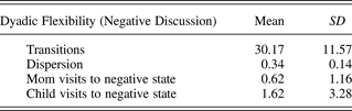
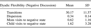
SSGs were constructed for each dyad. One dyad member's coded behavior was plotted on the x axis, and the other member's behavior was plotted on the y axis. Each x–y coordinate represented a period of time in a particular state for the dyad. The dyad's trajectory (sequence of behavioral states) was plotted as it proceeded in real time on a grid representing all possible behavioral combinations between mother and child (Granic et al., Reference Granic, Hollenstein, Dishion and Patterson2003; Granic & Lamey, Reference Granic and Lamey2002). To quantify different aspects of mother–child flexibility, two parameters were calculated from the grids: (a) transitions (TRANS), which is a count of the number of movements between cells on the grid, where a higher value on this measure indicates more frequent changes of dyadic behavioral states and therefore more flexibility; and (b) dispersion (DISP), which is the sum of the squared proportional durations across all cells corrected for the number of cells and inverted so that values range from 0 (no dispersion at all–all behavior in one cell) to 1 (maximum dispersion), where higher values indicate more flexibility. In all our analyses, we controlled for the total number of times parents and/or children were coded as “negative” (i.e., whining, angry, or contemptuous), in other words, the total times they visited the negative region of the state space. Table 1 provides the means and standard deviations of the flexibility measures and the mean number of times parents and children were coded as visiting the negative region of the state space.
Means and standard deviations for all measures derived from the state space grids
EEG go/no-go paradigm
Task
The emotion induction go/no-go task used in the present study was partly adapted from a task developed by Garavan, Ross, and Stein (Reference Garavan, Ross and Stein1999). The task was presented using E-Prime software (Psychological Software Tools, Pittsburgh, PA). In a standard go/no-go paradigm, participants are instructed to press a button as fast as possible given a particular category of stimuli (“go” condition) and to withhold responding given another category of stimuli (“no-go” condition). Participants in this study were instructed to click a button for each letter presented but to avoid clicking when a letter repeated a second time in succession. The task was divided into three blocks, and different pairs of similarly shaped letters were used for each block to enhance novelty without modifying the level of difficulty (Block A: x, y; Block B: o, p; Block C: u, d). The error rate for no-go trials was maintained at roughly 50% ± 10% by dynamically adjusting the stimulus duration (and thus the intertrial interval). Stimulus duration was increased with each erroneous response made on no-go trials, and it was decreased when a correct no-go trial followed a correct go trial. This constraint was incorporated to prevent stimulus time adjustments because of chronic nonresponding. The dynamic adjustment of stimulus time was intended to provide the same level of challenge to all participants across all ages and to obtain a sufficient number of correct no-go trials for ERP averaging. Because there may still have been slight variations in task difficulty between age groups and blocks even with this dynamic adjustment of stimulus duration, we also entered mean stimulus presentation time as a covariate for all analyses.
Children were presented with a practice block followed by three blocks of trials (Blocks A, B, and C). Blocks A and C were structurally identical, each consisting of 200 trials, including 66 no-go trials, in pseudorandom sequence. In Blocks A and C, children gained points steadily. In Block B, children steadily lost points because of changes in the point-adjustment algorithm. By the end of Block B, children had lost all or almost all of their points. The loss of points was intended to induce negative emotions such as anxiety, anger, or sadness. To limit the duration of children's distress, Block B only had 150 trials, including 40 no-go trials. With a return to the more generous algorithm in Block C, children regained their points to win the big prize. In each block, accumulated points were displayed every 5 to 25 trials in the center of the computer screen. The points were displayed in red if points were lost since the last reward window and in green if points were gained. In addition, if points were lost, an unpleasant buzzer sound was delivered to highlight that performance was poor. If points were gained, a pleasant tinkling sound was delivered to highlight that performance was good. Points were added for correct no-go responses and deducted for response errors on both go and no-go trials. Children were reminded at the beginning of the task and the onset of each block that a high number of points were required to win the big prize.
EEG data collection and analyses
The EEGs were recorded using a 128-channel Geodesic Sensor Net and sampled at 250 Hz using Electrical Geodesic, Inc. software (Electrical Geodesic, Inc., Eugene, OR). Data acquisition was started after all impedances for all EEG channels were reduced to below 50 kΩ. All channels were referenced to Cz (channel 129) during recording and were later rereferenced against an average reference (Bertrand, Perrin, & Pernier, Reference Bertrand, Perrin and Pernier1985; Tucker, Liotti, Potts, Russell, & Posner, Reference Tucker, Liotti, Potts, Russell and Posner1994). Eyeblink and eye movement artifacts (70-µV threshold), signals exceeding 200 µV, and fast transits exceeding 100 µV were removed during averaging. Data were filtered using a far infrared bandpass filter with a low-pass frequency of 30 Hz and a high-pass frequency of 1 Hz. Correct no-go trials were segmented into epochs 400 ms before stimulus onset to 1000 ms after stimulus onset and baseline corrected for the 400 ms preceding the stimulus. Correct no-go trials that did not have a correct go trial preceding and following them were removed, because they most likely reflected attentional lapses or chronic nonresponding and would thus pollute the averaged ERP. Only children with trial counts ≥10 were included in the subsequent analyses. Further, to avoid the confounding effect of trial count on amplitude values, analyses were conducted with trial count as a covariate. The no-go N2 was scored as the largest negative deflection with mediofrontal topography between 200 and 500 ms poststimulus. Two independent coders scored all ERP data, and discrepancies were inspected and adjusted by a third coder.
Source-space analysis
We used a distributed inverse model, a model that uses the change in activation from one electrode to another (in this case 128 electrodes), to calculate the source-space activation, because this type of algorithm estimates activation voxel-by-voxel and sample-by-sample and the user does not have to “fit” any dipoles. We were therefore able to limit the influence of user bias. Specifically, we utilized an algorithm called local autoregressive average or LAURA, a constraint applied to the minimum-norm method that minimizes the discrepancy between the values of adjacent voxels (to achieve the most realistic model) within the GeoSource interface (Electrical Geodesic, Inc.; for a review of these constraints and other minimum norm solutions, see Michel et al., Reference Michel, Murray, Lantz, Gonzalez, Spinelli and Grave de Peralta2004). Furthermore, we used a regularization constant (indicating how much noise is modeled) of 10−3. This amount of regularization revealed current flow patterns that matched (visual inspection) the grand-averaged scalp topography (collapsing across all blocks and groups) better than other levels.
Morphology-based ROIs were generated using the Montreal Neurological Institute average adult magnetic resonance imaging. The ventral prefrontal ROI approximates activation in the vmPFC, OFC, and rostral ACC. The dorsomedial prefrontal ROI approximates activation in the dACC. Each ROI comprised a subset of dipoles (or voxels). Source waveform amplitudes (nA) for all dipoles within an ROI were extracted for 400 ms before stimulus onset to 500 ms after stimulus onset and baseline corrected using the 400 ms before stimulus onset. To ensure that each participant's maximal activation was analyzed, we chose the voxel and moment in time (within the 100-ms time window corresponding to the average latency of the N2) that showed the most activation for each ROI.
Results
Behavioral analyses
Flexibility analysis
Means and standard deviations for flexibility measures are presented in Table 1. To test our first hypothesis, we ran Pearson correlations examining the relation between flexibility measures and externalizing CBCL scores. As expected, the results revealed a significant negative relation between DISP and the broadband externalizing scores (r = −.32, p = .05), indicating that children who exhibited more parent–child flexibility in their conflict interactions had fewer externalizing behavior problems. We also ran correlations with the subscale scores of the externalizing scale and found that DISP was significantly negatively correlated with rule break (r = −.40, p = .01), but not with attention problems (r = .06, ns) or aggression (r = −.17, ns). Although results with the TRANS measure and externalizing scores (and subscale scores) were in the right direction, they failed to reach significance (r = .07 to –.32, ns).
ERP analyses
All analyses presented here were conducted for the emotion induction block (Block B) because we wanted to evaluate the activations underlying ER while children were emotionally challenged. N2 amplitudes were extracted for correct no-go stimulus-locked waveforms for electrode FCz (where grand averaged ERPs revealed maximal scalp activation for the N2). We then examined the amplitudes in relation to the flexibility scores: TRANS and DISP.
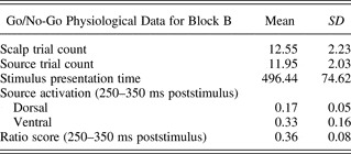
Means and standard deviations for the go/no-go measures are presented in Table 2. Data were entered into a three-step linear regression model, where the dependent measure was N2 amplitudes at site FCz. The first step of the regression model controlled for age, gender, ERP trial count, mean stimulus presentation time, and medication, because these variables may all affect N2 amplitudes. The second step in the regression model controlled for the number of parent–child visits to a negative state. This was important because flexibility scores capture a dyad's movement around the state space (representing emotional flexibility), and movement in and out of negative regions of the state space could inflate flexibility estimates excessively. Moreover, emotional flexibility, not negative emotion, was the variable hypothesized to be linked with frontal regulatory mechanisms. In the third step, each of the two dyadic flexibility measures (TRANS and DISP) was entered in turn. After this preliminary analysis, the regression was repeated with the nonsignificant predictor variables removed. Next, influential cases (cases with Studentized deleted residuals ≥ 2) were removed from the analysis and the regression model was repeated. The results revealed no significant relation between dyadic flexibility scores and children's N2 amplitudes.
Means and standard deviations for go/no-go measures

Source-space analyses
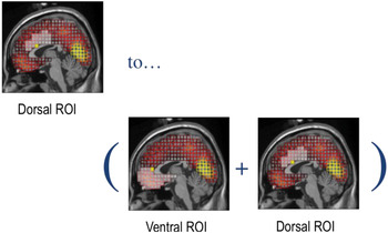
Source analyses were performed on spatially reduced data in the 100-ms time window corresponding to the peak latency of the N2 on the grand-averaged waveform (250–350 ms). From the sample of 65 clinical children, individuals with low source segment counts (<10) were excluded, leaving 42 children. We assumed that overall frontal activation was an individual-difference variable with little significance for our analysis. Therefore, consistent with past approaches (for a review, see Drevets & Raichle, Reference Drevets and Raichle1998), we targeted the contribution of dorsomedial activation in relation to ventral activation by calculating the ratio of dorsomedial activation to total prefrontal (dorsal + ventral) activation. ROIs are shown in Figure 1. As indicated in the figure, the ratio score was calculated by dividing the maximal dorsal source activation by the total (maximal dorsal and maximal ventral) source activation [D/(D + V)] in the 100-ms time window containing the average peak N2 amplitude (250–350 ms).
The dorsal and ventral regions of interest superimposed on a standard magnetic resonance imaging head map. Voxel activation was recorded in each of these regions, from which maximum activation values were computed and ratio scores derived. [A color version of this figure can be viewed online at http://journals.cambridge.org/dpp]

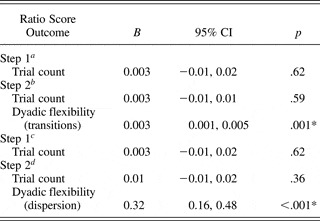
The ratio measure was entered as the dependent variable in a three-step linear regression model. As before, the first step controlled for gender, age, medication, mean stimulus presentation time, and trial count. The second step controlled for the number of parent–child visits to a negative state. In the third step, each of the two dyadic flexibility measures were entered in turn. As before, the regression analysis was repeated with the nonsignificant predictor variables and highly influential cases (cases with Studentized deleted residuals ≥ 2) removed. Table 3 presents the results. Both dyadic flexibility measures were significantly associated with the ratio of dorsal to ventral source activation. Thus, in support of our second hypothesis, greater dyadic flexibility predicted a higher ratio score (more dorsomedial to ventral activation), suggesting that children from more flexible dyads recruited relatively more dorsomedial activity.
Regression results showing dyadic flexibility scores are significantly associated with dorsomedial to ventral source activation

aR 2 = .08, F (1, 37) = 0.25, p = .62.
bR 2 = .56, F (2, 36) = 6.63, p = .004.
cR 2 = .08, F (1, 37) = 0.25, p = .62.
dR 2 = .52, F (2, 36) = 8.37, p = .001.
*p < .05.
Discussion
The main goal of the current study was to examine the link between two very different levels of ER processes in a sample of children referred for treatment for externalizing behavior problems. The first level was behavioral manifestations of parent–child flexibility in the context of a dyadic problem-solving task. The second level was the neurocognitive processes that subsume ER. Families referred for behavior problems showed a wide range of parent–child flexibility. In partial support of our first hypothesis, one of the two measures of parent–child flexibility, dispersion, was negatively associated with severity of externalizing symptoms. These results are consistent with past studies that have found a relation between parent–child rigidity and the subsequent development of externalizing behavior problems (Hollenstein et al., Reference Hollenstein, Granic, Stoolmiller and Snyder2004; Lunkenheimer et al., Reference Lunkenheimer, Olson, Hollenstein, Sameroff and Winter2011), and they are in line with previous findings that showed improvements in externalizing problems corresponding with increases in parent–child flexibility over the course of treatment (Granic et al., Reference Granic, O'Hara, Pepler and Lewis2007). It should be noted that the correlations were weak, however, and only held for one of the two flexibility measures. Thus, parent–child flexibility seems to be related to behavior problems for some children, but certainly not all. Where emotional rigidity is a factor, it may be either a cause or a consequence of behavior problems. If we assume that pathologies such as childhood aggression are built into temperament (e.g., Stadler et al., Reference Stadler, Sterzer, Schmeck, Krebs, Kleinschmidt and Poustka2007), then it makes sense that they would precede and predict ineffective emotional communication in the family as a whole. This perspective is consistent with findings of structural brain anomalies in aggressive children (Sterzer et al., Reference Sterzer, Stadler, Krebs, Kleinschmidt and Poustka2005; van Goozen et al., Reference van Goozen, Fairchild, Snoek and Harold2007) and adults (Blair, Reference Blair2001; Hoptman, Reference Hoptman2003). However, if we assume that parenting problems precipitate aggressive behavior in children (e.g., Dumas & LaFreniere, Reference Dumas and LaFreniere1993; Dumas, LaFreniere, & Serketich, Reference Dumas, LaFreniere and Serketich1995; Patterson, Reference Patterson1982; Patterson, Reid, & Dishion, Reference Patterson, Reid and Dishion1992; Snyder & Patterson, Reference Snyder and Patterson1995), then the causal direction would be reversed. Our preferred model of the association between low dyadic flexibility and child psychopathology highlights a feedback relation in which rigid parenting leads to behavior problems that further shut down parent–child reciprocity and flexibility (Granic & Patterson, Reference Granic and Patterson2006).
The larger purpose of this research was to look for associations between emotional flexibility in parent–child interactions and the relative activation of two prefrontal regions assumed to mediate ER: the ventral prefrontal region associated with immediate, reactive regulation and the dorsomedial region associated with more deliberate, strategic control. We hypothesized higher dorsal (relative to ventral) activation for more flexible dyads and, when comparing dorsal/ventral activation values, this hypothesis was confirmed. Greater dyadic flexibility (measured both by state transitions and overall dispersion of behavior) predicted a higher ratio of dorsomedial to ventral activation. This association was robust, accounting for more than 25% of the variance.
Of course, this study was cross-sectional; therefore, causal directions cannot be established. However, it seems important to consider the various possibilities. These associations can be interpreted according to the imputed causal direction between child psychopathology and parent–child interactions but also with reference to the specific nature of behavior problems in our sample. If dispositional differences in brain structure and function precede interaction style, then children who regulate emotions reactively and spontaneously may set up dyadic patterns that are habitual and inflexible. Because of biological predispositions, perhaps these children have a narrow response repertoire when emotionally challenged, so their parents eventually reciprocate with an equally narrow repertoire. However, aggressive individuals with an impulsive, risk-taking style often show less rather than more ventral prefrontal activation (Blair, Reference Blair2001; Davidson, Putnam, & Larson, Reference Davidson, Putnam and Larson2000; Hoptman, Reference Hoptman2003) as did the infamous Phineas Gage (Damasio, Reference Damasio1994). In contrast, high ventral activation is associated with childhood anxiety (McClure et al., Reference McClure, Monk, Nelson, Parrish, Adler and Blair2007; Monk et al., Reference Monk, Nelson, McClure, Mogg, Bradley and Leibenluft2006), consistent with the elevated internalizing scores for most of our sample. In two previous studies with similarly referred children for aggressive behavior problems, children actually turned out to have clinically elevated levels of internalizing symptoms as well (e.g., anxiety); through treatment, these children showed initially high levels of ventral prefrontal activation that decreased when clinical symptoms declined (Lewis et al., Reference Lewis, Granic, Lamm, Zelazo, Stieben and Todd2008; Woltering et al., Reference Woltering, Granic, Lamm and Lewis2011). Thus, relatively high vPFC activation appears characteristic of this population, and it appears not to be biologically fixed.
It seems unlikely that internalizing symptoms and related patterns of cortical activity precede the formation of dyadic habits. On the contrary, it may be that high ventral to dorsal ratios, corresponding to a vigilant style of appraisal and biased attention to threat, result from parenting practices that are sometimes harsh and inconsistent (e.g., Bögels & Brechman-Toussaint, Reference Bögels and Brechman-Toussaint2006; Dadds, Barrett, Rapee, & Ryan, Reference Dadds, Barrett, Rapee and Ryan1996) but have settled into an uneasy truce, marked by defensive avoidance of emotional expression. Thus, we posit the development of a feedback cycle in which children's aggression and anxiety emerge from insensitive parenting, resulting in further decrements in flexible dyadic interactions. Future longitudinal studies will be needed to track brain and behavioral interactions as they proceed over development for aggressive children. Yet the present results build on our previous findings, suggesting that these children show a specific neural signature of ER, with greater ventral relative to dorsomedial prefrontal activation. The present results also go a step further, indicating that this neural signature corresponds with rigidity in the emotional transactions between externalizing children and their parents.
Limitations and future directions
A number of problems limit the conclusions that can be drawn from this study. First, most but not all clinically referred children were comorbid for internalizing and externalizing problems. With a larger group of participants, and perhaps with finer instruments than the CBCL, we could have partitioned our sample into pure-aggressive and anxious/aggressive children. Given our model, only the anxious/aggressive children should show high ventral activation associated with dyadic rigidity. Future studies with more refined measures and/or larger samples would help us to disentangle and identify subgroup differences at the neural and behavioral levels. Second, this study was cross-sectional, limiting any sort of causal explanations. We speculated about feedback processes between parent–child behavioral and brain manifestations of ER, but it is critical to follow this study up with a longitudinal one that can help establish whether parent–child flexibility results from or contributes to neurocognitive styles of ER and vice versa. Third and finally, EEG source analysis remains a controversial method for drawing conclusions about anatomical locations in the cortex. Because the ventral and dorsal prefrontal generators were identified and ROIs were selected a priori, we were not as concerned with error as we would be if regions were simply extracted from the source-space data. Nevertheless, functional magnetic resonance imaging methods would allow us far more certainty in comparing the activation values of anatomically defined structures.
Despite these concerns, the present study is the first to attempt to integrate observational data on parent–child interactions with neural markers of ER, it did so specifically with children referred for serious behavior problems, and it was based on hypotheses derived from a research program investigating the neural correlates of ER in aggressive children. As such, we trust that it can help expand the repertoire of approaches for linking diverse kinds of data in the study of child psychopathology.