Introduction
Recent technological advances, excellent picture resolution and greater availability have made high-resolution computed tomography (CT) imaging of the temporal bone an essential tool in the management of ear disorders. High-resolution CT has made it possible for clinicians to study middle- and inner-ear pathology prior to undertaking surgery. This is particularly useful when the disease process has altered the anatomy and/or resulted in complications such as a lateral canal fistula and/or facial canal or tegmen erosion.Reference Tatlipinar, Tuncel, Öğredik, Gökçeer and Uslu1
Although there have been several studies reporting the value of high-resolution CT for investigations of cholesteatoma, temporal bone tumours and congenital anomalies, studies on the prevalence of normal anatomical variations of the temporal bone are lacking.Reference Jackler, Dillon and Schindler2–Reference Brennan and Walsh4 As the use of high-resolution CT imaging for temporal bone investigations is now widespread, we feel it is necessary for the investigating otologist to be aware of common anatomical variations and their clinical implications.
This study aimed to identify the prevalence of normal variations of temporal bone anatomy on high-resolution CT imaging and discuss their clinical importance.
Materials and methods
A retrospective review was conducted of high-resolution temporal bone CT imaging performed at NHS Greater Glasgow and Clyde (Glasgow, UK) over an eight-year period (2006–2014). The presence of five variants was determined. These variants were: a high dehiscent jugular bulb, an anteriorly located sigmoid sinus, a deep sinus tympani, an enlarged cochlear aqueduct and an enlarged internal auditory meatus (IAM).
A GE LightSpeed 16, four-slice, helical CT scanner (Fairfield, Connecticut, USA) was used for all patients. The scan parameters were as follows: 1 mm slices, smart Ma (50–250 Ma), 120 kV, 0.8-second rotation time, 0.562:1 pitch factor and 5.62 mm rotation speed. All scans covered an area from 1 cm inferior to the mastoid tip to 1 cm superior to the petrous temporal bone.
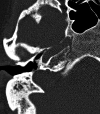
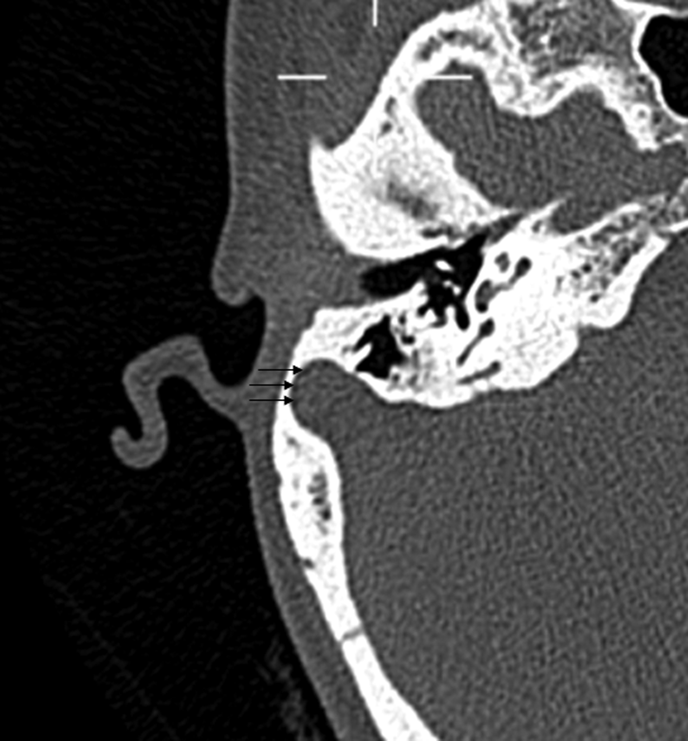
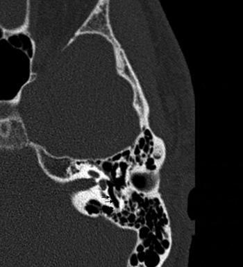
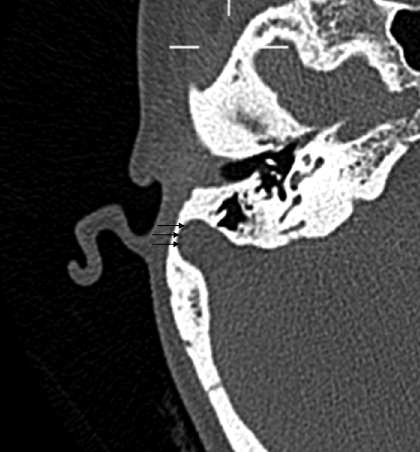
The following five criteria were used to identify and record the frequency of each variation. A high dehiscent jugular bulb was defined as extension of the jugular bulb into the middle ear (Figure 1), with vertical extension above the inferior bony annulus.Reference Subotić5 An anteriorly located sigmoid sinus was reported when there was no anteroposterior distance between the posterior wall of the bony external auditory canal and sigmoid sinus (Figure 2).Reference Potter6 A deep sinus tympani (Figure 3) was determined when the depth of the sinus tympani visualised on the axial plane was over 6 mm.Reference Saito, Igarashi, Alford and Guilford7 An enlarged cochlear aqueduct was reported when the anteroposterior diameter of the entire duct (from its medial to lateral extent) was over 2 mm.Reference Mukherji, Baggett, Alley and Carrasco8 An enlarged IAM was defined as an IAM measuring over 8 mm in its mid portion.Reference Ebenius9
High-resolution axial plane computed tomography image demonstrating a high dehiscent jugular bulb on the right side.

High-resolution axial plane computed tomography image demonstrating an anteriorly located sigmoid sinus on the right temporal bone (arrows).
High-resolution axial plane computed tomography image of the left temporal bone demonstrating a deep sinus tympani (arrows).

Results
A total of 186 patients were identified and 372 high-resolution temporal bone CT images were examined. Patients who had a history of middle- or inner-ear surgery, tumours or congenital anomalies involving the temporal bone were excluded from our study. Altogether, 33 scans were excluded; hence 339 images were eligible for inclusion in this study.
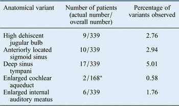
Although the degree of pneumatisation varied from diseased and normal temporal bones within the same patient, our study primarily recorded the prevalence of five normal variants of the temporal bone. In this regard, there was no difference in the prevalence of anatomical variations studied between the right or left temporal bones. The different anatomical variants and their frequencies are summarised in Table I.
Incidence of normal anatomical variants on temporal bone computed tomography imaging

*Cochlear aqueduct visualised convincingly only in 168 patients out of 339.
Discussion
High-resolution temporal bone CT imaging has become a key investigation in the management of ear disorders. Although the primary responsibility of reporting on CT images lies with a head and neck radiologist, it is nevertheless important for the practising otologist to be aware of common anatomical variants and their clinical relevance.
High dehiscent jugular bulb
The jugular bulb is the point where the sigmoid sinus feeds into the jugular vein. It is normally located below the posterior part of the middle-ear floor. The incidence of a high dehiscent jugular bulb in our series was 2.76 per cent. Other published series report an incidence of up to 34 per cent.Reference Moore10, Reference Koesling, Kunkel and Schul11 A dehiscent jugular bulb is susceptible to injury during tympanomastoid surgery, which can result in unexpected haemorrhage.Reference Ball, Elloy, Vaidhyanath and Pau12 In the event of an injury, surgery should be discontinued; the patient should be placed in a head-down position and the ear packed.
Anteriorly located sigmoid sinus
An anteriorly located sigmoid sinus limits the amount of space for a postaural approach to the mastoid antrum. Ten patients in our series demonstrated no anteroposterior space between the most anterior aspect of the sigmoid sinus wall and the posterior aspect of the external auditory canal wall. Prior to undertaking mastoidectomy, it is important to study the location of the sigmoid sinus in order to avoid unnecessary injury.
Deep sinus tympani
The sinus tympani is a recess that lies medial to the pyramidal eminence and facial canal. Located in its vicinity are the round window, facial nerve canal and posterior semicircular canal. The sinus tympani is frequently involved in cholesteatoma cases, and knowledge of its depth in the axial plane helps the otologist to plan the surgical approach. A deep sinus tympani appears to be the most common anatomical variant, with some studies reporting an incidence of up to 30 per cent.Reference Niemczyk, Bruzgielewicz, Wysocki, Nitek and Olesinski13 In addition, Nitek et al. reported that the presence of deep sinus tympani correlated with a prominent facial canal.Reference Nitek, Wysocki, Niemczyk and Ungier14
Enlarged cochlear aqueduct
The cochlear aqueduct is a narrow bony canal that connects the scala tympani to the subarachnoid space of the posterior fossa. An enlarged cochlear aqueduct can be associated with congenital hearing loss and a risk of perilymphatic gusher following stapes surgery. We observed two patients with an enlarged cochlear aqueduct. The cochlear aqueduct can be a difficult anatomical structure to visualise because of its small diameter. The cochlear aqueduct was visualised in only 168 patients in our series (49 per cent). Jackler and Hwang reviewed over 100 CT images, and reported that only 31 per cent of patients had a cochlear aqueduct that could be visualised in its entirety.Reference Jackler and Hwang15
• High-resolution computed tomography imaging is a key investigation in temporal bone disorder management
• Normal anatomical variations are not uncommon; the investigating otologist should be aware of such variants
• A deep sinus tympani was the most common variant noted in our series (5.1 per cent)
• Knowledge of such variations is crucial prior to undertaking surgery to avoid unnecessary and avoidable complications
Enlarged internal auditory meatus
There has been some debate regarding the normal size of the IAM. Ebenius reported that the diameter of the porus portion of IAM ranged from 4 to 8 mm (average of 5.7 mm).Reference Ebenius9 Valvassori reported that the diameter of the lateral end of the canal ranged from 2 to 8 mm (average of 4 mm).Reference Valvassori16 Six patients in our series demonstrated an IAM of over 8 mm at its mid portion. An enlarged canal differs from an abnormally dilated IAM secondary to a vestibular schwannoma by the presence of an intact cortex and crista falciformis on coronal images. None of our six patients demonstrated any abnormalities of the cortex or transverse crest. Some authors have reported a correlation between an enlarged IAM and sensorineural hearing loss, but the significance of this is questionable.Reference Migirov17, Reference Weinberg, Kim and Gore18
Conclusion
Normal anatomical variations of the temporal bone are not uncommon. It is vital for the investigating otologist to be aware of such variations when considering differential diagnoses of temporal bone disorders and also prior to undertaking surgery. Furthermore, it is crucial that the investigating radiologist is aware of such variations and flags them as necessary to aid effective patient care.
Acknowledgement
We sincerely thank and acknowledge Dr Deepak Vishwanath (Department of Radiology, Southern General Hospital, Glasgow, UK) for helping us access the images and interpret the relevant findings.