Introduction
While percutaneous transcatheter closure is generally regarded as safe and effective compared to surgical repair, it is associated with rare complications, one of which is haemolysis, occurring in 1% to 3% of all cases.Reference Amoozgar, Soltani and Edraki1 Although there is substantial literature addressing haemolysis following ventricular septal defect and patent ductus arteriosus closures, information on post atrial septal defect closure is limited.Reference Amoozgar, Soltani and Edraki1 In the context of hemodynamics associated with ASD, a low-pressure gradient between the left atrium and right atrium is unlikely to result in haemolysis, even in the presence of a residual shunt. This report details our experience with a case of significant haemolysis following atrial septal defect device closure in a 10-year-old female, highlighting the encountered dilemma, a stormy postoperative course, with a brief discussion on potential aetiology and management strategies.
Case report
Presentation
A 10-year-old female weighing 29 kg and height 143 cm, hailing from a tribal community of remote North-East India, was referred to us for management of ASD detected during health check-up. She presented with a 1-year history of shortness of breath on exertion and palpitations, which have worsened over the last 2 months (NYHA III) despite diuretic therapy. The second heart sound was prominent, and a grade 2/6 systolic murmur was heard at the left midsternal border during physical examination. Baseline SpO2 recorded was 98%.
Investigations
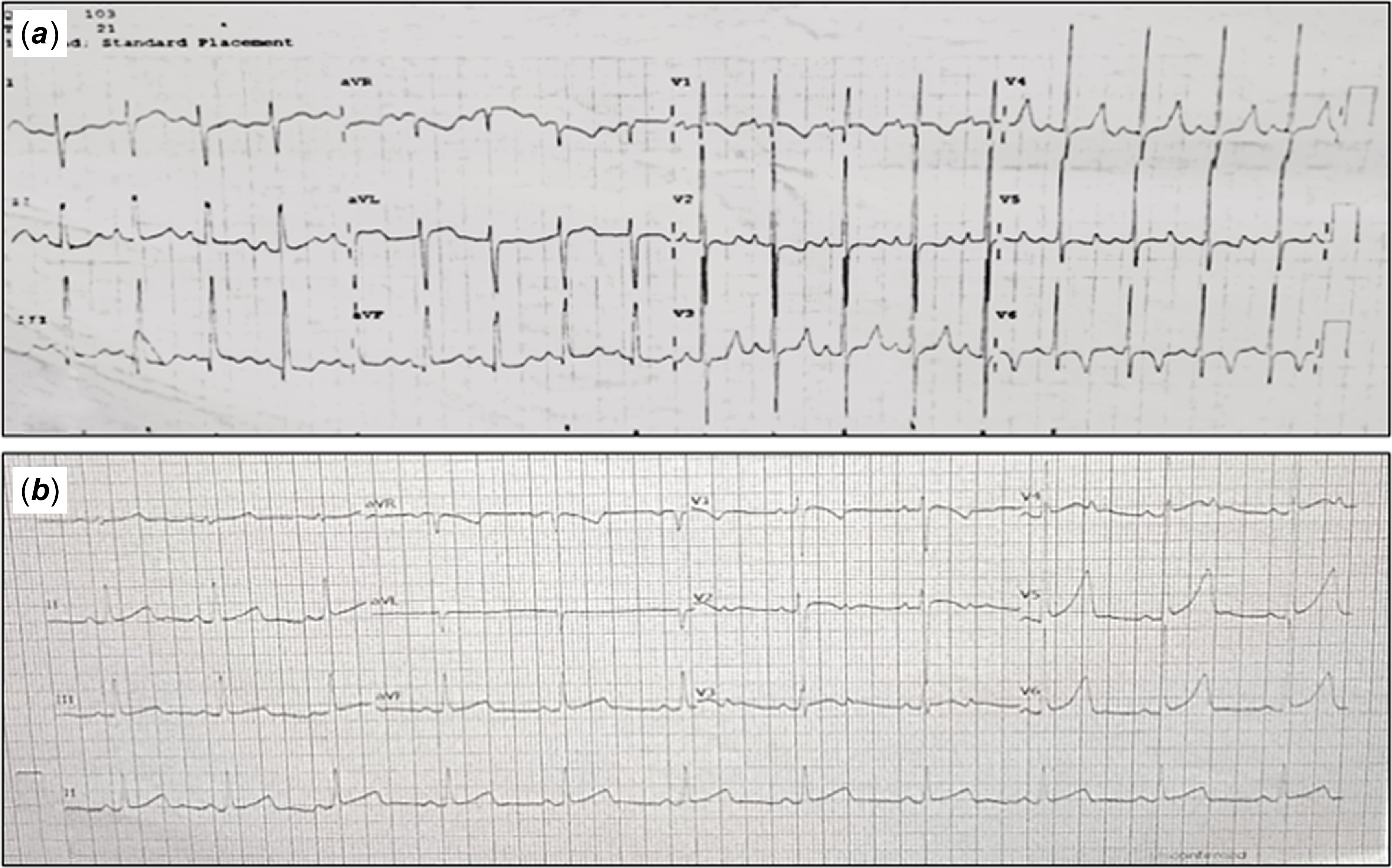
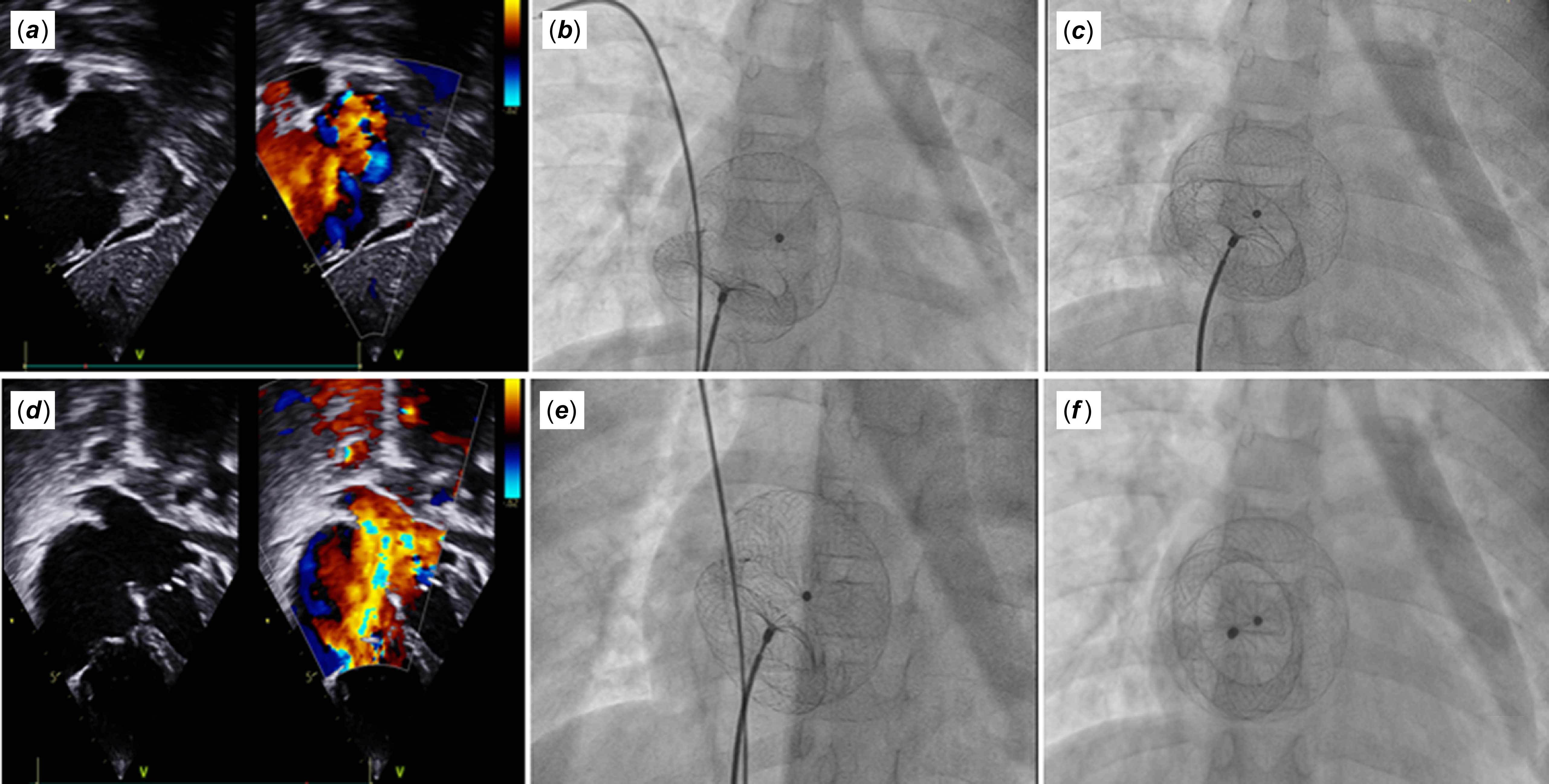
Electrocardiogram showed sinus rhythm with features of right ventricle overload with incomplete right bundle branch block (Figure 1a), and echocardiography revealed the presence of a large ostium secundum-type ASD measuring 34 mm with a small posterior rim 4 mm and inferior caval vein rim around 5 mm with floppy in nature, all other rims were adequate and were more than 6 mm. The total atrial septum measures 49 mm in length. She also had mild mitral valve prolapse with Grade I mitral regurgitation and Grade II tricuspid regurgitation with a peak gradient of 42 mmHg, with mild to moderate pulmonary artery hypertension with dilated right-sided heart chambers on echocardiography (Figure 2a and b).
( a ) Preoperative ECG showing sinus rhythm with RV forces with right atrial enlargement. ( b ) Postprocedure ECG showing dominant LV forces with left atrial enlargement.

Figure 1. Long description
Panel A: Preoperative ECG showing sinus rhythm with right ventricular (RV) forces and right atrial enlargement. The graph displays multiple leads labeled AVR, AVL, AVF, V1, V2, V3, V4, V5, and V6. The waveform patterns indicate the electrical activity of the heart before the procedure. Panel B: Postprocedure ECG showing dominant left ventricular (LV) forces and left atrial enlargement. This graph also displays multiple leads labeled AVR, AVL, AVF, V1, V2, V3, V4, V5, and V6. The waveform patterns indicate the electrical activity of the heart after the procedure.
( a ) and ( b ) Echocardiographic images depicting ASD measuring 34 mm with a small posterior rim. ( c ) and ( d ) Placement of a 40 mm amplatzer atrial septal occluder via the assisted catheter technique. ( e–f ) Showcase final device position before and after release.

Procedure
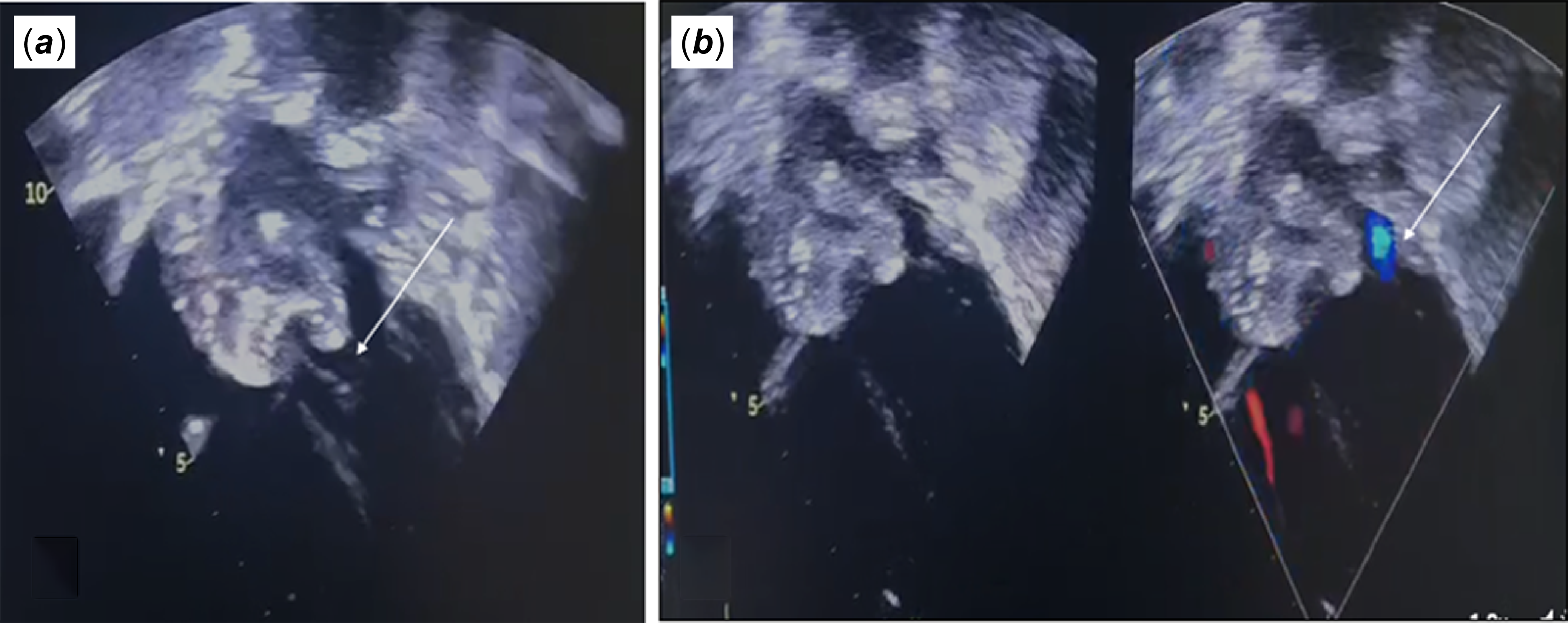
Considering two borderline ASD rims and pulmonary artery hypertension, parents were offered surgical closure of atrial septal defect; however, parents were unwilling to undergo surgical closure due to some ethnic and social stigma attached to cardiac surgery. As her symptoms were worsening despite anti-failure medications with the onset of pulmonary artery hypertension, we opted for atrial septal defect device closure after the parents’ consent. After obtaining consent and preoperative preparation, diagnostic catheterisation was performed, revealing a mean pulmonary artery pressure of 32 mmHg with Qp: Qs 2.4: 1 and baseline PVRi of 1.8 Woods units.m2. With two borderline rims and a floppy nature of the IVC rim, a 40 mm Amplatzer TM atrial septal defect occluder (original defect + additional 6 mm) was selected with sizing of 117% to the original defect and 137% device/weight ratio and was implanted via right femoral venous approach through a 12F Amplatzer delivery sheath using LUPV-assisted technique with additional catheter support from left femoral vein (Figure 2c–f). Post-implantation fluoroscopy showed a stable device position. Intraoperative echocardiography before release showed the acquisition of all rims by the occluder, a small intra-device residual shunt with mild increase in previous mitral regurgitation and tricuspid regurgitation, and separation of the device and valves during diastole was observed (Figure 3). Post-procedure mPAP came down to 18 mmHg. After the procedure, the patient was treated with heparin and transferred to the paediatric ICU with continuous ECG monitoring.
Intra-procedure Echo post deployment of device: ( a ) showing dynamic interaction of device withmitral valve with separation during diastole, ( b ) colour Doppler showing GradeI-II MR post deployment on table assessment.

Postoperative course
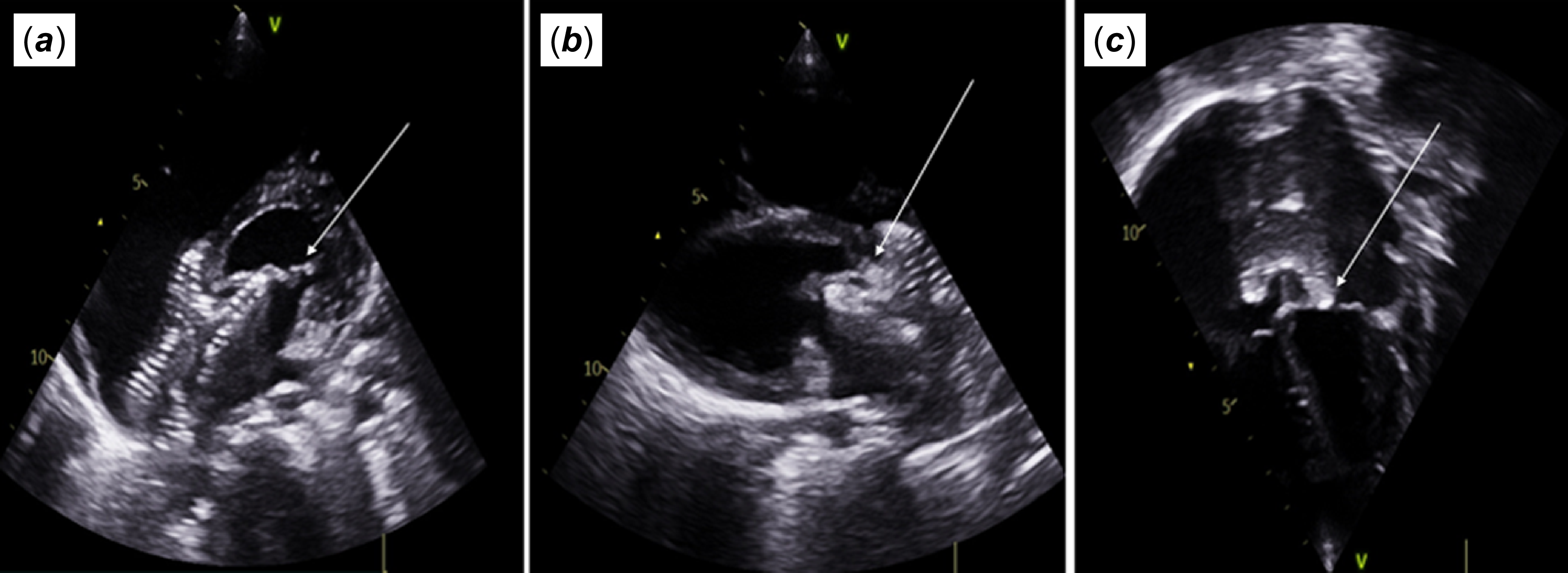
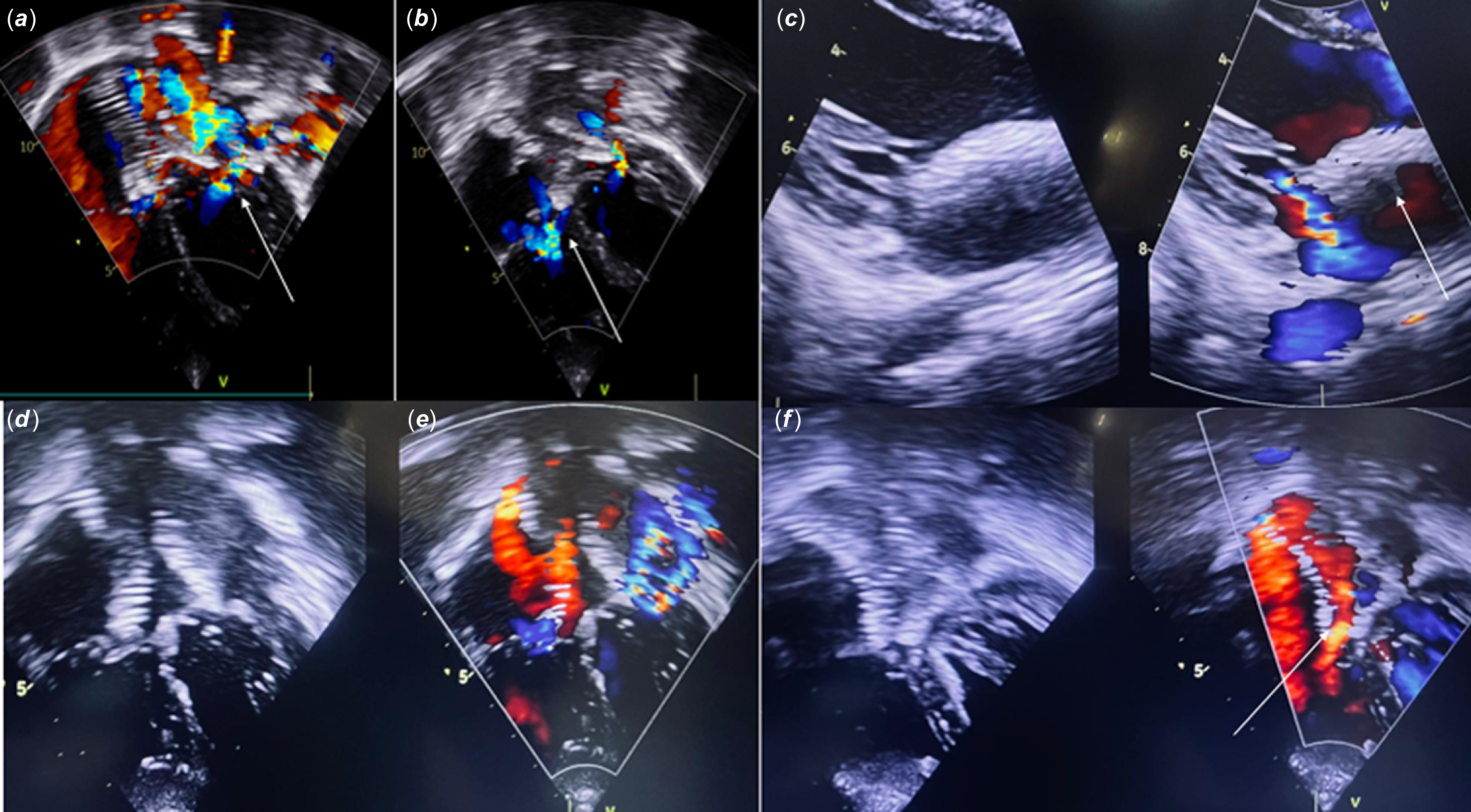
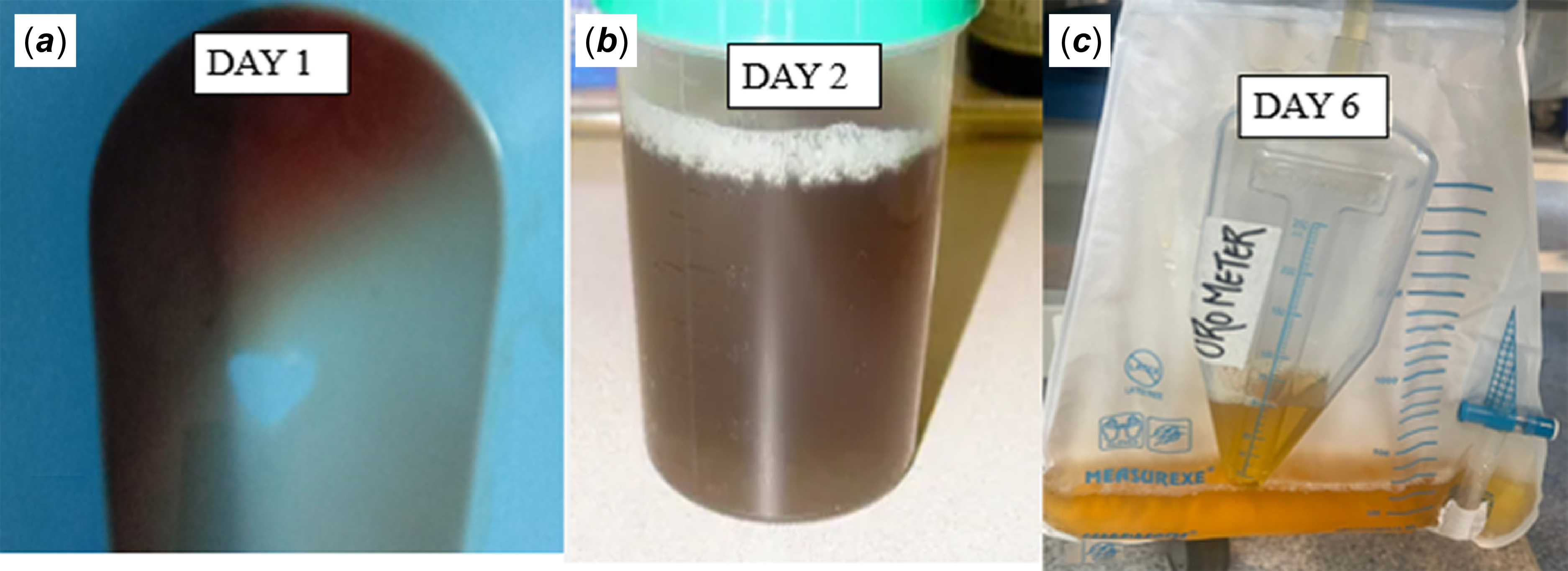
On the next day after the operation, the patient developed severe haemolysis marked by dark cola coloured urine, and her haemoglobin fell from 11 g/dL to 8 g/dl. Anticoagulation was ceased, while sodium bicarbonate for urinary alkalisation was immediately instituted with intravenous fluid infusion. Echocardiography showed a persistent residual shunt, but with a significant increase in the severity of mitral regurgitation, which was not present previously and was initially attributed to increased left ventricular end-diastolic pressure due to shunt closure and the patient was started on IV furosemide with ACE inhibitors. Workup for haemolysis showed a reticulocyte count of 2.5%, lactate dehydrogenase 1883 U/l, with normal PT, APTT and INR. With this, there was an increase in serum urea (48 mg/dl) and creatinine level (1.2 mg/dl). Other investigations, including the Coombs test and ANA profile, were negative. On the second day, her haemoglobin dropped to 7.8 g/dL for which 1 unit of PRBC was transfused, but her vitals were stable and the colour of urine started to turn lighter, Next day, detailed echocardiography revealed shift in configuration of device with persistence of intra-device flow across the atrial septal defect device, and the left disk seen to impinge on the anterior mitral leaflet during systole, causing increase in intensity of mitral regurgitation and jet being directed towards the disk of device. Dynamic interaction of the right disk and the septal leaflet of the tricuspid valve was also observed during systole (Figure 4). There was a change in the configuration of the device compared to immediately post-procedure, and the mechanical exposure of the atrial septal defect device due to the blood flow from atrioventricular valve regurgitation with increased erythrocyte fragility was attributed to haemolysis (Figure 5). Prompt surgical removal of the device was recommended, but the family declined and requested more time for discussion despite repeated counselling. With continuous afterload reduction and diuresis and conservative treatment, the urine colour changed gradually from cola coloured to normal on day 6, with post-transfusion Hb 9.2 g/L, with a decreasing trend of lactate dehydrogenase to 683 U/l (Figure 6). With further management, a gradual improvement was achieved in haemolysis parameters, renal function, and haemoglobin levels. The patient was successfully discharged on the 6th day post-procedure on antifailure and iron therapy. Danger signs, close follow-up. and future retrieval of the device were explained to guardians.
2D Echocardiography images showing impingement of the ASD device disk over AML and STL leaflets of the mitral and tricuspid valve.

( a–b ) Arrow highlighting mitral and tricuspid regurgitation resulting from mechanical interferencepost ASD device, ( c–d ): severe mitral regurgitation with 2 jets in four chamber and long axis view, ( e ): depictsintra-device shunting due to high velocity MR jet hitting ASD device.

Post-procedure urine appearance, ( a – b ) dark cola coloured urine on days 1 and 2, ( c ): lighter appearance of urine on day 6 with conservative measures.

Follow-up
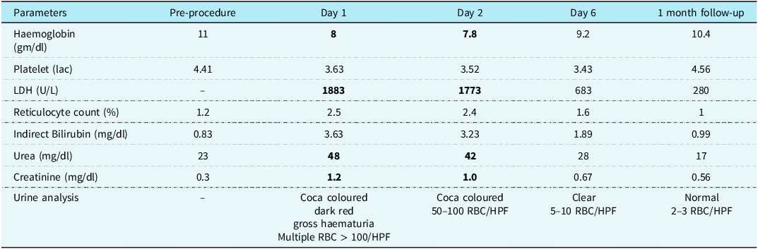
Regular follow-up at 14th day and 1month after the closure showed her haemoglobin increased to 10.6 g/dL, and the urine test showed no evidence of subclinical haemolysis with lactate dehydrogenase level 280 U/l in the latest follow-up (Table 1). Echo showed a persistence of residual shunt across the device, and her mitral valve regurgitation decreased from severe to moderate, which may have resulted in cessation of haemolysis with sinus rhythm in ECG (Figure 1b). However, as there is still ongoing mitral valve and device mechanical interference and associated future risks, guardians were counselled to undergo surgical retrieval of the device and mitral valve repair for the benefit of the patient; however, the patient was lost to follow-up thereafter despite various communication efforts.
Laboratory parameters of the case from pre-procedure to 1 month follow-up

Table 1. Long description
A table comparing laboratory parameters over time. The table has 8 rows and 6 columns. The columns are labeled Pre-procedure, Day 1, Day 2, Day 6, and 1 month follow-up. The rows are labeled with different parameters: Haemoglobin (gm/dl), Platelet (lac), LDH (U/L), Reticulocyte count (%), Indirect Bilirubin (mg/dl), Urea (mg/dl), Creatinine (mg/dl), and Urine analysis. Row 1: Haemoglobin (gm/dl), Pre-procedure 11, Day 1 8, Day 2 7.8, Day 6 9.2, 1 month follow-up 10.4. Row 2: Platelet (lac), Pre-procedure 4.41, Day 1 3.63, Day 2 3.52, Day 6 3.43, 1 month follow-up 4.56. Row 3: LDH (U/L), Pre-procedure -, Day 1 1883, Day 2 1773, Day 6 683, 1 month follow-up 280. Row 4: Reticulocyte count (%), Pre-procedure 1.2, Day 1 2.5, Day 2 2.4, Day 6 1.6, 1 month follow-up 1. Row 5: Indirect Bilirubin (mg/dl), Pre-procedure 0.83, Day 1 3.63, Day 2 3.23, Day 6 1.89, 1 month follow-up 0.99. Row 6: Urea (mg/dl), Pre-procedure 23, Day 1 48, Day 2 42, Day 6 28, 1 month follow-up 17. Row 7: Creatinine (mg/dl), Pre-procedure 0.3, Day 1 1.2, Day 2 1.0, Day 6 0.67, 1 month follow-up 0.56. Row 8: Urine analysis, Pre-procedure -, Day 1 Coca coloured dark red gross haematuria Multiple RBC > 100/HPF, Day 2 Coca coloured 50-100 RBC/HPF, Day 6 Clear 5-10 RBC/HPF, 1 month follow-up Normal 2-3 RBC/HPF.
Bold values represents variation from normal range.
Discussion
The incidence of haemolysis subsequent to atrial septal defect device closure is exceedingly rare and predominantly documented in three case reports as per the literature review.Reference Butera, De Rosa and Chessa2 Manli Yu et alReference Lambert, Belli, Piot, Planche and Losay3 and Lambert V et alReference Yu, Shan, Bai, Qin and Zhao4 reported an analogous case of acute severe haemolysis following atrial septal defect device closure.
The mechanism of intravascular haemolysis associated with intracardiac mechanical devices is primarily linked to high flow rates and residual shunt jets, commonly seen after the closure of ventricular septal defect and patent ductus arteriosus.Reference Cannata, Cantoni, Sciortino, Bruschi and Russo5 Rapid acceleration–deceleration velocities from residual leaks, along with mal-aligned device configurations, can create significant shear stress on red blood cells, resulting in haemolysis, which is rare after atrial septal defect device closures.Reference Cannata, Cantoni, Sciortino, Bruschi and Russo5–Reference Lu, Lai and Liu7 Our case highlights an unusual residual shunt associated with the large atrial septal defect device, characterised by severe mitral regurgitation and a high-velocity jet directed towards the device, which contributes to increased erythrocyte fragility and shear and ultimately leads to erythrocyte rupture.Reference Blackshear, Fung, Perrone and Anliker6,Reference Lu, Lai and Liu7 Though the primary mechanism remains mechanical, other causes, such as autoimmune haemolytic anaemia, should also be considered in these situations.Reference Lu, Lai and Liu7
We postulated that inadequate rims of the ASD, along with device oversizing resulting in abnormal configuration of the device, along with smaller left atrial size attributed to a large atrial septal defect, malaligned defects, and pre-existing mitral regurgitation are the potential risk factors that contribute to mechanical interference between the device and the atrioventricular valve, ultimately leading to haemolysis.Reference Lambert, Belli, Piot, Planche and Losay3,Reference Cannata, Cantoni, Sciortino, Bruschi and Russo5,Reference Sallam and Hwang8 Second, ASD, being a dynamic shunt reorientation of the device post-deployment within 24 hours, especially with small rims, can also affect the device’s mechanical interaction with valves, as highlighted in our case. Furthermore, in cases of larger defects, thrombosis within the device occurs at a comparatively slower rate, leading to the persistence of a residual shunt that promotes haemolysis.
Management strategies for haemolysis are contingent upon the clinical presentation. In ventricular septal defect and patent ductus arteriosus cases, subacute haemolysis often resolves with conservative measures, while acute severe haemolysis requires intensive care and close monitoring and device retrieval.Reference Mulvaney and Grech9,Reference Spence, Thomson, Weber and Qureshi10 In exceptional cases, post-atrial septal defect device haemolysis necessitates the prompt surgical retrieval of the device. Key laboratory evaluations—such as lactate dehydrogenase, reticulocyte count, and urine analysis—are essential for assessing ongoing haemolysis, and monitoring renal function is crucial in these cases.Reference Spence, Thomson, Weber and Qureshi10 Conservative management involving intravenous fluids, sodium bicarbonate, dexamethasone, blood transfusions, and medications for heart failure may be utilised temporarily as a bridge to definitive repair. Nevertheless, the surgical removal of the device remains the preferred treatment and should be conducted at the earliest opportunity. Discontinuing aspirin and heparin can promote fibrin and platelet adhesion to the device, forming an adhesive tissue layer that reduces red blood cell interaction with the nickel–titanium fabrics and alleviates haemolysis.Reference Godart, Rodes and Rey11,Reference Anil, Sivakumar, Philip, Francis and Kumar12 In instances of severe renal failure, dialysis or plasmapheresis may be necessary.
Although haemolysis has been successfully managed and the patient remains asymptomatic in our case report, we anticipated that, for ongoing mechanical interference between the device and the mitral valve, device retrieval is necessary. We believe that the reduction in left ventricular end-diastolic pressure following heart failure treatment, combined with an increase in left atrial and left ventricular dimensions, the shifting of the interventricular septum towards the right ventricle and right auricle and right ventricle remodelling, promotes alignment of the oversized device. Manli Yu et al.Reference Lambert, Belli, Piot, Planche and Losay3 reported that the oversized closure device is not well aligned to the wall initially but flattens out on the first day or two. This alignment may decrease the severity of mitral regurgitation, leading to a reduction in residual shunt and alleviation of haemolysis, but persistent mechanical interference should be subjected to surgical repair to prevent future valve damage, as seen in our case, where mechanical interaction persists.
An important aspect highlighted by this case is the influence of socio-economic, geographic, and cultural factors on treatment decisions in resource limited and remote settings. Despite clear recommendations for surgical closure in patients with large atrial septal defects and unfavourable septal anatomy, some families remain reluctant to accept open-heart surgery because of fear of surgical risks, financial constraints, limited access to specialised cardiac surgical centres, and prevailing social beliefs. These factors may lead to the selection of higher-risk transcatheter interventions despite suboptimal anatomy. Although such real-world challenges should not alter evidence-based management, they underscore the importance of comprehensive pre-procedural counselling, shared decision-making, and multidisciplinary Heart Team discussions to ensure that patients and families make informed treatment choices.
Through this case report, we aim to emphasise certain limitations with transcatheter closure of large atrial septal defects as follows:
-
1. Despite adherence to standard transcatheter sizing guidelines for evaluating deficient rims in atrial septal defect closure, catastrophic haemolysis may still occur with oversizing. Oversizing presents considerable risks, despite instances where oversizing has been reported at levels ranging from 150% to 200% of the original defect. This approach should not be applied indiscriminately; rather, it should be customised based on individual circumstances.
-
2. Large atrial septal defects necessitate thorough evaluation, with particular attention to the tricuspid, mitral, and aortic valves both before and during the procedure. Even a modest increase in valvular regurgitation following device placement must be subjected to careful evaluation, as this aspect is often overlooked, as demonstrated in our case. Three-dimensional echocardiography offers critical insights into the dynamic interactions between the device and surrounding structures, which was a limitation noted in our case report.
-
3. Reorientation of the device post-release can occur after post-deployment, more with larger defects, deficient and malaligned rims.
-
4. We emphasise the limitations of the conservative wait-and-see approach in specific cases, particularly due to the societal stigma surrounding cardiac surgery in remote tribal regions, where the risk of loss to follow-up is evident, as demonstrated in our case.
-
5. Finally, the immediate success of conservative treatment as seen in our case against the long-term risk of valve damage should be considered only as bridge therapy. Any mechanical interference between the device and the valves should warrant immediate retrieval. A low threshold for surgical closure should be maintained if any complications are anticipated.
Conclusion
Acute haemolysis is a rare but dreadful complication of transcatheter occlusion of atrial septal defect and a nightmare for cardiologists. Contributing factors may include device oversizing, inadequate rims (especially mitral), device reorientation, pre-existing valvular regurgitation, and residual shunts. Our case highlights complex haemodynamic and altered-anatomical interactions leading to haemolysis post-percutaneous atrial septal defect closure. Our clinical experience offers insights into preventive measures and management strategies for such rare cases.
Financial support
This research received no specific grant from any funding agency, commercial or not-for-profit sectors.
Ethical standard
All procedures performed in the study were in accordance with the ethical standards of the institutional research committee and with the 1964 Helsinki Declaration and its later amendments. The authors certify that they have obtained all appropriate patient consent forms.
Consent for publication
In the patient consent form, the guardians have given their consent for images and other clinical information to be reported in the journal. The guardians understand that names and initials will not be published and due efforts will be made to conceal identity, but anonymity cannot be guaranteed.