Introduction
Insect haemocytes play an important role in the immune system and are capable of fighting pathogens and parasites that invade the haemocoel (Lavine and Strand, Reference Lavine and Strand2002). The cellular defence mechanism may involve several haemocyte-mediated responses such as phagocytosis, encapsulation and nodule formation (Pech and Strand, Reference Pech and Strand2000; Schmidt et al., Reference Schmidt, Theopold and Strand2001). In addition, haemocytes are carriers of growth hormone and actively involved in biological processes such as coagulation, wound repair, formation of connective tissue and basement membrane, synthesis of proteins, glycoproteins and amino acids, and metabolism of proteins, carbohydrates, lipids, amino acids and phenols (Gupta, Reference Gupta and Gupta1979, Reference Gupta, Kerkut and Gilbert1985). The classification of insect haemocytes is mainly based on morphology, functions and staining, or histochemical reactions. Various researchers have used different terminologies for the same type as haemocytes acquire various shapes and sizes under different physiological conditions (Essawy et al., Reference Essawy, Maleville and Brehelin1985). Gupta (Reference Gupta and Gupta1979, Reference Gupta, Kerkut and Gilbert1985) classified haemocytes into seven main types based on their morphological characteristics as reported in various insects: prohaemocytes; plasmatocytes; granulocytes; spherulocytes; adipohaemocytes; oenocytoids; coagulocytes. However, the picture continues to be confusing and intriguing. Several authors have reported different types of haemocytes in the haemolymph of dipteran insects. For instance, these are prohaemocytes, adipohaemocytes, granulocytes, plasmatocytes, oenocytoids and thrombocytoids in the haemolymph of Aedes aegypti (Araujo et al., Reference Araujo, Cavalcanti, Santos, Alves and Brayner2008); prohaemocytes, spherulocytes, adipohaemocytes, oenocytoids, plasmatocytes and granulocytes in Culex quinquefasciatus (Brayner et al., Reference Brayner, Araujo, Cavalcanti, Alves and Peixoto2005); prohaemocytes, oenocytoids and granulocytes in Anopheles gambiae and A. aegypti (Castillo et al., Reference Castillo, Robertson and Strand2006); granulocytes, oenocytoids, adipohaemocytes and thrombocytoids in A. aegypti (Hillyer and Christensen, Reference Hillyer and Christensen2002); plasmatocytes, thrombocytoids and oenocytoids in Calliphora erythrocephala (Zachary and Hoffmann, Reference Zachary and Hoffmann1973); prohaemocytes, plasmatocytes and crystal cells in Drosophila (Brehelin, Reference Brehelin1982; Meister and Lagueux, Reference Meister and Lagueux2003); prohaemocytes, plasmatocytes, granulocytes and oenocytoids in Sarcophaga bullata (Rivers et al., Reference Rivers, Ruggiero and Hayes2002); prohaemocytes, plasmatocytes, granulocytes, oenocytoids, adipohaemocytes and spherulocytes in Anastrepha obliqua (Silva et al., Reference Silva, Boleli and Simoes2002). The vast diversity in the presence of different haemocytes in different dipteran insects is possibly linked to defence responses, such as phagocytosis, encapsulation, wound healing and coagulation (Lavine and Strand, Reference Lavine and Strand2002; Falleiros et al., Reference Falleiros, Bombonato and Gregorio2003).
The flesh fly Sarcophaga ruficornis Fab. (Diptera: Sarcophagidae), the blowfly Chrysomya megacephala Fab. (Diptera: Calliphoridae) and the housefly Musca domestica L. (Diptera: Muscidae) are of medical and veterinary importance. These flies are synanthropic, pestiferous scavengers and coprophagic, acting as vectors and being responsible for the mechanical transmission of pathogenic organisms (Graczyk et al., Reference Graczyk, Knight and Tamang2005; Sukontason et al., Reference Sukontason, Bunchoo, Khantawa, Piangjai, Rongsriyam and Sukontason2007). Insect haemocytes have a bacteriostatic effect, besides playing a significant role in resistance to disease, wound healing, immunity and detoxifying poisons (Gillott, Reference Gillott2005). Because of this unique property of insect haemocytes, although being the carriers of harmful pathogens, insects remain unaffected. The present study describes a comparative study of haemocytes in these three cyclorrhaphous dipteran flies.
Materials and methods
Insect rearing
Sarcophaga ruficornis
The flesh fly is a larviparous insect. Adult flies were captured from the wild and reared in the laboratory at 27 ± 1 °C, 75 ± 5% relative humidity (RH) and a 10 h light–14 h dark (10L:14D) photoperiod in 1 × 1 × 1′ cages with mesh netting and a cloth sleeve on one side to allow the flies to be gently handled. The adults were fed with powdered sugar and water. Fresh pieces of goat's liver were provided for larviposition, for feeding the larvae, and also to constitute a standard protein source. Liver pieces were kept in cages and checked each hour to get first-instar larvae from a single batch. There were three larval instars. After feeding, third-instar larvae entered a wandering phase during which they stopped feeding, emptied their gut and entered the pupal stage. This lasted 8–10 days after which adult flies emerged from the puparium.
Chrysomya megacephala
Adults were captured from the wild and reared in the laboratory at 27 ± 1 °C, 75 ± 5% RH and a 10L:14D photoperiod to get a pure line colony. The adult flies were kept in 1 × 1 × 1′ cages as done for S. ruficornis and fed with powdered sugar and water. The females laid elongated, slender eggs, shaped like rice grains, in batches of 200–300. Fresh pieces of goat's liver were provided for oviposition and also to constitute a standard protein source. The eggs from a single batch were collected, and after hatching, the larvae were reared on goat's liver to get a stock batch. There were three larval instars, after which pupation occurred. The pupal stage lasted 4 days. Adult flies emerged after breaking the puparium.
Musca domestica
Adult houseflies were captured from the wild, reared in the laboratory at 27 ± 1 °C, 75 ± 5% RH and a 10L:14D photoperiod in 1 × 1 × 1′ cages as done for S. ruficornis, and fed with powdered sugar and water. The females laid 75–150 small, white, elongated eggs in batches. Fresh pieces of goat's liver were provided for oviposition, for feeding the larvae, and also to constitute a standard protein source. Eggs from a single batch were reared separately to obtain a homogeneous batch of larvae. There were three larval instars, after which pupation occurred. The pupal stage lasted 4–5 days. Adult flies emerged by breaking the puparium using the ptilinum.
Haemolymph extraction and characterization of haemocytes
The last-instar larvae of the three species studied were immobilized by cold (0 °C, 5 min) and then washed thoroughly with 70% ethanol. The haemolymph from the last-instar larvae was drawn, and the blood film was prepared, fixed and stained according to the method described by Arnold and Hinks (Reference Arnold, Hinks and Gupta1979). A drop of formalin (5%) was placed on a clean slide. A sharp incision was made at the posterior end of the third-instar larva to draw the haemolymph. The drop of blood oozing out was poured on the fixative and stirred well; then, the mixture was spread on the slide and air-dried. The glass slide with the air-dried blood film was immersed in Giemsa solution for 10–15 min (one drop of concentrate/ml of distilled water). The slide was thoroughly rinsed in distilled water and then immersed briefly in water containing a few drops of lithium carbonate to differentiate the red staining structures. It was again rinsed in distilled water containing a few drops of HCl to differentiate the blue staining structures. The slide was again rinsed in distilled water, dried and mounted in DPX. Haemocytes were photographed using the Nikon Trinocular Fluorescence Research Microscope 80-i with a camera control unit DS-U2 and NIS Elements F Software (Nikon Corporation, Tokyo, Japan) for image acquisition, and identified according to the method described by Gupta (Reference Gupta and Gupta1979, Reference Gupta, Kerkut and Gilbert1985).
Total and differential haemocyte counts
For the determination of total and differential haemocyte counts (THCs and DHCs), the haemolymph was drawn from last-instar larvae (0–4, 24, 48, 72 and 96 h old) of the three flies studied (n= 8). The larvae were immobilized by cold treatment (0 °C, 5 min) and then washed thoroughly with 70% ethanol. A sharp incision was made at the posterior end of the third-instar larvae to draw the haemolymph. Similarly, haemolymph was drawn from the white puparia (6 and 16 h old) of the three flies (n= 8) by making an incision towards the head region of the puparium. The total haemocyte count (THC) was determined according to the standard procedure using an improved Neubauer haemocytometer. The DHCs were determined by counting 100 cells and the percentage of different cell types recorded. The data obtained from the THC and DHC were analysed by the one-way analysis of variance (ANOVA) followed by Bonferroni's post hoc test (P≤ 0.05).
Results
The different types of haemocytes observed in the larval haemolymph of the three flies of interest are listed as follows:
-
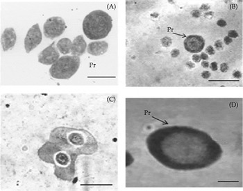
Prohaemocytes. These are the smallest haemocytes found in the three species. In S. ruficornis, these may be round, oval or elliptical cells that are about 2–10 μm in diameter. The large, centrally located nucleus almost completely fills the cytoplasm. The cells may contain a few vacuoles, droplets and granules (Fig. 1A). In M. domestica, the prohaemocytes are spherical or ovoid cells, about 5–12 μm long and 8–16 μm wide. The large, centrally located nucleus occupies almost the whole cell. The cytoplasm is thin, and there are many vacuoles, droplets and granules (Fig. 1B). The division of prohaemocytes can also be observed in the haemolymph (Fig. 1C). In C. megacephala, these cells are spherical, about 5–15 μm in diameter. The large, centrally located nucleus, about 2–10 μm in diameter, fills the whole cell so that the cytoplasm occupies a narrow area around it. The plasma membrane is smooth but vesiculation can be observed in some cells (Fig. 1D).
-
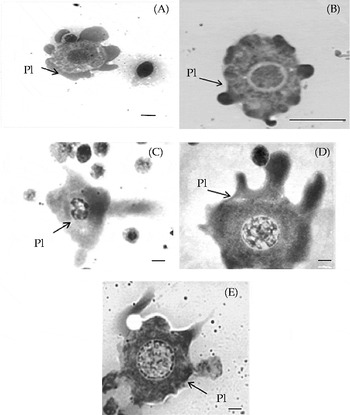
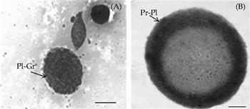
Plasmatocytes. These cells are highly polymorphic in nature, varying in shape and size in the three species studied. In S. ruficornis, the cells may be round, oval or fusiform, about 11–32 μm in diameter. The nucleus is spherical or oval, about 5–19 μm in diameter. The plasma membrane shows many filopodia and pseudopodia and vesicular invaginations. The cytoplasm contains fine granules, vacuoles and droplets (Fig. 2A and B). Some cells showing intermediate characteristics between plasmatocytes and granulocytes can be observed (Fig. 3A). In M. domestica, these cells, about 15–22 μm long and 16–32 μm wide, may be round, ovoid, elongated or spindle-shaped, with many filopodia and pseudopodia. The nucleus is round, centrally located, about 8–18 μm in diameter. The cytoplasm is homogeneous and agranular (Fig. 2C and D). In C. megacephala, plasmatocytes are round or oval cells, about 15–30 μm in diameter. The nucleus is round, punctuate and centrally located, about 7–18 μm in diameter, and contains scattered chromatin. Several vacuoles and fine granules are found in the cytoplasm. One or two filopodia and several long pseudopodia are visible (Fig. 2E). Cells having the characteristics of prohaemocytes and plasmatocytes can also be observed (Fig. 3B).
-
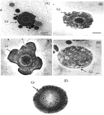
Granulocytes. In S. ruficornis, these cells are round or oval in shape, about 15–22 μm in diameter, with the cytoplasm containing characteristic dark blue granules. The nucleus, about 9–10 μm in diameter, occupies a central position (Fig. 4A). In M. domestica, these are large cells, about 15–26 μm in diameter, round or ovoid, with vesicular invaginations in the plasma membrane, or else are very large with pseudopodia. The nucleus is round, centrally situated, about 5–13 μm in diameter. The cytoplasm contains many fine or coarse granules and vacuoles (Fig. 4B–D). In C. megacephala, these are round cells, about 20–35 μm in diameter. The nucleus is round, centrally situated and about 8–13 μm in diameter. The cytoplasm contains characteristic granules of several types that give a characteristic granular appearance to the cells. The plasma membrane shows pinocytotic vesicles or vesicular invaginations (Fig. 4E).
-
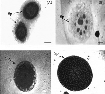
Spherulocytes. In S. ruficornis, spherule cells measure about 4–18 μm and are found in variable forms, such as round or oval. They contain many prominent spherules obscuring the nucleus that is about 4–7 μm in diameter. The plasma membrane shows vesicular invaginations (Fig. 5A). In M. domestica, these cells are spherical or oval in appearance, about 13–26 μm in diameter. The cytoplasm contains characteristic spherules of many sizes, vacuoles and droplets obscuring the nucleus (Fig. 5B and C). In C. megacephala, these are spherical or oval cells, about 14–24 μm in diameter. The cytoplasm is thick and homogeneous, filled with spherical granules obscuring the nucleus (Fig. 5D).
-
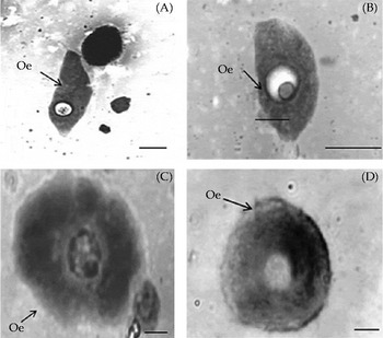
Oenocytoids. In S. ruficornis, these cells are ovoid or spindle-shaped, about 20–25 μm in diameter. The nucleus is round and eccentric, about 3–7 μm in diameter. The cytoplasm is smooth, homogeneous and agranular (Fig. 6A and B). In M. domestica, these cells are round or oval, about 30–45 μm in diameter. The plasma membrane has no vesicular invaginations, filopodia or pseudopodia. The nucleus is centrally situated, about 5–10 μm in diameter. The cytoplasm is thick, homogeneous and agranular with a few vacuoles (Fig. 6C). In C. megacephala, these are round cells, about 35–43 μm in diameter. The nucleus, about 4–8 μm in diameter, is small compared with the cell size. The cytoplasm is thick and homogeneous (Fig. 6D).
-
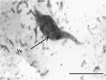
Vermicytes. These appear as fusiform cells, about 6–10 μm in diameter, with tail-like extremities and are found only in the haemolymph of S. ruficornis (Fig. 7).
-
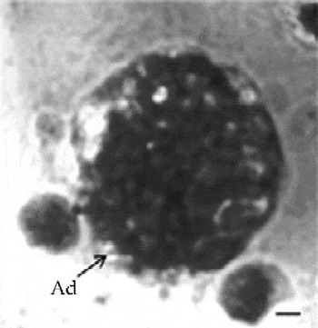
Adipohaemocytes. These are round or ovoid cells found in the haemolymph of M. domestica. They measure about 30–35 μm in diameter with a small, round, centrally located nucleus. The cytoplasm contains characteristic refringent fat droplets and vacuoles that fill the entire cell and sometimes obscuring the nucleus (Fig. 8).
Prohaemocytes (Pr) of Sarcophaga ruficornis (A), Musca domestica (B, C) and Chrysomya megacephala (D) (scale bar 10 μm).

Plasmatocytes (Pl) of Sarcophaga ruficornis (A, B), Musca domestica (C, D) and Chrysomya megacephala (E) (scale bar 10 μm).

Intermediate between plasmatocytes and granulocytes (Pl-Gr) of Sarcophaga ruficornis (A); intermediate between prohaemocytes and plasmatocytes (Pr-Pl) of Chrysomya megacephala (B) (scale bar 10 μm).

Granulocytes (Gr) of Sarcophaga ruficornis (A), Musca domestica (B–D) and Chrysomya megacephala (E) (scale bar 10 μm).

Spherulocytes (Sp) of Sarcophaga ruficornis (A), Musca domestica (B and C) and Chrysomya megacephala (D) (scale bar 10 μm).

Oenocytoids (Oe) of Sarcophaga ruficornis (A, B), Musca domestica (C) and Chrysomya megacephala (D) (scale bar 10 μm).

Vermicyte (Ve) of Sarcophaga ruficornis (scale bar 10 μm).

Adipohaemocyte (Ad) of Musca domestica (scale bar 10 μm).

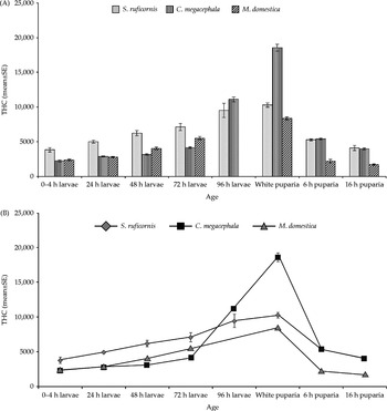
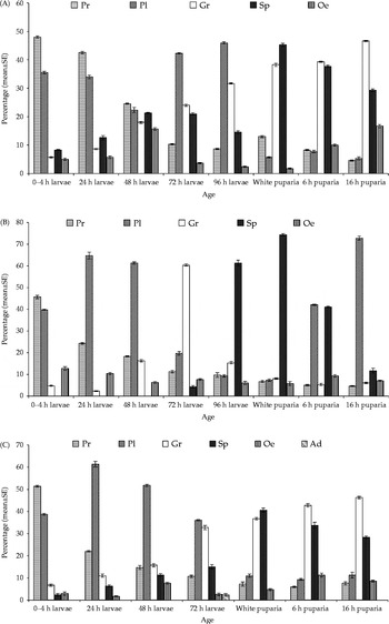
Throughout the larval stage, the THC registered a progressive increase, attaining a peak value in the white puparium stage and then declining in S. ruficornis (F= 24.5; degree of freedom (df) = 7, 56; P≤ 0.05), M. domestica (F= 173.07; df = 6, 49; P≤ 0.05) and C. megacephala (F= 551.3; df = 7, 56; P≤ 0.05) (Fig. 9A and B). The DHC showed a decline in the number of prohaemocytes and an increase in the number of the other cell types, especially plasmatocytes, granulocytes and spherulocytes, in S. ruficornis (F= 1769.25; df = 4, 25; P≤ 0.05; Fig. 10A), C. megacephala (F= 1792.9; df = 4, 25; P≤ 0.05; Fig. 10B) and M. domestica (F= 582.3; df = 5, 30; P≤ 0.05; Fig. 10C).
(A, B) THC of Sarcophaga ruficornis, Chrysomya megacephala and Musca domestica (mean ± SE, n= 8; one-way ANOVA followed by Bonferroni's post hoc test; P≤ 0.05).

DHC of Sarcophaga ruficornis (A), Chrysomya megacephala (B) and Musca domestica (C) (mean ± SE, n= 8; one-way ANOVA followed by Bonferroni's post hoc test; P≤ 0.05): Pr, prohaemocytes; Pl, plasmatocytes; Gr, granulocytes; Sp, spherulocytes; Oe, oenocytoids; Ad, adipohaemocyte.

Discussion
Prohaemocytes in the three species studied show uniform characteristics, being small in size with a large nucleus:cytoplasm ratio as reported in other insects (Yamashita and Iwabuchi, Reference Yamashita and Iwabuchi2001; Giulianini et al., Reference Giulianini, Bertolo, Battistella and Amirante2003; Jalali and Salehi, Reference Jalali and Salehi2008). In the present study, prohaemocytes were frequently observed to be dividing, confirming that these are stem cells (Yamashita and Iwabuchi, Reference Yamashita and Iwabuchi2001; Brayner et al., Reference Brayner, Araujo, Santos, Cavalcanti, Alves, Souza and Peixoto2007) and give rise to other cells by the process of division and differentiation. Many researchers have reported the mitotic activity of prohaemocytes (Jones, Reference Jones1962; Arnold and Hinks, Reference Arnold and Hinks1976). In S. ruficornis, the injection of a mitotic stimulator, phytohaemagglutinin P, induces mitosis only in prohaemocytes but not in the other cell types (Srivastava and Varma, Reference Srivastava and Varma1979). In the present study, the observation of many cell types showing intermediate characteristics between prohaemocytes and plasmatocytes, and between plasmatocytes and granulocytes supports the contention that prohaemocytes are stem cells and they are converted into plasmatocytes (Beaulaton and Monpeyssin, Reference Beaulaton and Monpeyssin1976). Yamashita and Iwabuchi (Reference Yamashita and Iwabuchi2001) observed that in Bombyx mori, plasmatocytes, granulocytes and spherulocytes are produced by the division and differentiation of prohaemocytes that are capable of self-renewing and pluripotent. Sohi (Reference Sohi1971) also reported the formation of plasmatocytes from prohaemocytes.
Plasmatocytes and granulocytes in the three species are highly polymorphic, displaying various shapes and sizes and possessing long filopodia and pseudopodia. These cells are involved in cellular defence responses in most insects except in Drosophila in which plasmatocytes and lamellocytes perform this function (Lavine and Strand, Reference Lavine and Strand2002). Plasmatocytes are professional phagocytes in Drosophila, whereas lamellocytes are concerned with encapsulation (Meister and Lagueux, Reference Meister and Lagueux2003). Granulocytes are potent phagocytes, as demonstrated in several insects such as Heliothis armigera (Essawy et al., Reference Essawy, Maleville and Brehelin1985), Galleria mellonella (Tojo et al., Reference Tojo, Naganuma, Arakawa and Yokoo2000), Manduca sexta (Nardi et al., Reference Nardi, Gao and Kanost2001) and Spodoptera littoralis (Costa et al., Reference Costa, Ribeiro, Girard, Zumbihl and Brehelin2005). In the present study, vermicytes were observed in the haemolymph of S. ruficornis. Similar cells have also been reported in the larvae of Diatraea saccharalis (Falleiros et al., Reference Falleiros, Bombonato and Gregorio2003) and Papilio demoleus (Jalali and Salehi, Reference Jalali and Salehi2008).
Oenocytoids were found in all the three species studied, although the cells showed different morphological characteristics, varying in shape and size. These cells are round in M. domestica and C. megacephala with a small, centrally placed nucleus and a large amount of thick cytoplasm, whereas the cells are spindle-shaped in S. ruficornis with an eccentric nucleus. Giulianini et al. (Reference Giulianini, Bertolo, Battistella and Amirante2003) observed that in Cetonischema aeruginosa, oenocytoids have short filopodia on their surface, and these cells and granulocytes are the only cell types actively involved in phagocytosis. The role of oenocytoids in phagocytosis and melanization involving the secretion of phenoloxidases responsible for darkening of the haemolymph has been reported in many insects (Essawy et al., Reference Essawy, Maleville and Brehelin1985; Hillyer and Christensen, Reference Hillyer and Christensen2002; Giulianini et al., Reference Giulianini, Bertolo, Battistella and Amirante2003; Hillyer et al., Reference Hillyer, Schmidt and Christensen2003; Ribeiro and Brehelin, Reference Ribeiro and Brehelin2006). In the present study, adipohaemocytes were observed in the haemolymph of M. domestica but not in the other two species studied. Adipohaemocytes have not been reported in other cyclorrhaphous dipteran insects, such as Calliphora (Zachary and Hoffmann, Reference Zachary and Hoffmann1973) and Drosophila (Brehelin, Reference Brehelin1982; Meister and Lagueux, Reference Meister and Lagueux2003); however, they have been found in other dipteran insects, such as the yellow fever mosquito A. aegypti (Araujo et al., Reference Araujo, Cavalcanti, Santos, Alves and Brayner2008; Hillyer and Christensen, Reference Hillyer and Christensen2002) and the filarial mosquito C. quinquefasciatus (Brayner et al., Reference Brayner, Araujo, Cavalcanti, Alves and Peixoto2005). Adipohaemocytes in the haemolymph of M. domestica are characterized by the presence of a large number of lipid droplets that may function as energy storage (Araujo et al., Reference Araujo, Cavalcanti, Santos, Alves and Brayner2008). Some authors have reported that these cells are similar to granulocytes or fat body cells and not a distinct cell type (Kaaya and Ratcliffe, Reference Kaaya and Ratcliffe1982; Brayner et al., Reference Brayner, Araujo, Cavalcanti, Alves and Peixoto2005). However, these cells have distinct morphological features in M. domestica and are certainly different from granulocytes and fat body cells. The reason for the absence of these cells in the other two species studied is not clearly understood. Spherulocytes in all the three dipteran flies are either spherical or oval cells containing spherules of variable sizes. These cells contain enzymes for the metabolism of tyrosine, the derivatives of which are subsequently used in tanning (Gillott, Reference Gillott2005). In all the three species studied, the number of spherulocytes increases throughout the larval stage, reaches the peak during the white puparium stage and then declines, showing the possible involvement of these cells in the sclerotization of the cuticle.
In the three flies studied, the THC increases progressively throughout the larval stage, attains a peak value during the white puparium stage and then sharply declines. This trend is similar to that reported in other insects, such as Ephestia larvae (Arnold, Reference Arnold1952), Sarcophaga (Jones, Reference Jones1956), Calliphora larvae (Zachary and Hoffmann, Reference Zachary and Hoffmann1973) and P. demoleus (Jalali and Salehi, Reference Jalali and Salehi2008). Thus, in most holometabolous insects, the THC shows an increasing trend during the larval stage, attains a peak value in the prepupal stage, and then sharply declines in the pupal stage. The increase in the number of the circulating cells may result from the production of new cells or increased mitotic activity during the period of active growth. The decrease in the number of cells may be due to the adherence of haemocytes to the tissues (Chapman, Reference Chapman2008). Haemocytes play an important role in intermediary metabolism, such as protein synthesis, transport of nutrients, metabolism of phenol, amino acids, carbohydrates and lipids (Gupta, Reference Gupta, Kerkut and Gilbert1985), carriers of growth hormone (Essawy, Reference Essawy1985) and growth stimulation. It is well known that hormones play a role in the regulation of insect haemogram during metamorphosis (Essawy et al., Reference Essawy, Gadelhak, El-karaksy and Idriss1997). It is quite natural that intermediary metabolism is higher during the period of active growth, requiring an enhancement in the production of haemocytes.
The DHC in the three species studied reveals that plasmatocytes, granulocytes and spherulocytes are the most abundant cell types. This supports the observation made by Akai and Sato (Reference Akai and Sato1971) reporting that well-differentiated plasmatocytes, granulocytes, spherulocytes and oenocytoids are present during the larval–pupal moult. The number of prohaemocytes in the three species shows a decrease with an increase in the age of the larva, whereas the number of granulocytes and spherulocytes shows a progressive increase throughout the larval stage and in the freshly formed white puparium stage. This suggests the differentiation of prohaemocytes into granulocytes and spherulocytes passing through plasmatocytes (Gupta, Reference Gupta and Gupta1979, Reference Gupta, Kerkut and Gilbert1985). Franchini et al. (Reference Franchini, Miyan and Ottaviani1996) reported that in dipteran insects, only prohaemocytes are capable of division and are the stem or progenitor cells giving rise to plasmatocytes (Tepass et al., Reference Tepass, Fessler, Aziz and Hartenstein1994). The number of plasmatocytes in the latter part of the larval stage of the three flies studied is larger than that of prohaemocytes, suggesting their differentiation from prohaemocytes. Sohi (Reference Sohi1971) also suggested that prohaemocytes give rise to plasmatocytes, which are subsequently transformed into other cell types, such as granulocytes, spherulocytes and oenocytoids (Gupta and Sutherland, Reference Gupta and Sutherland1966). The appearance of certain cells exhibiting the characteristics of both prohaemocytes and plasmatocytes, and of both plasmatocytes and granulocytes suggests the lineage of differentiation. The increase in the number of plasmatocytes and granulocytes during feeding larval stages and also in the haemolymph obtained from the puparia (non-feeding stage) suggests that these cells are actively involved in phagocytosis, encapsulation, nodule formation and foreign body recognition (Gupta, Reference Gupta, Kerkut and Gilbert1985). During metamorphosis, these phagocytic haemocytes invade larval tissues and cause their rapid histolysis (Gillott, Reference Gillott2005), which may be associated with the increase in the number of these cells during the course of the larval–pupal transition. Moreover, the increase in the number of spherulocytes is attributed to the fact that these cells contain enzymes responsible for the metabolism of tyrosine, the derivatives of which are subsequently used in the darkening or tanning of the cuticle (Gillott, Reference Gillott2005) as the newly formed white puparium subsequently undergoes tanning in these insects.