Trans-oesophageal echocardiography has become routine in the diagnosis and care of adults and children with CHD. It has been especially valuable in the operating room where it is used pre-operatively to delineate anatomy and structural details that may have remained ill-defined by transthoracic imaging, and used post-operatively to evaluate surgical repair, assess residual lesions, and estimate cardiac function.Reference Bettex, Schmidlin and Bernath 1 – Reference Muhiudeen, Roberson, Silverman, Haas, Turley and Cahalan 3 The need to return to bypass after initial surgical intervention is a serious decision that is influenced not only by the intra-operative trans-oesophageal imaging but also by direct visualisation and assessment by the surgeon and post-operative cardiovascular haemodynamics. Early identification of findings requiring further intervention prevents re-operation at a later date, further morbidity and mortality, and higher costs; however, the frequency of returning to bypass and the resulting outcomes in our institution are unknown. Therefore, we endeavoured to evaluate the importance of post-bypass trans-oesophageal echocardiography for assessing additional operative needs before separation from cardiopulmonary bypass and to evaluate where trans-oesophageal echocardiography provides the greatest positive yield.
Materials and methods
We retrospectively reviewed from our database all trans-oesophageal echocardiograms performed on children and adults with CHD in the operating room between 1 October, 2005 – the first date of our current reporting system – and 31 December, 2010 at Lucile Packard Children’s Hospital. All trans-oesophageal echocardiograms were performed by paediatric cardiologists using Siemens® Acuson C512, rev 12.0 (Siemens® Medical Solutions Inc., Mountain View, California, United States of America). We performed complete trans-oesophageal and trans-gastric conventional scans using 5–7 MHz probes. We identified all children requiring trans-oesophageal echocardiography, and excluded those performed in the cardiac catheterisation laboratory or ICUs and those without a clear indication. We categorised these patients based on primary lesion/initial diagnosis. To elucidate where trans-oesophageal echocardiograms led to further surgical repair, we further refined the review to those patients who required a second cardiopulmonary bypass run for residual or additional problems discovered by the first post-operative trans-oesophageal echocardiogram. We evaluated these patients based on primary lesion and findings on the initial echocardiogram that necessitated return to bypass. Finally, we identified all patients who left the operating room but needed to return within 1 week for additional surgical intervention. Details regarding surgical decisions and post-operative clinical management were obtained from the electronic health record. Each patient’s date of birth and gender were also obtained from Stanford Translational Research Integrated Database Environment (STRIDE), a research and development project at Stanford University formed to create a standards-based informatics platform supporting clinical and translational research. The project described was supported by the National Center for Research Resources and the National Center for Advancing Translational Sciences, National Institutes of Health, through grant UL1 RR025744. The content is solely the responsibility of the authors and does not necessarily represent the official views of the National Institute of Health.Reference Lowe, Ferris, Hernandez and Weber 4 The study was approved by the Stanford Institutional Review Board (protocol# 21195).
Statistical calculations were carried out using SAS Enterprise Guide version 4.2 (SAS Institute Inc., Cary, North Carolina, United States of America) and Microsoft Excel®. All values are expressed as mean±standard deviation. A Student’s t-test was used to compare the ages and length of hospital stay for patients who did and did not require a second post-operative trans-oesophageal echocardiogram. Standard χ2 analysis was used to compare gender, mortality, and trans-oesophageal echocardiography usage between surgeons. A χ2 r×c table compared the percentage of trans-oesophageal echocardiograms performed each year. Finally, a Fisher exact test was used to compare the percentages of single and second post-operative cases returning to the operating room within 1 week. For all tests, a p-value <0.05 was considered statistically significant.
Results
We reviewed a total of 1748 intra-operative trans-oesophageal echocardiograms. Ages ranged from 1 day to 64.0 years (average 4.6±7.8 years) that fit our selection criteria. There were 766 females and 982 males. During the study period, all trans-oesophageal echocardiograms performed on a patient in a single operative setting were considered as one complete echocardiographic study.
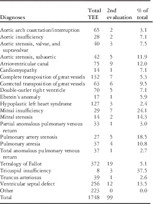
There were 99 (5.66%) cases that required second post-operative trans-oesophageal echocardiograms (Table 1). The group was composed of 48 females and 51 males whose ages ranged from 1 day to 40.5 years (5.2±8.2 years). When comparing these patients with those only requiring a single post-operative assessment, gender (p=0.42) and age (p=0.47) were not significantly different. In addition, there was no statistical difference in length of hospital stay or mortality within 90 days of admission between those with a second versus single post-operative assessment (20.2±29.6 versus 20.0 days±29.7, p=0.46; 5.05 versus 3.26%, p=0.25).
Pre-operative diagnoses requiring second post-operative trans-oesophageal echocardiographic evaluations (TEE).

Of the total group of trans-oesophageal echocardiograms, those diagnoses that had the highest frequency of second post-operative studies relative to the number of operating room cases for that diagnosis included mitral valve disease (20.9%), tricuspid valve disease (16.0%), and atrioventricular valve stenosis or regurgitation (amongst atrioventricular canal defects) (12.0%) (Fig 1). In addition, 14.1% of pulmonary valve repairs required second studies.
Percentage of total lesion-specific cases requiring second post-operative trans-oesophageal echocardiographic evaluations (raw numbers of cases within each column).

Within the 99 operations requiring second post-operative trans-oesophageal echocardiograms (Fig 1), underlying diagnoses of tetralogy of Fallot (19 cases) and disease of the atrioventricular valves (22 cases) – tricuspid, mitral, and atrioventricular canal – accounted for the highest number of studies.
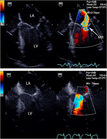
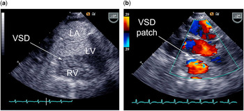
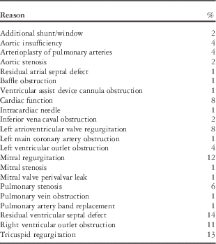
Reasons for return to bypass varied by case, but were based upon a combination of initial trans-oesophageal echocardiographic findings and surgical and haemodynamic concerns (Table 2). The most common reasons cited included residual ventricular septal defect (14%), residual tricuspid regurgitation (13%), residual mitral regurgitation (12%), right ventricular outlet obstruction (11%), and residual left atrioventricular valve regurgitation (8%). These included a 5.5-year-old boy with transposition of the great vessels who was found to have a cleft mitral valve with significant atrioventricular valve regurgitation after arterial switch operation (Fig 2), and a 21-month-old girl with complex infundibular muscular narrowing who underwent muscle resection, but was found to have a residual ventricular septal defect by color Doppler and left atrial contrast echocardiography (Fig 3).
Post-operative trans-oesophageal echocardiogram in a 5.5-year-old boy with transposition of the great vessels, demonstrated moderate-to-severe residual mitral regurgitation (MR)( a ). After return to bypass, a mitral valve cleft was discovered, and after repair was no longer insufficient ( b ). LA=left atrium; LV=left ventricle.

Twenty-one-month-old girl with complex infundibular muscular narrowing underwent muscle resection, found to have a residual ventricular septal defect (VSD) by left atrial contrast echocardiography ( a ) with left-to-right shunting. Further intra-cardiac repair with a ventricular septal patch alleviated the inter-ventricular shunt ( b ) by color Doppler. LA=left atrium; LV=left ventricle; RV=right ventricle.

Indications for second post-operative trans-oesophageal echocardiographic evaluations.

There was no temporal difference in the frequency of return to bypass in any year.
All the patients who returned to the operating room within 1 week were further evaluated. In total, 14 patients (4 females and 10 males, ages 5 days to 32 years, average 7.02 ±8.75 years), or 0.8% of the total trans-oesophageal echocardiograms, returned to the operating room for surgical revision. Statistically, patients who had a second trans-oesophageal echocardiogram were slightly more likely than those with a single trans-oesophageal echocardiogram to return to the operating room at a later date for revision (0.7 versus 3.0%, p=0.04).
Of the patients who returned to the operating room within 1 week, three had progressive atrioventricular valve or semilunar valve regurgitation and two had cyanosis suggesting baffle leak after atrial switch operations. In addition, there were two patients with progressive left ventricular systolic dysfunction associated with pulmonary artery banding, and two patients with Fontan palliation requiring fenestration closure or reduction. All these findings were changes from the initial post-operative evaluation.
Discussion
Intra-operative imaging in many centres has become a mainstay in the evaluation of surgical repair of patients with CHD. Previous studies have demonstrated the safety,Reference Stevenson, Sorensen, Gartman, Hall and Rittenhouse 5 , Reference Daniel, Erbel and Kasper 6 utility,Reference Bengur, Li, Herlong, Jaggers, Sanders and Ungerleider 7 , Reference Peng, Sun, Hanley, Olson and Punn 8 and cost-effectivenessReference Randolph, Hagler and Connolly 9 of its routine use. Except for patients weighing <2 kg and select patients with tracheal and oesophageal anomalies,Reference Ayres, Miller-Hance and Fyfe 10 routine intra-operative trans-oesophageal imaging in combination with surgical and haemodynamic evaluation provides immediate assessment of surgical results that can prompt further intervention.Reference Guzeltas, Ozyilmaz and Tanidir 11
Our evaluation is the largest study to characterise patients who require further surgical intervention based on intra-operative trans-oesophageal echocardiographic findings. Although imaging aids the surgical repair of nearly all forms of CHD, we found that the evaluation of the atrioventricular valves benefits from it most frequently. Nearly 20% of all mitral and tricuspid valve repairs and 12% of all common atrioventricular canal repairs required return to bypass after imaging-identified residual problems, with residual tricuspid, mitral, and left atrioventricular valve regurgitation being the most commonly cited reasons. Furthermore, even with attentive intra-operative assessment, atrioventricular valve regurgitation proved to be a finding that, in our study, caused 2/14 second operations within 1 week. Similar findings were described by Randolph et al.Reference Randolph, Hagler and Connolly 9 Ultimately, the repairs may benefit from the use of more advanced imaging modalities such as three-dimensional echocardiography to evaluate the architecture and function post-operatively.Reference Shanks, Delgado and Ng 12
Cases that involved relief of pulmonary valve stenosis frequently required additional post-operative imaging, and right ventricular outflow tract obstruction was the top reason for returning to bypass. As with atrioventricular valve regurgitation, trans-oesophageal echocardiography remains the most commonly used tool for assessment; however, stenotic lesions can also be evaluated with three-dimensional echocardiographyReference Khaw, von Bardeleben and Strasser 13 or can be directly measured.
The previous study by Guzeltas et alReference Guzeltas, Ozyilmaz and Tanidir 11 found post-operative imaging to be very useful in patients with tetralogy of Fallot. Although this diagnosis did make up a large percentage of the total repeat trans-oesophageal echocardiograms (Table 1), we felt this was skewed by the multitude of complex tetralogy patients who underwent repair at our institution. In fact, only 5% of all tetralogy of Fallot patients who underwent repair during the study period required further imaging, which was relatively less than many of the other diagnoses.
Intraoperative trans-oesophageal echocardiography was extremely successful at identifying the vast majority of lesions needing acute revision. This is unquestionably beneficial for the patient’s overall course. We also confirmed that additional imaging after surgical revision is safe for patients and does not confer any increased risk of mortality or increased hospital length of stay.
For the 5% in whom trans-oesophageal echocardiographic imaging was not considered safe, because of low weight, airway compromise with probe insertion, or inherent tracheal and oesophageal abnormalities, assessment was unfortunately limited. Among those who could safely be evaluated, in ~5.7% of the cases, post-operative trans-oesophageal echocardiographic imaging provided information that led to bypass return immediately for further surgical correction. Routine intra-operative imaging provides better outcomes for patients with CHD. In our institution, trans-oesophageal echocardiograms are performed in over half of all congenital cases and 95% of all congenital cases requiring bypass.
As a retrospective review, this study is subject to inherent limitations. It represents a single institution’s experience and may not reflect other institutions’ reliance upon intra-operative imaging in decision making, spectrum of CHD, or surgical skill.
We did not evaluate patients who returned to bypass but did not have a second post-operative study while still not completely separated from cardiopulmonary bypass. It is assumed that the number of these patients is low, given that intra-operative imaging helped in the original decision to return to bypass, and would likely be requested to assess outcomes after further surgical intervention.
We did not investigate those patients who returned to bypass when that decision was not based on imaging. Again, it is assumed that this number would be small given our high frequency of trans-oesophageal echocardiographic usage in congenital cases.
It is difficult to speculate whether there were any intra-operative imaging clues that could have prevented a second surgery later in the hospitalization. Ventricular dysfunction after banding (two patients) and excessive right-to-left shunting with Fontan fenestration (two patients) are both diagnoses that often require several post-operative days to be adequately assessed. Progressive valve regurgitation (three patients) also may develop with time and physiological changes during recovery. It is, therefore, not surprising that these patients needed to return to the operating room at a later date, and most likely their course could not have been predicted at the time of initial surgery.
In conclusion, routine trans-oesophageal echocardiographic imaging is helpful in the surgical repair of CHD in children and adults. This has been particularly valuable in patients with mitral, tricuspid, and common atrioventricular valve stenosis and regurgitation. The vast majority of patients with a second post-operative evaluation are unlikely to return to the operating room at a later date. In fact, from routine post-operative imaging and haemodynamic assessment, 99 patients benefited from immediate identification of surgical need, and 96 (5.5%) benefited from avoiding possible long-term complications associated with re-operations, redo-sternotomies, and potentially longer lengths of hospital stay. Those patients who do have to return to the operating room often have progressive disease that requires time and serial evaluations to be assessed. Overall, routine repeat imaging successfully identifies patients requiring further interventions, does not confer additional risk of mortality or prolong length of hospital stay, and can greatly influence the operative and post-operative course of a substantial number of patients.
Acknowledgements
None.
Financial Support
This research received no specific grant from any funding agency, commercial, or not-for-profit sectors.
Conflicts of Interest
None.