Percutaneous balloon pulmonary valvuloplasty represents a major milestone in the field of interventional cardiology, and the technique has been applied with varying success to the dilatation of many other congenital and acquired cardiovascular lesions.Reference Rocchini, Beekman, Shachar, Benson, Schwartz and Kan1
Since its use was first reported in 1982 by Khan et al., balloon pulmonary valvuloplasty has replaced surgery as the initial treatment of choice for patients of all ages with moderate to severe pulmonary valve stenosis and an intact ventricular septum. The procedure continues to be performed as originally described with few variations, including the use of double balloons or oversized balloons.Reference Khan, Youssef and Mullins2
The aim of this work was to determine the outcome of transcatheter balloon dilatation of the pulmonary valve in the neonatal period, early infancy, and childhood. The predictors of the outcome of balloon pulmonary valvuloplasty and its complications during and early after the intervention were studied.
Materials and methods
This was an observational retrospective cross-sectional study reporting the data of patients who were referred to the cardiac catheterisation unit in the paediatric cardiology department of Cairo University for balloon pulmonary valvuloplasty over the last 10 years. The study was approved by the research committee of paediatric department in Cairo University.
Patient data were collected from the medical reports and the electronic medical records from 2007 to 2016.
We collected the data of 1287 patients. Thirteen patients underwent angiography without proceeding to balloon valvuloplasty as the pressure gradients measured by catheterisation were less than that indicated for the procedure. Seventy-four patients were excluded from the study group due to insufficient data in their medical records. Therefore, the records of 1200 patients were reviewed and included in the study.
Echocardiography: All patients were assessed and diagnosed by echocardiography.
Balloon valvuloplasty was recommended to all patients with isolated pulmonary valve stenosis from birth to 12 years old. Simple associated abnormalities were not excluded from the study (e.g., patent foramen ovale and patent ductus arteriosus with pulmonary valve stenosis). Pre-term infants and patients with complex CHDs were excluded from the study.
The recorded data include the following:
1. Age: classified into subgroups including: (a) neonates and early infants: first 90 days of life; (b) infants: from 90 days of life to 2 years of life; and (c) children: from 2 years to 12 years of life.
2. Sex: males and females
3. Pulmonary valve annulus: measured by echocardiography and by catheterisation, and the correlation between both results was evaluated.
4. Pressure gradient across the pulmonary valve: measured by echocardiography and by catheterisation before and after balloon pulmonary valvuloplasty.
5. Associated cardiac anomalies: (A) pulmonary regurgitation: graded as mild, moderate, or severe; (B) tricuspid regurgitation: graded as mild, moderate, or severe; (C) tricuspid valve: normal tricuspid valve or a dysplastic valve (thickened, immobile, and formed from myxomatous tissue); (D) right ventricle: normal, dilated, hypertrophied or dilated, and hypertrophied; and (E) presence or absence of all the following: infundibular stenosis of the pulmonary valve, supravalvular in association with valvular pulmonary stenosis, mitral regurgitation, ventricular septal defect, patent ductus arteriosus, atrial septal defect, and patent foramen oval.
6. Right ventricular pressure: measured by catheterisation before and after balloon pulmonary valvuloplasty.
7. Pulmonary artery pressure: measured by catheterisation before and after balloon pulmonary valvuloplasty.
8. Type of balloon: (Tyshak II, Powerflex, Cordis, Wanda, and Z-med II).
9. Size of the balloon: based on multiple studies regarding the immediate and follow-up results; balloon size recommendations were 1.2 to 1.4 times larger than the pulmonary valve annulus.Reference Rao3
10. Success is considered achieved when the pressure gradient across the pulmonary valve drops to less than or equal to 50% of the baseline measurements.Reference Saad, Roushdy and Elsayed4
11. Repeat balloon pulmonary valvuloplasty: if it was indicated again at a later time point.
12. Surgery: if the patient was referred for surgery after failure of more than one balloon pulmonary valvuloplasty.
13. Complications during or immediately after balloon pulmonary valvuloplasty: (A) perforation; (B) minimal pericardial effusion; (C) massive pericardial effusion; and (D) cardiorespiratory arrest.
Balloon pulmonary valvuloplasty
It was performed at the children’s hospital in the Cairo University catheterisation laboratory using a biplane INTEGRIS ALLORA (Philips) system as follows: informed written consent was obtained from the parent after explaining the risks and benefits of the procedure. A full diet was given until 5–6 hours before the procedure, and clear fluids are given until 4–5 hours before the procedure.Reference Lock, Lock, Keane and Perry5 All the procedures were performed on our patients under general anaesthesia. Femoral vein access was achieved and cannulated by an introducer sheath according to the size and the age of the patient. Haemodynamic measurements such as the right ventricular and pulmonary artery pressures were recorded (pre- and post-balloon valvuloplasty). Antero-posterior and straight lateral angiograms were performed. Lateral projection usually provides the optimal image of the valve hinge points at which the pulmonary annulus is measured using the catheter diameter as an internal reference.Reference Cheatham, Park and Troxler6 Balloon dilatation: in young children, we use 2-cm-long balloons to prevent damage to the right ventricular free wall, branch pulmonary arteries, or tricuspid valve, while in older children, 3–5 cm long balloons are used to facilitate balloon positioning. The balloon is centred across the valve annulus with the help of a road map in most cases.Reference Bridges, O’Laughlin, Mullins, Allen, Clark and Gutgessell7
The patient was transferred to the intermediate care unit to be observed for 6–12 hours, during which the vital signs were monitored, the puncture site was inspected for bleeding or haematoma maintenance IV fluids and parenteral antibiotics, usually cefotaxime 100 mg/kg/day in divided doses, were given.
Statistical analysis
Data were entered and statistically analysed on the Statistical Package of Social Science Software program, version 23 (IBM SPSS Statistics for Windows, Version 23.0. Armonk, NY: IBM Corp.). Data are presented as the range, median, mean, and standard deviation for quantitative variables and as the frequency and percentage for qualitative variables. Comparison between groups was performed using the chi-square test for qualitative variables. For quantitative variables, comparison between groups was conducted using independent sample t-tests (if normally distributed) or the Mann–Whitney test (if not normally distributed). Multivariate logistic regression models were used to explore the predictors of outcome variables. p Values less than 0.05 were considered statistically significant.
Results
We collected the data of 1287 patients. Thirteen patients underwent angiography without proceeding to balloon valvuloplasty, as the pressure gradients measured by catheterisation were less than those indicated for the procedure. A total of 74 patients were excluded from the study group, as the data in their medical records were insufficient; therefore, 1200 patients were included in the study. The number of patients per year ranged from 97 to 173, with an average of 120 patients per year.
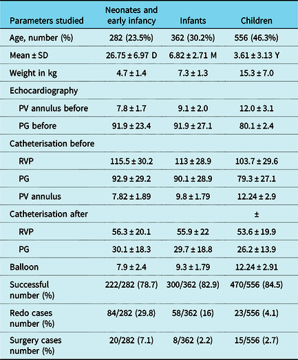
Demographic data and parameters studied by both echocardiography and catheterisation before and after valvuloplasty are shown in Table 1.
Demographic data and parameters studied by both echocardiography and catheterisation before and after valvuloplasty

PG = pressure gradient; PV = pulmonary valve; RVP = right ventricular pressure.
Echocardiographic findings associated with pulmonary valve stenosis in the studied group before balloon pulmonary valvuloplasty included infundibular stenosis in 316 patients (26.3%), supravalvular stenosis in 212 patients (17.7%), both infundibular and supravalvular stenoses in 110 patients (9.2%), and dysplastic tricuspid valve in 156 patients (13%); in addition, 38.8% of patients had right ventricular hypertrophy and/or dilatation.
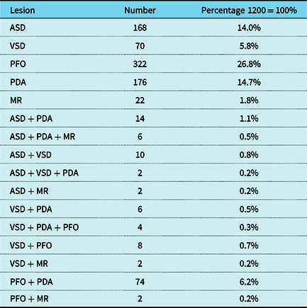
The associated cardiac anomalies obtained by echocardiography before balloon pulmonary valvuloplasty of the studied group are listed in Table 2.
Associated cardiac anomalies obtained by echocardiography before balloon pulmonary valvuloplasty of the studied group

ASD = Atrial septal defect, VSD = Ventricular septal defect, PFO = Patent foramen ovale, PDA = Patent ductus arteriosus, MR = Mitral regurgitation.
The diameter of the pulmonary valve annulus determined by catheterisation was strongly correlated with the measurements made by echocardiography in all age groups. In most of our patients (81.3%), the mean diameter of the annulus measured by echocardiography was equal to or 2 mm shorter than the mean diameter measured by angiography (976 patients). The catheterisation pressure gradient readings before balloon pulmonary valvuloplasty in 54% of our patients were 20% less than those obtained by echocardiography, while in 16.8% of our patients, these readings were 30–50% lower than those obtained by echocardiography. The difference was greater than 50% in only 3.7% of patients.
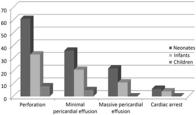
The most common complication was perforation of the right ventricular outflow tract, which usually closes spontaneously or causes minimal pericardial effusion, and this complication was most common in the first 3 months of life. Massive pericardial effusion occurred either due to right ventricular outflow tract perforation or rupture of the pulmonary annulus and was managed on the procedure table; none of the patients were referred for urgent surgery. Cardiac arrest or near cardiac arrest was most common in patients with severe pulmonary valve stenosis and no patent ductus arteriosus.
Descriptive data of complications during or immediately after balloon pulmonary valvuloplasty in each age group are shown in Figure 1.
Descriptive data of complications during or immediately after balloon pulmonary valvuloplasty in each age group.

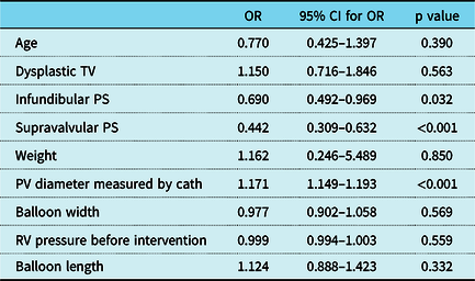
Multivariate analysis revealed that only infundibular pulmonary stenosis (odds ratio 0.69, p value 0.032), supravalvular in association with valvular pulmonary stenosis (odds ratio 0.44, p value <0.001), and pulmonary valve diameter by angiography (odds ratio 1.17, p value <0.001) were significant predictors of success, as shown in Table 3.
Multivariate logistic regression model to explore predictors of success of catheterisation intervention

CI = confidence interval; OR = odds ratio; PS = pulmonary stenosis; PV = pulmonary valve; TV = tricuspid valve.
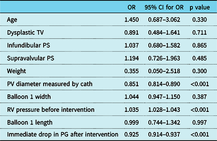
Multivariate analysis also revealed that a small pulmonary valve annulus measured by angiography, a minor immediate drop in the pressure gradient after intervention, and high right ventricular pressure before intervention were significant predictors of repeat catheterisation, as shown in Table 4.
Multivariate logistic regression model to explore predictors of re-intervention:

CI = confidence interval; OR = odds ratio; PG = pressure gradient; PS = pulmonary stenosis; PV = pulmonary valve; RV = right ventricle; TV = tricuspid valve.
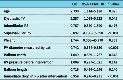
The age of the patient, the presence of dysplastic TV and supravalvular in association with valvular pulmonary stenosis, the pulmonary valve annulus diameter, and an immediate drop in the pressure gradient after transcatheter intervention were the most significant predictors for the need for surgical valvotomy in multivariate analysis, as demonstrated in Table 5. z Score of the pulmonary valve was not a predictor of need of surgical valvotomy with the odds ratio of 1.106 and 95% confidence interval for odds ratio of 1.097–1.123, and p value of 0.152 in multivariate regression logistic model.
Multivariate logistic regression model to explore predictors of need for surgical valvotomy

CI = confidence interval; OR = odds ratio; PG = pressure gradient; PS = pulmonary stenosis; PV = pulmonary valve; RV = right ventricle; TV = tricuspid valve.
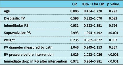
In the multivariate analysis, the presence of supravalvular in association with valvular pulmonary stenosis, low weight, high right ventricular pressure before intervention, and a minor immediate drop in the pressure gradient post-intervention were found to be the most significant predictors for the occurrence of complications, as shown in Table 6.
Multivariate logistic regression model to explore predictors of complications

CI = confidence interval; OR = odds ratio; PG = pressure gradient; PS = pulmonary stenosis; PV = pulmonary valve; RV = right ventricle; TV = tricuspid valve.
Discussion
The incidence of pulmonary valve stenosis is approximately 0.6 to 0.8 per 1000 live births. The incidence is approximately 50% in patients with CHD when associated with other congenital cardiac defects.Reference Botto, Correa and Erickson8
A total of 1200 patients were included in the study, with 710 males representing 59.2% and 490 females representing 40.8% of the studied patients. This cohort was the largest number of patients studied in this field, as our hospital is the largest tertiary referral centre in Egypt.
Age categorisation
The studied patients were categorised into three age groups: neonates and early infants (282 patients, 23.5%), infants (362 patients, 30.2%), and children (556 patients, 46.3%). Similar studies also use this age categorisation, as there is a wide range of variation in the outcome of balloon pulmonary valvuloplasty within each age group. Holzer group classified their population to neonatal and post-neonatal groups.Reference Holzer, Gauvreau and Kreutzer9 Ghaffari group also used age categorisation in their study.Reference Ghaffari, Ghaffari, Ghaffari and Sgafy10
In our study, the smallest baby was 2.4 kg, while the maximum body weight was 40.7 kg, with a mean of 10.1 ± 8.9 kg. These results are comparable to those of Behjati et al., who reported a mean weight of 12.2 ± 4.7 kg (age ranged from 0.4 to 8 years).Reference Behjati, Seyed and Shahrokh11
Transvalvular gradient
Initially, most haemodynamic data of patients with pulmonary valve stenosis were obtained from echocardiography. Gielen et al. mentioned that the use of Doppler echocardiography allows a reliable assessment of stenosis in all age groups.Reference Gielen, Daniels and van Lier12
Regarding the echocardiographic data obtained before balloon pulmonary valvuloplasty, the mean pressure gradient across the pulmonary valve in all age groups was approximately 86.43 ± 25.66 mmHg (minimum pressure gradient 45 mmHg and maximum pressure gradient 188 mmHg).
Balloon pulmonary valvuloplasty was attempted in all patients with a peak-to-peak systolic gradient ≥45 mmHg at catheterisation; this criterion is in accordance with most published data, which considers a peak-to-peak gradient of approximately 50 mmHg as an absolute indication for balloon pulmonary valvuloplasty.Reference Handoka and El-Eraky13
In 13 patients, this gradient was lower when determined by pullback pressure gradient assessment, and thus, balloon dilatation was not indicated. Those patients were followed up regularly and none of them required intervention.
Associated cardiac lesions occurred in 50.2% of our patients (602 patients) (Table 2).
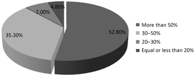
Concerning the pressure gradient across the pulmonary valve assessed by catheterisation, in approximately 54% of our study group, readings were 20% less than those obtained by echocardiography, while in approximately 16.8% of the patients, the pressure gradients assessed by catheterisation were 30–50% less than those obtained by echocardiography. This finding reveals that the pressure gradient measurements by echocardiography should be re-evaluated by catheterisation before balloon pulmonary valvuloplasty, even if the readings obtained by echocardiography were indicative of balloon pulmonary valvuloplasty, especially in borderline cases (Figure 2).
Details of the pressure gradient difference before and after balloon pulmonary valvuloplasty.

Balloon size
Concerning the selection of the balloon size, at first, we started using balloons that are 1.5 times the size of the pulmonary valve annulus, as we progressed further we are currently using balloons that are 1.2 times the size of the pulmonary valve annulus which was in concordance with most of the published studies in this field. Rao also recommended this balloon size on the basis of immediate as well as follow-up results.Reference Rao3 Balloons larger than 1.5 times the pulmonary valve annulus can damage the right ventricular outflow tract; furthermore, these balloons do not have an advantage beyond that produced by balloons that are 1.2–1.4 times the annular size, which is why such large balloons should not be used. However, large balloons with a balloon/annulus ratio of 1.4:1.5 can still be used when dilating dysplastic pulmonary valves. The balloon/annulus ratio in the patients whose right ventricular pressure dropped to more than half was 1.23 ± 0.24 mm, while it was 1.27 ± 0.25 mm in other patients.
Recommendations for the use of 20-mm-long balloons in neonates, 30-mm-long balloons in infants, and 30–40 mm long balloons in children were followed. Rao also used similar balloons in each age group. However, Rao reported that there are no data supporting that a given length balloon is more successful than other lengths in relieving an obstruction. With short balloons, it is difficult to maintain the balloon centre across the pulmonary valve annulus during balloon inflation. However, long balloons may impinge upon the tricuspid valve, causing tricuspid insufficiency, or affect the conduction system, causing heart block.Reference Rao3
The mean diameter of the pulmonary valve annulus, as assessed by echocardiography before balloon pulmonary valvuloplasty in most of our patients) 81.3%), was ≤2 mm shorter than the mean diameter measured by angiography (976 patients). This finding suggests that we can depend on echocardiographic measurements to select the proper balloon size for the procedure.
Handoka and El-Razky mentioned that the balloon size was determined according to the maximum internal diameter of the pulmonary valve from hinge point to hinge point during systole as measured in the lateral projection of the right ventricular angiogram with corrections for magnification.Reference Handoka and El-Eraky13
In our study group, sequential balloon valvuloplasty using two consecutive balloons were used in 30.8% of the total number of patients (370/1200), most of whom were children (146 patients). Saad et al. reported the use of two consecutive balloon inflations in 56% of infants in their study group.Reference Saad, Roushdy and Elsayed4
Rao mentioned that Tyshak II balloons are most commonly used by interventional cardiologists, as they have the widest range of balloon sizes and diameters available in this line of catheter.Reference Rao3 In our study group, Tyshak II balloons were used in 58.5% (702/1200) of patients as the first balloon, and 57.3% (212/370) of patients when two balloons were used.
Outcome of balloon valvuloplasty
Regarding the outcome of balloon pulmonary valvuloplasty, in approximately 52.8% of our study group, the pressure gradient across the pulmonary valve dropped by more than 50% of the pre-catheterisation values (634 patients), while in approximately 35.3% of our study group, the pressure gradient dropped to 30–50% of the pre-catheterisation values (424 patients). We considered balloon pulmonary valvuloplasty as successful when the pressure gradient across the pulmonary valve drops to half or more than that of the pre-catheterisation values. In addition, Rao defined the intervention as effective if it decreased the stenosis by at least 50% of the initial value.Reference Rao3 Other authors consider the intervention to be successful if the transvalvular peak gradient dropped to <36 mmHg.Reference Handoka and El-Eraky13
In this study, patients had a significant reduction in the right ventricular systolic pressure from 107.81 ± 30.88 mmHg to 54.92 ± 20.74 mmHg as well as in right ventricular systolic pressure – pulmonary artery pressure from 86.43 ± 25.66 mmHg to 28.15 ± 16.68 mmHg immediately after balloon pulmonary valvuloplasty. This finding is in accordance with that of Behjati et al. who reported a reduction in the peak-to-peak systolic pressure gradient across the pulmonary valve from 83.28 ± 32 mmHg to 19.3 ± 14.2 mmHg.Reference Behjati, Seyed and Shahrokh11
Our success rate is approximately 82.7% (992/1200), with the best success rates within the children age group (84.5%). Our success rate is comparable to that of Ahmadi and Sabri, who reported an immediate dilation success rate of 77%.Reference Ahmadi and Sabri14 The success rate was reported to be 88.9% in the study by Liu et al.,Reference Liu, Wu and Huang15 while a success rate of 90% was reported in the study of Jaing et al.Reference Jaing, Hwang, Lu, Hsieh and Meng16 Our findings and the results of previous studies indicated the effectiveness of balloon pulmonary valvuloplasty in the treatment of pulmonary valves.
However, Larry reported that the initial success does not guarantee that the pressure gradient will remain the same, as most of the studies reported that approximately 15–30% of patients with an initially successful balloon pulmonary valvuloplasty may show evidence of significant residual or recurrent right ventricular outflow tract obstruction within 1–2 years. Repeated balloon pulmonary valvuloplasty is successful in many of these patients. However, up to 10% of patients may eventually require surgical right ventricular outflow tract resection or pulmonary valve resection. This finding confirms that close follow-up of the successful cases should be performed.Reference Larry17
In our study group, approximately 13.7% of patients (165 patients) underwent another balloon dilation by repeat catheterisation, 3.6% of patients were referred for surgical valvotomy (43 patients), and 8 patients underwent both repeat catherisation and surgery after balloon pulmonary valvuloplasty. This finding is comparable to that of Peterson et al. who reported the development of restenosis in 13 patients who underwent balloon pulmonary valvuloplasty (14.1%); 6 patients were re-dilated and 7 patients required surgery (108 patients with a mean age of 3.6 ± 3.9 years were included in their study).Reference Peterson, Schilthuis, Dodge-Khatami, Hitchcock, Meijboom and Bennink18
Complications of balloon valvuloplasty
Regarding the complications of balloon pulmonary valvuloplasty, we reported a total percentage of approximately 8.5% of complications (102 patients) during the catheterisation procedure. Some of these patients had multiple complications. The most common complication was perforation of the right ventricular outflow tract that resulted in either minimal pericardial effusion (usually resolved spontaneously or after pericardiocentesis) or massive pericardial effusion. Cardiac arrest and near arrest occurred in 10 patients (0.8% of the studied group) and were resolved with successful resuscitation trials. This rate is in close agreement with that reported by Tggart et al.; in that report, 11% of the 89 patients in the paediatric study group had periprocedural complications (10 patients), and perforation/pericardial effusion was the most common complication (3 patients).Reference Tggart, Cetta, Cabalka and Hagler19
Larry reported that a wide variety of complications has been observed over the years in attempted treatment of pulmonary valve stenosis with balloon pulmonary valvuloplasty. In early reports from the 1980s, the available balloon valvuloplasty catheters were considerably larger and stiffer than the present generation of catheters. Vascular complications, including retroperitoneal haematomas and avulsions of the iliac vein, were previously encountered, but these complications should be extremely rare with the new generation of valvuloplasty catheters.Reference Larry17
Balloon valvuloplasty in neonatal age group
Regarding balloon pulmonary valvuloplasty in the neonatal and early infancy age group, approximately 23.5% of our studied patients (282/1200 patients) were neonates with a mean age of approximately 26.7 ± 6.97 days and a mean weight of approximately 4.7 ± 1.4 kg (the minimum weight was 2.4 kg).
The mean pressure gradient across the pulmonary valve by echocardiography before balloon pulmonary valvuloplasty was 91.9 ± 23.4 mmHg.
Bernstein reported that when severe pulmonary valve stenosis occurs in a neonate, markedly decreased right ventricular compliance may lead to right-to-left shunting through a patent foramen ovale, a condition termed critical pulmonary stenosis. The mean pulmonary valve annulus diameter was 7.8 ± 1.7 mm,Reference Bernstein, Behrmen, Kliegman and Jenson20 which is in accordance with that in the study group of Holzer et al., which had a median diameter of 6.9 mm (4–9.5 mm).Reference Holzer, Gauvreau and Kreutzer9
A total of 282 neonatal patients underwent balloon pulmonary valvuloplasty in our study. The most commonly used balloon was the Tyshak II balloon, and approximately 16 of those patients needed another consecutive balloon dilation.
Holzer et al. reported a success rate of around 96% (92 patients) in his <1-month study group (96 patients). Holzer attributed this slightly high procedural success rate to the fact that in neonates with critical pulmonary stenosis, a large patent ductus arteriosus is often present, leading potentially to increased pulmonary artery pressure and thus a reduced transvalvular gradient after the procedure.Reference Holzer, Gauvreau and Kreutzer9 Our success rate within this age group was approximately 78.7% (222/282 patients). This difference in outcome could be partially explained by the large number of patients in our study compared to other studies.
Repeat balloon pulmonary valvuloplasty was performed in approximately 29.8% of the neonates (84/282 patients), while Holzer et al. reported that 6% of his study group underwent re-intervention during the study period. The neonatal age group has a higher incidence of re-intervention in this study than in similar studies; their re-intervention rate is also higher than that of other age groups in this study.Reference Holzer, Gauvreau and Kreutzer9
The pressure gradient across the pulmonary valve dropped after balloon pulmonary valvuloplasty to more than half of the pre-catherisation values in approximately 61.7% of the studied patients within neonatal age group (174/282 patients). The pressure gradient across the pulmonary valve dropped from 92.9 ± 29.2 mmHg to 30.1 ± 18.3 mmHg, while the right ventricular systolic pressure dropped from 115.5 ± 30.2 mmHg to 56.3 ± 20.1 mmHg immediately after balloon pulmonary valvuloplasty.
Complications during balloon pulmonary valvuloplasty occurred in approximately 21.6% of the studied group (61/282 patients). The most common complication was perforation. Transient cardiopulmonary arrest occurred in six patients of our study group but resolved with immediate and effective resuscitation efforts. This perforation rate was in close agreement with that in the study by Holzer et al., who reported complications in approximately 19% of his study group, but no deaths occurred. This high rate of complications can be partially explained by high haemodynamic vulnerability scores, increased use of vasoactive drugs, and severe pulmonary stenosis in this age group, especially in the early neonatal period. This finding suggests that this age group needs much more care during balloon pulmonary valvuloplasty and close follow-up immediately after the procedure.Reference Holzer, Gauvreau and Kreutzer9
Predictors of success and complications
The work of Handoka and El-Eraky showed that the age of patients, associated cardiac lesions, and pre-balloon pulmonary valvuloplasty haemodynamic parameters (right ventricular systolic pressure, systolic pulmonary artery pressure, right ventricle to pulmonary artery pressure gradient) were not significant predictors of successful balloon pulmonary valvuloplasty in the studied patients.Reference Handoka and El-Eraky13 This result is consistent with that reported by Mendelsohn et al., who found no significant difference in these parameters between successful and unsuccessful balloon pulmonary valvuloplasty groups by comparing the age or weight. However, Mendelsohn et al. observed that the successful balloon pulmonary valvuloplasty group had significantly lower pre-balloon pulmonary valvuloplasty gradients and lower systolic pressure ratios than the unsuccessful balloon pulmonary valvuloplasty group.Reference Mendelsohn, Banerjee, Meyer and Schwartz21
In our study, the absence of infundibular and supravalvular stenosis and a large pulmonary valve diameter were the most significant predictors of success. A minor immediate drop in pressure gradient across the pulmonary valve post-intervention was found to be a significant predictor for repeat catheterisation, the need for surgical valvotomy, and the occurrence of complications. This result is in agreement with that of McCrindle who reported that a high gradient immediately after the procedure predicted a suboptimal outcome.Reference McCrindle22 A higher right ventricular pressure pre-intervention was also found to be a significant predictor for repeat catheterisation and the occurrence of complications.
In addition, Silvilairat et al. observed that patients who achieved incomplete relief of the obstruction immediately after valvuloplasty had a higher baseline pressure gradient, higher RA pressure, and higher right ventricular systolic pressure than patients whose obstruction was immediately relieved.Reference Silvilairat, Pongprot, Sittiwangkul and Phornphutkul23
In the multivariate analysis, the presence of supravalvular in association with valvular pulmonary stenosis, low weight, high right ventricular pressure before intervention, and a minor immediate drop in pressure gradient post-intervention were found to be the most significant predictors of the occurrence of complications.
Conclusion
Balloon pulmonary valvuloplasty is the treatment of choice for pulmonary valve stenosis in all age groups. The pressure gradient measurements by echocardiography should be re-evaluated by catheterisation before balloon pulmonary valvuloplasty, even if the readings obtained by echocardiography indicated balloon pulmonary valvuloplasty, especially in borderline cases. However, we can depend on echocardiographic measurements to select the proper balloon size for balloon pulmonary valvuloplasty. We found that the highest percentage of complications were in the neonatal and early infancy age group, so we recommend close follow-up during and after balloon pulmonary valvuloplasty by an experienced operator. The absence of infundibular and supravalvular stenosis and large pulmonary valve diameter were the most significant predictors of success.
The presence of supravalvular in association with valvular pulmonary stenosis, low weight, high right ventricular pressure before intervention, and a minor immediate drop in pressure gradient post-intervention were found to be the most significant predictors for the occurrence of complications.
Acknowledgements
The authors thank the whole catheter laboratory team in Cairo University children’s hospital.
Financial support
This research received no specific grant from any funding agency, commercial or not-for-profit sectors.
Conflicts of interest
None.