Introduction
Inflammatory pseudotumours have been reported as arising from many different regions of the human body.Reference Narla, Newman, Spottswood, Narla and Kolli1–Reference Patana, Sevrukov, Elsayes, Viswanathan, Lubner and Menias3 Typical histopathological and radiological features have recently been identified.Reference Patana, Sevrukov, Elsayes, Viswanathan, Lubner and Menias3, Reference Moran and Abbondanzo4 Commonly affected anatomical areas include the abdomen and orbit. Reports of tumours arising from the head and neck region are relatively rare in comparison.Reference Patana, Sevrukov, Elsayes, Viswanathan, Lubner and Menias3, Reference Aboulafia5–Reference Segawa, Yasumatsu, Shiratsuchi, Tamae, Noda and Yamamoto8 Human immunodeficiency virus (HIV) and other conditions causing immunocompromise can be associated with many atypical infections and neoplastic processes;Reference Wang, Silverberg and Abrams9 however, a definite association with inflammatory pseudotumour has not been proven.Reference Murdoch, Venter, Van Rie and Feldman10
We present an unusual and rare case of an inflammatory pseudotumour presenting in the head and neck of a patient with a background of HIV and hepatitis C.
Case report
A 49-year-old female with known HIV and hepatitis C presented to the ENT department with a 3-month history of a left-sided neck lump, odynophagia, fatigue and weight loss. She was diagnosed with HIV in 1996 and began treatment with antiretroviral therapy (stavudine and lamivudine) in 1997. Her cluster of differentiation 4 counts and viral loads had been maintained at around 500 cells/mm3 and less than 50 copies/ml respectively since the initiation of highly active antiretroviral treatment (HAART), and were stable when she presented to the ENT department. In 2008, however, there was a brief period of treatment non-compliance, with a transitory increase in viral load. She contracted hepatitis C in 2008, but as she has been unable to commit to the compliance required for drug therapy, she remains under periodic monitoring in the out-patient setting.
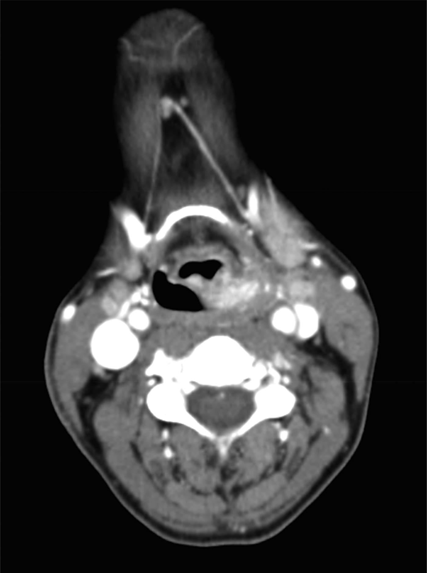
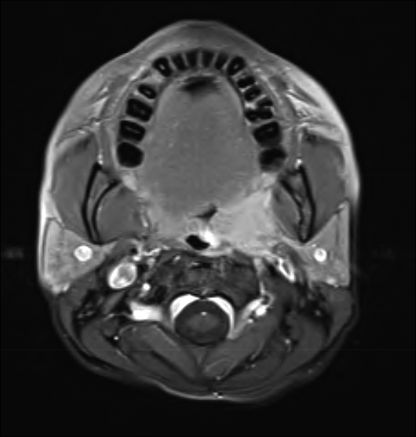
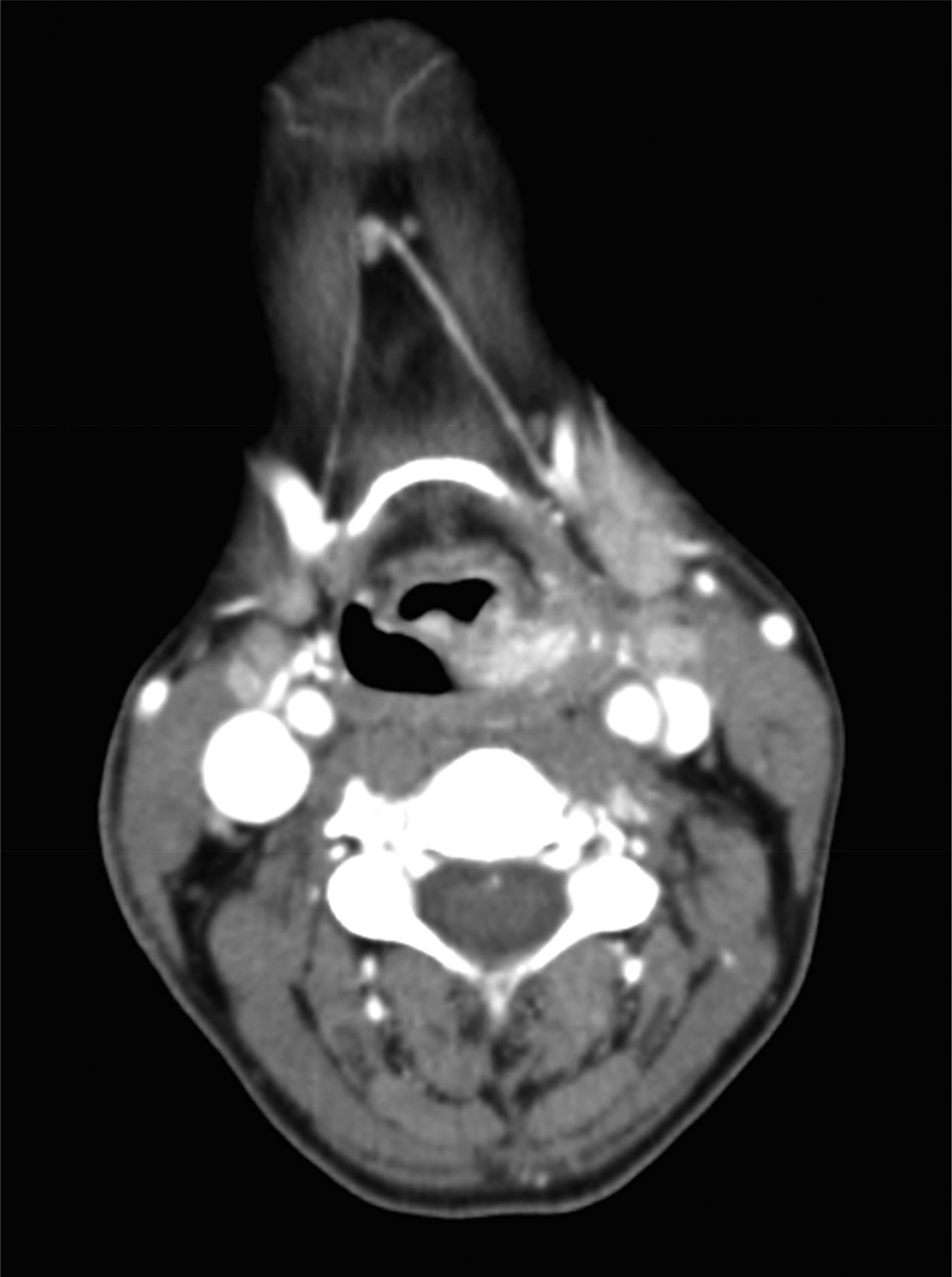
On clinical examination, there was an ulcerated hard mass arising from the left tonsil, and an ipsilateral, level II, firm and palpable neck lump that was 3 cm in size. The clinical findings were suspicious of a malignant process and imaging was therefore arranged. Both computed tomography and magnetic resonance imaging revealed an enhancing infiltrative mass within the left tonsil, with probable involvement of the tongue base and vallecula. In addition, bilateral cervical lymphadenopathy was observed. The radiological report suggested a high likelihood of a primary tonsillar squamous cell carcinoma (SCC), and a provisional tumour–node stage of T4N2c was given (Figures 1 and 2).
Axial computed tomography scan with contrast, showing a mass in the left tonsillar fossa approaching the left pyriform fossa.
Axial, T1-weighted magnetic resonance imaging scan with gadolinium, showing a lesion in the left tonsillar fossa.

As the patient was needle phobic, fine needle aspiration cytology (FNAC) of the left-sided cervical lymph node was undertaken under general anaesthetic, alongside panendoscopy and biopsy of the abnormal left tonsil. Histological evaluation was carried out by the regional head and neck multidisciplinary team (MDT), and it was concluded that there was no evidence of malignancy. The evaluation revealed a fibrovascular stroma covered with non-keratinising, stratified squamous epithelium with evidence of atypia, and florid tonsillitis. Unfortunately, the FNAC sample was inadequate for analysis. Despite these results, a potential diagnosis of malignant SCC was still suspected; therefore, a wide local excision left tonsillectomy was undertaken. The lymph node was left in situ to prevent contamination of the field for future treatment. Further histological evaluation of the entire tonsil did not reveal malignancy, but again the fibro-inflammatory features previously identified were observed. At this point, the decision was made to monitor the patient.
Subsequently, the patient was monitored in the out-patient setting. The operative site had healed satisfactorily, and there was no mucosal abnormality seen within the oropharynx. Additionally, the patient's symptom of odynophagia had resolved. However, the left level II cervical lymph node was still palpable and it was decided to monitor it through serial ultrasound scanning. After two scans, the lymph node architecture remained abnormal, and there was a marginal increase in size on the second scan. There were no other abnormal lymph nodes identified at this stage. An ultrasound FNAC was not possible because of the patient's extreme anxiety, and so the lymph node was excised under a general anaesthetic.
The histological specimens were stained using the periodic acid-Schiff, Ziehl–Neelsen and Grocott's methenamine silver stain protocols for micro-organisms. Immunohistochemistry for immunoglobulin light chains confirmed a polyclonal population of plasma cells. Stains for human herpesvirus-8 and cluster of differentiation 31 were also carried out given the patient's HIV status, but these were negative. The findings were reported as a lymph node with a structure that was largely obliterated by a densely sclerotic fibro-inflammatory process, extending into the perinodal soft tissue. This was the first confirmation of a local inflammatory pseudotumour. A second opinion from a tertiary hospital on both the lymph node and retrospectively on the tonsil specimens confirmed the same.
The patient currently remains well, and has had no further symptoms, cervical lymphadenopathy or mucosal abnormality on clinical examination.
Discussion
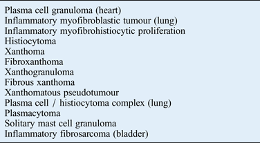
Inflammatory pseudotumours were first described in 1939Reference Narla, Newman, Spottswood, Narla and Kolli1 and named in 1954.Reference Umiker and Iverson2 Since then, many synonymous terms have been used to describe these lesions, which are outlined in Table I.Reference Narla, Newman, Spottswood, Narla and Kolli1 Inflammatory pseudotumours remain important as they have been found in many anatomical sites, and pose a dilemma in every medical and surgical specialty.Reference Patana, Sevrukov, Elsayes, Viswanathan, Lubner and Menias3
Various nomenclature for inflammatory pseudotumours
Case reports of such lesions affecting the head and neck are rare in comparison to other anatomical sites.Reference Patana, Sevrukov, Elsayes, Viswanathan, Lubner and Menias3, Reference Segawa, Yasumatsu, Shiratsuchi, Tamae, Noda and Yamamoto8 A literature search of relevant databases such as Medline and PubMed was conducted using the Population, Intervention, Comparator and Outcomes (‘PICO’) structure, and Medical Subject Heading terms. This yielded no cases in the English-language literature of inflammatory pseudotumours presenting in the tonsillar region with anatomically separate lesions mimicking a metastatic process. Thus, the case presented here is unique.
It is well known that HIV is associated with many unusual neoplastic lesions, with some classed as non-autoimmune deficiency malignancies,Reference Wang, Silverberg and Abrams9 and these can appear clinically similar to lymphoma.Reference Aboulafia5 Inflammatory pseudotumours of the lymph nodes characteristically demonstrate a fibro-inflammatory process in the tissues,Reference Moran and Abbondanzo4 which is reflected in the case we have presented. Despite this, there are very few reports in which an inflammatory pseudotumour has developed in the presence of HIV positivity.Reference Aboulafia5–Reference Sasindran, Ravikumar, Senthil and Somu7 In two of the cases reported previously, the paranasal sinuses were identified as the site of development.Reference Chan-Tack, Chengappa, Wolf, Kao and Reisler6, Reference Sasindran, Ravikumar, Senthil and Somu7
The pathogenesis of inflammatory pseudotumours is unclear, but, in terms of immunohistology, B- and T-cell subpopulations vary widely, differentiating it from lymphoma which consists of clonal populations of the two cells.Reference Patana, Sevrukov, Elsayes, Viswanathan, Lubner and Menias3 There is a small body of evidence suggesting possible categories of predisposing or causative factors. These include local injury or surgery, infection, and immune-autoimmune mechanisms. It is thought that organisms that may have been locally present and remain dormant during a period of immunosuppression become unmasked once cluster of differentiation 4 counts rise and viral loads decrease after starting HAART. The result is a paradoxical clinical deterioration in the context of an improving immune system.Reference Murdoch, Venter, Van Rie and Feldman10 This particular sequence of events is termed ‘immune reconstitution inflammatory syndrome’, and offers a potential link between the development of inflammatory pseudotumours in HIV-positive patients. One report describes an inflammatory pseudotumour of the paranasal sinuses developing 20 weeks after the initiation of aggressive HAART.Reference Chan-Tack, Chengappa, Wolf, Kao and Reisler6
Immune reconstitution inflammatory syndrome can occur at any cluster of differentiation 4 count, and there have been cases of immune reconstitution inflammatory syndrome presenting several years after reconstitution of the immune system. Manifestations that occur after seven months (deemed late manifestations) are often atypical, with the clinical picture varying depending on the specific underlying illness.11 A definite temporal relationship between inflammatory pseudotumours and an immune reconstitution inflammatory syndrome manifestation is not well established, which in itself adds further complexity to the diagnostic dilemma we have presented.
In the ENT clinic, a neck lump is a common presenting symptom, and a potential malignancy should be excluded. As part of the standard screening of neck lumps, blood tests for particular infective conditions and for general health are conducted, as well as relevant imaging and cytology sampling. Suspicion of a head and neck tumour may result in invasive and painful biopsy methods, such as a tonsillectomy in this case. A fine balance must be struck between using the least invasive investigative methods possible and the possibility of overlooking a malignant condition. Furthermore, in the presence of HIV positivity, it is important to limit any invasive procedures as much as possible because of the additional potential hazard of accidental inoculation.
• Inflammatory pseudotumours of the head and neck mimicking metastatic disease are rare
• Human immunodeficiency virus and other immunocompromising conditions increase the likelihood of inflammatory pseudotumour
• This paper reports a patient successfully treated with surgical excision of lesions
• Multidisciplinary teams are important in the investigative and diagnostic process
It is felt that the surgical intervention in our case was as minimal as possible. In the absence of radiological or histological suggestions of an inflammatory pseudotumour, a patient could be subject to unnecessary invasive investigations or surgery. Therefore, inflammatory pseudotumour should be considered as one of the differential diagnoses of an immunocompromised individual presenting with symptoms and signs suggestive of malignancy, particularly in the HIV-positive subgroup, so that unnecessary surgical interventions can be limited. In our case, the period of time between the initial presentation and the first diagnosis of an inflammatory pseudotumour was six months. This highlights the need to continue the diagnostic process when the radiological and histological findings do not match the clinical picture. To maintain patient safety, a robust and efficient MDT process is also necessary. This ensures a clinical consensus between specialties, which will help guide decisions on diagnosis, treatment and surveillance in challenging cases.