





Introduction
Bacteria have become a valuable medium in Biodesign due to their living properties, particularly their ability to produce metabolites and respond to environmental changes (Karana et al. Reference Karana, Barati and Giaccardi2020; Venil et al. Reference Venil, Zakaria and Ahmad2013). Among these capabilities, bacterial pigment production is a promising bio-based alternative to conventional petroleum-based dyes (Gomes and Soares Reference Gomes and Soares2023; Huang et al. Reference Huang, Gan, He, Jiang and He2024). However, existing practices typically rely on a limited number of well-studied strains, such as Janthinobacterium lividum and highly absorbent, smooth textiles as substrates (Devi et al. Reference Devi, Ramakrishnan, Deka and Parasar2024; Shirata et al. Reference Shirata, Tsukamoto, Yasui, Hata, Hayasaka, Kojima and Kato2000). This restricts both the chromatic range and the dimensionality of the resulting designs, while the interaction between bacterial growth dynamics and substrate surface properties remains largely unexplored. These limitations suggest an opportunity to investigate new pigment-producing bacteria and to explore their application on non-textile, three-dimensional materials. Previous studies have explored the potential of using bacteria as dyes across a variety of materials, generally following two main pathways: the extraction of static pigments or the cultivation of pigments directly onto the substrate. This study focuses on the latter, treating dyeing as a “living performance” where color emerges as a visible record of metabolic activity. This processual approach has been exemplified by works such as (Werth Reference Werth2023) bacterial printing machine, which utilizes live bacterial baths to create temporal pigment depositions. Beyond functional coloring, such methods offer rich aesthetic possibilities for intentional visual control. Designers have previously employed external stimuli, such as sound frequencies in the work of (Kukka 2023) or material-based interventions like the fiber manipulations in Growing Patterns (Moser et al. Reference Moser, Fleck, Posch, Heinzel, Dumitrescu, Tomico and Robertson2023), to shape bacterial growth. However, these precedents remain largely confined to planar textile surfaces.
In this pictorial, we shift the focus from two-dimensional patterns to the surfaces of three-dimensional materials, investigating how material texture and form can actively shape the aesthetic outcomes of bacterial pigmentation. Through a liquid staining method, we identified bisque-fired ceramic as an optimal substrate due to its high water absorption and strong pigment adhesion. By examining the influence of surface texture on coloration, this research culminates in a series of sculptural works that highlight the dynamic interaction between microbial growth and three-dimensional form.
Bacteria, cultivation and staining
Considerations in bacterial selection
-
1. Material Accessibility: The bacterial strain should be obtainable through established biological suppliers, allowing the organism to function as an accessible living material rather than a highly restricted laboratory specimen. This supports reproducibility and transparency within the design process.
-
2. Cultivation as a Design Parameter: The organism should grow reliably under achievable laboratory conditions, enabling pigment production to be modulated through controllable environmental variables (e.g., medium composition, temperature, incubation time). Stable growth and chromogenic expression allow biological processes to be integrated into iterative material experimentation.
-
3. Biosafety and Responsible Material Practice: The strain should meet BSL-1 requirements, ensuring safe handling within a design-oriented laboratory setting. Sterilization procedures should effectively terminate biological activity without generating toxic residues, supporting responsible material cycles and laboratory waste management.
-
4. Chromatic Legibility and Visual Differentiation: Prior research has predominantly focused on Janthinobacterium lividum owing to its reliable pigment production and the high visual recognisability of its characteristic violet hue. However, the chromatic singularity of this organism imposes aesthetic limitations when deployed as a sole colorant within a designed artifact. To expand the visual vocabulary of the system, candidate bacteria selected for this project must be capable of producing a visually perceptible and materially expressive pigment. Critically, the resulting coloration must be clearly distinguishable from the violet of J. lividum, thereby enabling chromatic contrast and narrative differentiation across the composite artifact.
Based on the material and experimental requirements of this project, Serratia marcescens (Figure 1) was selected as the biological agent. It is well documented for producing the red pigment prodigiosin, a secondary metabolite studied for its coloration and biotechnological relevance (Han et al. Reference Han, Xiang, Li, Wang and Wang2021). Biologically, S. marcescens is a Gram-negative, facultatively anaerobic bacterium that grows readily on standard nutrient media and exhibits visible pigment formation under moderate conditions (Andreyeva and Ogorodnikova Reference Andreyeva and Ogorodnikova2015; Haddix and Shanks Reference Haddix and Shanks2018; Rjazantseva et al. Reference Rjazantseva, Andreeva and Ogorodnikova1994). These characteristics allow the bacterium to serve as an active material contributor, integrating biological growth and pigment production into iterative material experiments. The lyophilized microorganism used in this project is LWCC1044 (CMCC(B)41002), purchased from SHANGHAI LUWEI MICROBIAL SCI.& TECH. CO. LTD. Laboratory and environmental strains are commonly handled under established biosafety protocols, supporting reproducibility and safe handling.
Growth of S. marcescens after 72 hours.

Cultivate and staining methods
In this project, microbial strains were cultivated using two different methods depending on the experimental objective: solid culture medium cultivation and a liquid staining method. Figure 2 summarizes this two-step workflow from solid-medium cultivation on NA plates to preparation of an NB-based staining medium and subsequent liquid staining. For terminological consistency in this paper, we use dye to refer to the intended color-bearing role of the biological system in design discourse; staining to refer to the experimental liquid-contact procedure applied to materials; pigmentation to refer to pigment production and/or visible pigment-related effects associated with microbial activity; and coloration to refer to the final observable chromatic outcome on/in the material. Throughout the project, we employed the liquid staining method, treating bacterial pigment deposition as a material behavior that emerges through processes of absorption, diffusion and deposition across different substrates. In this paper, “coloration” refers to the visible chromatic result produced by a combination of dissolved/precipitated bacterial pigments, pigment-bearing medium transport and (where applicable) localized microbial growth and associated residues. In both approaches, the bacteria were cultivated in Nutrient Agar (NA) and Nutrient Broth (NB), respectively. Here, NA refers to the solid nutrient agar plates used for strain activation and passaging (serving as a stable source of pigmented biomass/colonies), whereas NB refers to the liquid nutrient broth used to prepare a pigment-bearing bacterial suspension for subsequent liquid staining. In practice, colonies grown on NA were gently scraped and re-suspended into NB to form the staining medium used across the material screening and object-based staining tests described later. Incubation was conducted in a constant temperature and humidity chamber at 28°C and 80% relative humidity. All cultures were grown under light-protected conditions to ensure stable pigment production.
Two-step workflow for cultivation and liquid staining (NA to NB).

Step 1
Lyophilized microbial strains were first activated using a solid culture medium. The medium was prepared by combining peptone, beef extract, sodium chloride and agar as a gelling agent in an Erlenmeyer flask to form a nutrient medium. After sterilization, the medium was poured into Petri dishes and allowed to cool and solidify. Microbial strains were then transferred from refrigerated slant cultures onto the solid medium using an inoculating loop. The inoculated plates were incubated in a controlled environment at 28°C and 80% relative humidity for 72 hours. This step ensured stable microbial growth and pigment production, providing a consistent biological basis for subsequent material experimentation. Representative NA plates after incubation are shown in Figure 3.
Passaging of S. marcescens on NA plates under controlled incubation.

Step 2
A liquid staining method was subsequently adopted to explore microbial pigments as an active material agent. Pigmented colonies were collected from NA plates and re-suspended into NB to prepare a pigment-bearing bacterial medium, and the objects to be stained were placed directly into the bacteria-containing solution, semi-immersed to enable bottom-up liquid uptake. The Petri dish was maintained in a partially enclosed, low-airflow cultivation environment and incubated under the same temperature and humidity conditions for the designated experimental duration, depending on the specific screening or staining test. During incubation, microbial growth led to the gradual release of pigments into the nutrient medium. These pigments, together with medium transport and possible microbial residues, were attached to the objects over time, allowing coloration to emerge through biological processes.
Multi-material screening
Broad exploration
An iterative material tinkering approach guided this initial screening: we used a research-through-making process to test multiple substrates under the same liquid staining conditions, selecting materials that could both absorb the pigment-bearing medium and produce legible surface coloration. This stage functioned as a broad screening process aimed at identifying substrates most suitable for staining, thereby informing subsequent, more focused experiments. A range of commonly available natural materials was collected. Given that the project seeks to replace synthetic, petroleum-based dyes with sustainable and environmentally friendly bacterial pigments, priority was given to materials that are likewise derived from natural sources, sustainably produced and non-toxic. The selected substrates included plant-based fibers, animal-derived surfaces and shells, as well as other naturally occurring inorganic materials such as clay and metal. Each material was cut into small samples and placed in labeled Petri dishes containing culture medium (Figure 4). All screening tests used the liquid staining method described above, using NB as the liquid staining medium prepared from NA-grown colonies. The pigment-producing bacterium S. marcescens was inoculated onto the samples and incubated under standard laboratory conditions. After 48 hours, the materials were evaluated according to two criteria: (1) liquid absorption rate and (2) the clarity and visibility of surface pigmentation.
Staining experiments of S. marcescens on surface of different materials by liquid staining method.

The screening results indicated that, in comparison to the bacterial films formed on metal sheets and animal shells, fiber-based materials, such as bamboo slices, feathers and wood, as well as inherently absorbent bisque-fired clay exhibited stronger liquid absorption capacities. On these substrates, the pigments produced by the bacteria diffused through the material along with the nutrient solution, resulting in more integrated and permeated coloration effects. Materials with volumetric mass, including uneven-surfaced clay blocks, coarse jute rope and wooden pieces, demonstrated more pronounced stratification and gradient coloration than fully submerged flat substrates such as feathers and silk. This effect can be attributed to their partial immersion during cultivation, which generated differential absorption zones and uneven pigment deposition across their surfaces. Based on comparative evaluation of absorption performance and visible pigmentation outcomes, bisque-fired ceramic and textured fabric were selected for two subsequent focused experiments. These investigations concentrated on the chromatic behavior of bacterial pigments on three-dimensional ceramic forms and on textured fibers of different components.
Porosity-driven gradients on ceramics
Following the NA to NB preparation described above, bisque-fired, unglazed ceramics were selected as the porous carrier for subsequent tests. Based on the screening results, these ceramics offered reliable liquid uptake and readable pigment registration at the material surface. In the liquid staining method, NA-grown colonies were re-suspended into NB to form a pigment-bearing culture solution. Each ceramic piece was then placed into the medium with its lower portion kept in contact with the liquid while the upper portion remained exposed to air, allowing the ceramic body to gradually draw the solution upward through capillary uptake during cultivation. This setup maintained a moist near-surface interface that supports bacterial adhesion and enables pigments (and associated residues) to accumulate and become visible as gradients and tide-like traces on the ceramic surface. Two bacterial colonization approaches and two absorption strategies were tested, resulting in three experimental groups. In the first (control) group, bacteria were inoculated into the culture medium at the bottom of the Petri dish. A bisque-fired teacup was placed upright in the dish and left undisturbed for 48 hours. In the second group, the colonization method remained unchanged; however, after 24 hours in the upright position, the teacup was inverted and left for the remaining 24 hours. This configuration enabled comparison with the first group, particularly to observe bacterial growth and pigment accumulation along the liquid boundary on the ceramic surface. In the third group, additional bacteria were directly and randomly inoculated onto the surface of the teacup. In contrast to the first group, this setup allowed examination of edge expansion dynamics and localized pigment intensification resulting from surface colonization.
Within 48 hours, the ceramic body gradually absorbed the liquid through capillary action, producing soft chromatic gradients, tide marks and stratified deposits. These traces visibly recorded the migration pathways of pigments within the porous matrix. In the first group, gradual diffusion of the solution generated progressively lighter chromatic edges across the cup surface. In the second group, more saturated pigmentation emerged at the intersection of liquid contact zones following inversion. In the third group, directly inoculated bacterial regions displayed noticeably deeper coloration than areas colored solely through absorption, with pigment intensity fading outward from the colony edges, suggesting the potential for controlled modulation of color density. These findings indicate that ceramic functions not merely as a passive surface for staining; rather, its porous texture and absorptive properties actively shape the formation of chromatic patterns and material expression.
In the ceramic-focused experiment, bisque-fired, unglazed ceramic pieces were used as porous carriers. Figure 5 compares multiple ceramic staining configurations and their resulting boundary conditions, including differences in liquid contact zones, exposure patterns and pigment accumulation at the ceramic surface. Under liquid staining, the pigment-containing medium remained in contact with the ceramic surface during cultivation. Within the incubation window, the ceramic body gradually absorbed liquid through capillary action, producing soft chromatic gradients, tide marks and stratified deposits. These traces visibly recorded pigment migration pathways within the porous matrix. We also tested different placement/colonization setups to compare how contact zones and exposure conditions modulate edge diffusion, saturation and localized intensification, revealing the ceramic surface as an active participant in pattern formation rather than a passive receiver of color.
Three sets of experiments on the staining of S. marcescens on the surface of ceramic materials by liquid staining method for 48 hours.

Absorption and distribution across textiles
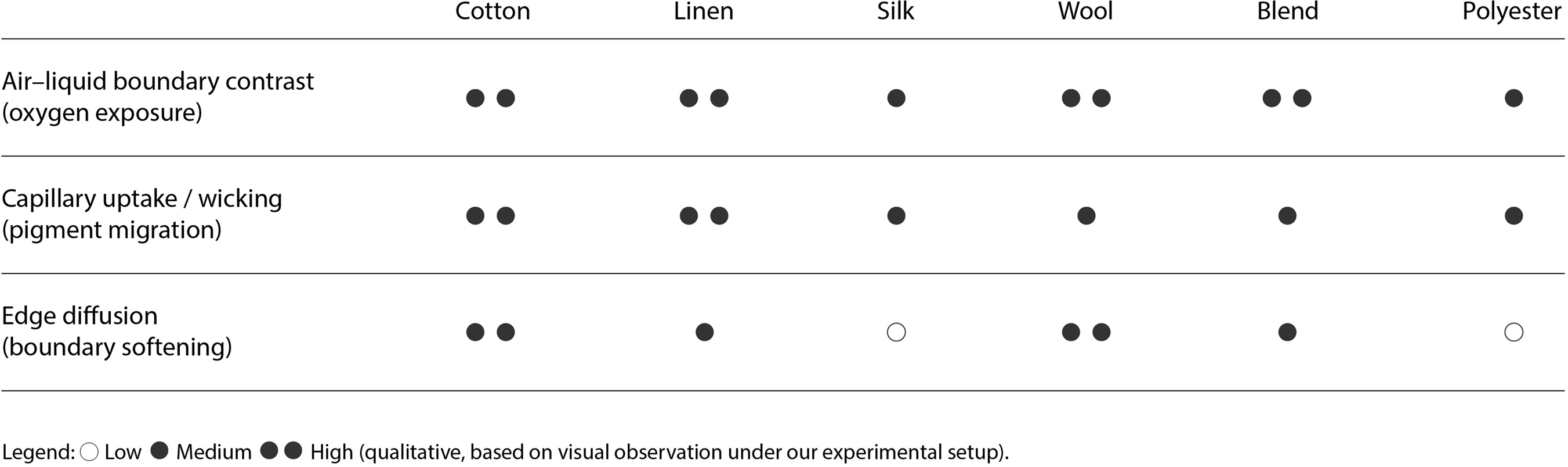
A series of staining tests was conducted on multiple fiber materials to examine differences in pigment distribution across varying textile compositions and structures. The samples included natural fibers such as cotton, linen, silk and wool, as well as blended fabrics and polyester. Each sample was positioned in a semi-immersed state within the culture medium. The glass containers were not fully sealed, allowing limited air circulation throughout the cultivation period. After 48 hours, coloration intensity, saturation and spatial distribution were documented and compared across the different textiles. Fabrics fully submerged in the liquid exhibited minimal visible pigmentation; instead, they primarily absorbed the culture medium. In contrast, the portions exposed to air demonstrated significantly stronger coloration (see Figure 6), revealing a clear air–liquid boundary contrast under semi-immersion (oxygen exposure). This outcome indicates that oxygen plays a critical role in the production of bacterial pigments. The liquid staining method proved to be effective in supporting object-based coloration. The semi-immersion strategy ensured sufficient nutrient availability from the medium while maintaining exposure to air, thus sustaining the bacterial metabolic activity responsible for pigment synthesis. Marked differences in staining results were observed between fiber types, often in correlation with fiber composition and weave structure. To summarize these observations in a compact way, Figure 7 provides a qualitative comparison across textile groups along three dimensions: (1) air–liquid boundary contrast (oxygen exposure), (2) capillary uptake/wicking (pigment migration) and (3) edge diffusion (boundary softening). Compared to ceramic substrates, fiber materials tended to achieve more uniform and saturated coloration, whereas ceramic surfaces more prominently exhibited diffusion gradients and sedimentary traces. Figure 7 provides a compact qualitative reference that makes these cross-textile differences easier to compare at a glance.
Experiments of S. marcescens on 18 textile types using liquid staining (48 hours).

Qualitative comparison of textile staining outcomes under semi-immersion (48 hours).

Design and realization
Textured ceramic sample making: printing, air-drying and bisque firing
After the material screening experiments, a set of comparable textured ceramic test samples was produced in order to systematically investigate how surface characteristics influence microbial coloration. The purpose of this step was to establish a controlled, repeatable and comparable set of textured surfaces, so that any differences observed under identical dyeing conditions could be clearly attributed to the mechanism of “texture–absorption–deposition.” These geometric samples were organized as a small “texture library.” Within similar scales and using the same material, surface topologies were intentionally varied – such as ridges, grooves, perforations and layered undulations. This approach provided a foundation for interpreting coloration results in relation to surface geometry, capillary absorption and temporal deposition, rather than allowing the outcomes to be dominated purely by sculptural form.
An open-source clay 3D printer was used in combination with the slicing software Ultimaker Cura. After slicing the digital models, support structures and internal configurations were adjusted according to different geometries to ensure printing stability while preserving the intended surface textures as much as possible. The samples were printed using high-white porcelain clay produced in Jingdezhen, Jiangxi, China. This is a standard ceramic clay which, after firing, yields a relatively fine and smooth surface without coarse sand particles. To ensure stable extrusion, the clay’s consistency and moisture content were carefully adjusted, and the material was compacted before being loaded into the cartridge for printing. Figure 8 primarily documents key moments of the printing process: nozzle extrusion, layer-by-layer deposition and preliminary test blocks. These layered traces are emphasized because they later function as readable surface features that actively participate in pigment absorption and deposition. After printing, external supports were removed, and minor surface imperfections were refined to ensure clarity of texture and comparability among samples. The formed green bodies were air-dried indoors until sufficiently strong, then bisque-fired at 800°C without glaze (Figure 9). After firing, the samples became white and hardened, while retaining the necessary microporous structure. This bisque-firing stage corresponds to a process of structural stabilization, preparing the porous body for subsequent wet dyeing experiments in which absorption and diffusion remain active.
Printing process: (a) Printer. (b) Printer in operation. (c) Close-up view of printing. (d) Side view of printing. (e) Textured layers of printed ceramic sample. (f) Textured layers of a 15cm ceramic sample.

The completed print is fired in a kiln at 800°C to solidify and finalize the ceramic structure.

In the Textured Ceramic Sample Making stage, the workflow followed: printing, air-drying, bisque firing (unglazed). This standardized set of textured samples provided a baseline for the next phase, enabling comparison of bacterial pigment migration and deposition under identical staining conditions.
Experimental dyeing on unglazed ceramics: expanded with dual pathways
Building on the previously developed standardized textured ceramic samples, experimental dyeing was conducted on bisque-fired, unglazed porous ceramics in order to preserve capillary absorption and diffusion as active mechanisms of pattern formation. At this stage, “coloration” was understood as an evolving process rather than a fixed, one-time coating. Meanwhile, the pigment-containing bacterial suspension/culture medium exhibited distinct phase characteristics after settling: pigment components formed a denser sediment layer at the bottom of the container (Figure 10a), further influencing pigment intensity and gradient formation near the contact zone.
Experimental dyeing on unglazed ceramics: (a) Pigment-bearing medium after standing. (b) Localized inoculation into selected grooves on the ceramic surface to guide site-specific pigmentation. (c) Incubation setup showing two pathways on one piece: absorption-driven staining at the bottom and localized pigmentation at the inoculated textures. (d) Time-series comparison documenting changes in intensity, diffusion boundaries, migration height, and deposits over time.

To explore how pattern formation could be modulated beyond passive absorption, we juxtaposed two complementary coloration pathways on the same ceramic sample. The first pathway relied on the capillary absorption of the ceramic: the base of the sample was kept in contact with the pigment-containing culture medium, allowing the liquid to gradually wick upward through the porous structure. This process generated soft gradients and tide-like marks along the printed layer lines and surface textures. The second pathway introduced localized inoculation: bacterial strains were “planted” directly onto selected textured areas of the model, such as grooves and cavities (Figure 10b). By leveraging the micro-topography’s ability to retain moisture, pigment deposition was guided toward specific sites, resulting in more concentrated and spatially defined coloration. In this way, under identical material and geometric conditions, it became possible to compare “absorption-driven migration” with “localized growth and deposition.”
During cultivation, the juxtaposition of these two pathways became clearly visible on the same model (Figure 10c): the lower region mainly exhibited absorption-driven gradients, while the upper region showed a more localized and directional pigment distribution in the inoculated textures. Finally, a time-series documentation was conducted (Figure 10d), observing and comparing how color intensity, boundary diffusion, upward migration height and deposition textures evolved over time. In general, this stage demonstrates how, without relying on a sealing glaze layer, the interplay of porous structure, surface texture and inoculation strategy collectively shapes microbial coloration in ceramics. The process enables both global gradients driven by liquid absorption and more “directional” texture markings achieved through localized colonization.
Results
We present Bactereature (Figure 11), a series of artworks that embody the results of our exploratory investigation. The images show the outcomes of liquid bacterial pigment staining on textured, unglazed ceramics. Although presented as artworks (Figure 12), the series also functions as material evidence, visualizing how porous structure, surface topology and exposure conditions translate microbial pigments into gradients, tide lines and sediment-like traces. Rather than aiming for uniform coloration, the results foreground capillary uptake and diffusion as active pattern-generating mechanisms. Printed layer lines, grooves and cavities guide where pigments accumulate and how boundaries emerge, producing differentiated surface expressions.
Bactereature: comparative set of textured, unglazed ceramic outcomes produced through liquid staining with bacterial pigments.

Final presentation view of the Bactereature setup, showing multiple ceramic samples cultivated in pigment-bearing medium under controlled conditions.

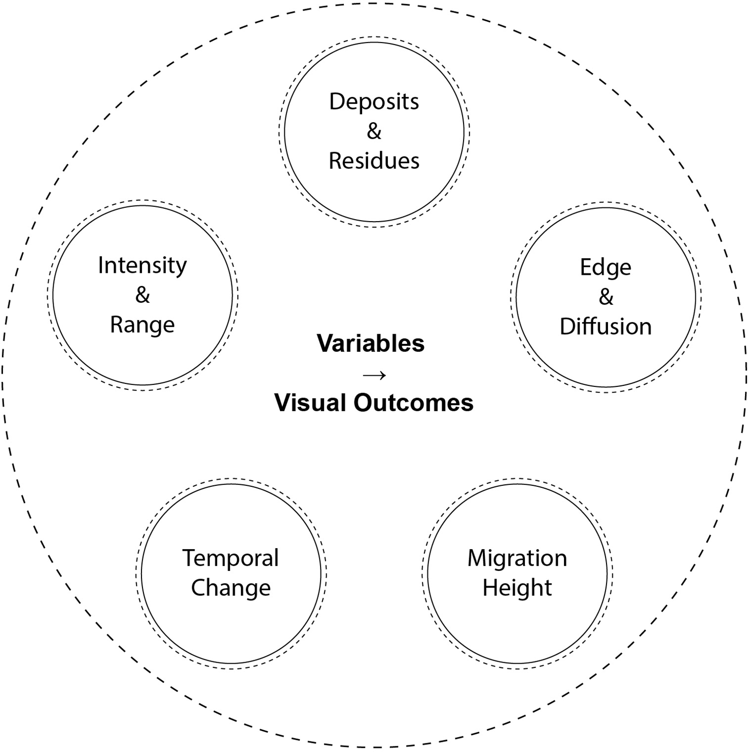
We summarize the link between experimental variables and visible outcomes through five observable dimensions (Figure 13): (1) Color intensity and chromatic range: how saturated or pale the coloration appears and whether the surface shifts between a narrow tone and a broader range of reds/pinks. This dimension makes differences in pigment load and deposition legibility comparable across samples. (2) Boundary sharpness and degree of diffusion: how clearly an edge is defined versus how softly it blooms into surrounding areas. Here, sharper boundaries often indicate constrained pathways, while softer boundaries suggest broader diffusion through pores and micro-relief. (3) Upward migration height: how far pigmentation travels against gravity from the liquid contact zone, registering the material’s capillary uptake and the availability of pigment-bearing medium over time. This provides a direct visual measure of absorption-driven transport. (4) Deposition and residue textures: the presence of tide marks, layered residues and sediment-like accumulation, including where pigments pool within grooves or settle along layer lines. This dimension emphasizes how micro-topology shapes where pigments become trapped or concentrated. (5) Temporal change: how the coloration evolves across time windows, such as shifts in saturation, edge softening or fading. This dimension frames coloration as a time-based record rather than a one-step finish.
A schematic mapping that links experimental and material variables to five observable dimensions in the final ceramic surfaces.

Taken together, the gradients and deposits can be read as material traces of microbial activity – evidence of coloration shaped by the interaction of porous matter, environment and time. Color here is not a fixed coating applied to an inert surface but a temporal record emerging from the interplay between material behavior and biological processes.
Proof-of-concept: porous ceramics as an active carrier for microbial pigmentation
Why porous ceramics were selected
In the preceding multi-material screening phase, comparing several ceramic bodies with fibers, wood, paper, leather and metal, we identified bisque-fired, unglazed porous ceramics as an effective non-textile substrate for bacterial pigment deposition. In contrast to other materials that showed insufficient absorption, weak or unstable coloration or poor pigment legibility, porous ceramics consistently demonstrated two key properties: (1) stable and repeatable capillary absorption and (2) visible pigment manifestation at or near the material surface. These observations justified the subsequent focus on ceramics as the primary substrate for textured sample experiments. Within this research context, the effectiveness of ceramics lies not merely in their ability to be stained but in their capacity to participate in a material-driven pattern formation process.
Mechanism: capillary uptake, pore network and deposition
Bisque-fired ceramics retain an interconnected pore network that draws liquid into the near-surface region through capillary action. When brought into contact with pigment-containing bacterial culture, color does not simply coat the exterior surface; rather, it migrates into the pore structure, redistributes within micro-textures and partially remains trapped. This creates a persistently moist interface in which pigments and residues associated with bacterial activity, accumulate, migrate and gradually become visible over time. Coloration is therefore not a superficial layer but a record embedded within the surface and subsurface through the combined effects of absorption pathways, local geometry and temporal progression, resulting in gradients, tide marks and deposition textures.
Parameter framework: variables and comparative indicators
This proof-of-concept phase further indicates that microbial coloration on porous ceramics should be understood as a parameter-driven patterning system rather than a single-step dyeing operation. The inoculation strategy (localized seeding, surface application or broader contact), which determines the origin and boundary formation of coloration; liquid availability, influencing migration height, saturation and deposition intensity; time, affecting saturation, diffusion, accumulation, stabilization and fading trajectories; environmental conditions such as temperature and humidity, which regulate liquid movement and bacterial activity; and surface topology and pore structure, including ridges, grooves, cavities and pore density, which modify capillary pathways and boundary definition. Importantly, these variables can be compared across samples using consistent visual criteria: chromatic range and intensity, boundary sharpness versus diffusion, gradient length or upward migration height, deposition and residue morphology and temporal change, including fading behavior. This framework moves the inquiry beyond whether coloration occurs and toward a mechanistic understanding of how porous ceramics actively shape microbial pigmentation.
Taken together, the proof-of-concept results show that porous ceramics are not passive carriers of bacterial pigment, but active participants in pattern generation. Their absorptive behavior and pore structure not only enable pigment infiltration, but also constitute part of the mechanism through which coloration forms, stabilizes and evolves. This provides a direct foundation for the subsequent chapter, which investigates how controlled textured ceramic samples modulate pigment migration and pattern formation.
Discussion
This project approaches bacterial coloration on porous, unglazed ceramics not merely as an alternative coloring technique, but as a pattern-generating system co-produced by material and biological processes. Liquid absorption, pore structure, surface texture and time collectively shape the final visual outcome. Based on the preceding material screening and proof-of-concept experiments, the significance and limitations of this project can be reflected upon from four perspectives: sustainability claims, material agency, temporality and exhibition context.
Alternative coloration as a proposition
This project proposes an alternative way to think about ceramic coloration: from chemically fixed surface finishes toward process-based coloration shaped by biological pigments and material absorption. Rather than presenting a sustainability outcome, the work positions microbial pigmentation as a material strategy that foregrounds low-temperature processing, visible material behavior and responsive color formation. In this sense, it can be discussed as an alternative finishing pathway within research and cultural contexts that value process legibility and time-based change. At the same time, the approach should not be interpreted as a direct replacement for industrial glazing or established ceramic finishing systems. The current workflow depends on controlled cultivation environments, laboratory protocols and significant time investment; moreover, color stability, repeatability and long-term maintenance remain open questions, especially beyond controlled settings. Accordingly, the contribution is best understood as a design-research proposition – articulating a different logic of coloration – rather than a validated, scalable production method.
Material agency
Bacteria in this context are not static pigments but evolving pigment systems. A central contribution of this research is the shift from understanding “bacteria as dye” to “bacteria as a temporal pigment system.” In ceramic substrates, color is not applied as a uniform coating on an inert surface; instead, it gradually emerges through the interaction of liquid transport, pore networks, surface relief, inoculation strategies and cultivation conditions. Bacteria thus function not only as alternatives to conventional dyes but as material agents within a pattern formation process. At the same time, the ceramic body is not a passive carrier. Its porosity and micro-topography actively regulate absorption, diffusion, deposition and boundary formation. For design practice, this reorients attention from selecting a final color toward designing the conditions under which color appears, migrates, accumulates and fades.
Temporality
Observed changes over time – such as shifts in saturation, softened diffusion boundaries, sediment accumulation and fading trajectories – raise a critical question: must microbial coloration be evaluated according to the industrial ideal of permanence, or can non-permanence itself constitute a meaningful material language? In this project, temporality functions as a generative dimension. Color evolves, weakens or transforms in response to temperature, humidity, light exposure and other environmental variables. This challenges the assumption that design outcomes must prioritize visual stability. At the same time, temporal variability introduces practical challenges related to preservation, reproducibility and public display. Rather than resolving this tension, the project foregrounds it: microbial coloration on ceramics may be better understood as a medium negotiated between ephemerality and legibility.
Exhibition context, biosafety and interpretive responsibility
Situated at the intersection of biological process and material result, the project requires careful framing in exhibition contexts. A crucial distinction concerns whether audiences encounter an ongoing live microbial process or the material traces left by prior microbial activity embedded in the ceramic surface. These two modes entail different temporal, maintenance and risk-management considerations. If presented as a live process, biosafety protocols, containment strategies, environmental controls and explanatory frameworks become integral components of the project rather than external technical support. Exhibition design must then address not only visual presentation but also procedural responsibility: what biological activity is being shown, under what conditions and how it is managed. If presented as post-cultivation material traces, the focus shifts toward reading coloration as evidence of process. This mode retains the project’s core material argument while potentially reducing logistical and safety complexities. The distinction has practical implications for future dissemination in artistic, design and public contexts, as it directly affects curatorial strategy, accessibility and interpretive pathways.
Limitations
Several limitations remain. First, color fastness and long-term stability are limited; the visual outcome is sensitive to environmental factors such as light, particularly UV exposure, temperature, humidity and other cultivation-related conditions. Second, the staining process currently relies on laboratory environments with appropriate biosafety measures, restricting broader implementation beyond research settings. Third, while qualitative relationships between pore structure, absorption behavior and visible pattern formation have been established, improved comparability and predictability will require more systematic parameterization, including pore size distribution, surface roughness, absorption rates and time-resolved color quantification.
Data availability statement
The data that support the findings of this study are available from the corresponding author upon reasonable request.
Author contributions
Conceptualization, J. Liang and W. Kuang; Methodology, Y. Qian, J. Liang and W. Kuang; Biological Cultivation and Staining, J. Liang; Material Experimentation, J. Liang, Y. Qian and W. Kuang; Hardware, Y. Qian; Software, Y. Qian, J. Liang and W. Kuang; Visualization and Graphic Development, J. Liang; Investigation, Y. Qian, J. Liang and W. Kuang; Formal Analysis, Y. Qian, J. Liang and W. Kuang; Writing – Original Draft, J. Liang and Y. Qian; Writing – Review & Editing, Y. Qian and J. Liang; Supervision, C. Ma and Y. Xiang.
Financial support
This research received no specific grant from any funding agency in the public, commercial or not-for-profit sectors.
Competing interests
The authors declare that there are no conflicts of interest.
Ethical standards
Ethical approval was not required for this study as it did not involve human or animal subjects.
AI statement
AI tools were used as a writing assistant for grammar correction and expression refinement only. All ideas, analysis and conclusions are the authors’ own and the author takes full responsibility for the content.





 Open access

