Introduction
Schistosomiasis is most commonly recognized as a tropical disease of humans. Yet, other mammals and aquatic birds harbour their own schistosome species, which are also implicated in causing disease and can cause cercarial dermatitis in humans. There are about 100 species of schistosomes that infect mammals and birds globally in both marine and freshwater environments. These trematodes have a two-host life cycle in which adult worms live in the venous (or rarely, arterial) system of their definitive host and release eggs, usually through the faeces of the host. Miracidia that hatch from the eggs penetrate an appropriate intermediate host, an aquatic snail, in which they undergo clonal reproduction. Cercariae are released from the snail and complete the life cycle by penetrating the mammal or bird host (Kuntz, Reference Kuntz1955; Malek, Reference Malek1961; Loker, Reference Loker1983).
Because of its medical and veterinary relevance, we know a great deal more about the mammal-infecting genus Schistosoma Weinland, 1858 than we do about other genera of mammalian schistosomes (the North American genera Schistosomatium Tanabe, Reference Tanabe1923 and Heterobilharzia Price, Reference Price1929, and the elephant schistosome Bivitellobilharzia Dutt & Srivastava, Reference Dutt and Srivastava1955). For the latter genus, we are largely ignorant of even the basics, including life cycles and patterns of definitive and intermediate host use, even though the mammalian hosts involved are large and conspicuous. In fact, studies to characterize the parasite fauna of these large hosts are difficult and typically opportunistic. Faecal samples are often the only window into their parasite infections, and identifications based on egg and larval morphology alone lack precision. Comparison of nucleic acid sequence data among different life cycle stages, often collected from disparate locations and times, provides an independent means to link adult stages with the larval stages or samples from different hosts. Therefore, inferences about the life cycle and transmission can be made without undertaking experimental infections or destructive sampling of the host (e.g. Brant et al., Reference Brant, Morgan, Mkoji, Snyder, Rajapakse and Loker2006).
With respect to Bivitellobilharzia, two species are known, both from the venous system of elephants. Bivitellobilharzia loxodontae Vogel & Minning, Reference Vogel and Minning1940 occurs in African forest elephants Loxodonta cyclotis Matschie, 1900 (Democratic Republic of Congo, Central African Republic). Bivitellobilharzia nairi (Mudaliar & Ramanujachary, Reference Mudaliar and Ramanujachary1945) Dutt & Srivastava, Reference Dutt and Srivastava1955 occurs in Asian elephants Elephas maximus Linnaeus, 1758 (India, Sri Lanka, Republic of the Union of Myanmar) (Vogel & Minning, Reference Vogel and Minning1940; Mudaliar & Ramanujachary, Reference Mudaliar and Ramanujachary1945; Rao & Hiregauder, Reference Rao and Hiregauder1953; Sundaram et al., Reference Sundaram, Padmanabha Iyer, Peter and Alwar1972; Agatsuma et al., Reference Agatsuma, Rajapakse, Kuruwita, Iwagami and Rajapakse2004; Kinsella et al., Reference Kinsella, Deem, Blake and Freeman2004; Brant et al., Reference Brant, Morgan, Mkoji, Snyder, Rajapakse and Loker2006). Thus far there have been no definitive reports of schistosomes from the African savanna elephant, Loxodonta africana (Blumenbach, 1797). Tiuria et al. (Reference Tiuria, Primawidyawan, Pangihutan, Warsito, Hariyadi, Handayani and Priosoeryarito2006) also reported a schistosome from the Javan rhino, Rhinoceros sondaicus Desmarest, 1822, which might be Bivitellobilharzia. In phylogenetic reconstructions, thus far based only on B. nairi, Bivitellobilharzia is basal to the Schistosoma+Orientobilharzia Dutt & Srivastava, Reference Dutt and Srivastava1955 clade of schistosomes that occur predominately in bovines, humans and rodents (Brant et al., Reference Brant, Morgan, Mkoji, Snyder, Rajapakse and Loker2006). There has been little work on Bivitellobilharzia, most of it published for B. nairi in Asia (Mudaliar & Ramanujachary, Reference Mudaliar and Ramanujachary1945; Dutt & Srivastava, Reference Dutt and Srivastava1961; Sundaram et al., Reference Sundaram, Padmanabha Iyer, Peter and Alwar1972; Agatsuma et al., Reference Agatsuma, Rajapakse, Kuruwita, Iwagami and Rajapakse2004; Brant et al., Reference Brant, Morgan, Mkoji, Snyder, Rajapakse and Loker2006). Since the original description of B. loxodontae by Vogel & Minning (Reference Vogel and Minning1940), this species has been reported once by Kinsella et al. (Reference Kinsella, Deem, Blake and Freeman2004). In this report we provide details on the phylogenetic position of B. loxodontae, the species that occurs in African forest elephants.
Materials and methods
Collections of fresh forest elephant faecal samples were made in the area surrounding Bai Hokou field station (2°51′34″N, 16°28′03″E) in the Dzanga Sector of Dzanga Ndoki National Park (NP) in south-western Central African Republic (CAR) in November and December 2008, 2009 and 2010 by K.J.P. and K.P. with assistance from local BaAka trackers. Faecal samples were immediately preserved in both 10% formalin and 96% ethanol. All preserved samples were shipped from CAR to the Department of Pathology and Parasitology at the University of Veterinary and Pharmaceutical Sciences in Brno for screening. A subsample of the ethanol-preserved samples was later sent to S.V.B. and E.S.L. at University of New Mexico for molecular analysis. Sixteen unsieved ethanol-preserved faecal samples (each about 10 ml) from L. cyclotis were examined for schistosome eggs. Faecal samples were spread out in water in a Petri dish and examined with the aid of a dissecting scope, and eggs were individually isolated and cleaned of debris.
Schistosome eggs were digested overnight in proteinase K and the DNA was extracted using the Qiagen DNeasy Blood and Tissue Kit (Valencia, California, USA). The nuclear ribosomal gene 28S and the mitochondrial cytochrome oxidase I (cox1) gene were amplified by polymerase chain reaction. Methods were as described in Brant et al. (Reference Brant, Morgan, Mkoji, Snyder, Rajapakse and Loker2006). There are only two known species of Bivitellobilharzia and to confirm that our sample was different at the species level from B. nairi, we compared cox1 pairwise sequences. This allowed us to also relate our results with species differences among closely related species of Schistosoma.
The 28S fragment was used for phylogenetic analysis. Phylogenetic analyses were performed on this dataset using maximum parsimony (MP), maximum likelihood (ML), and minimum evolution (ME), which were carried out in PAUP* version 4.0b10 (Swofford, Reference Swofford2002), and Bayesian inference (BI) with the use of MrBayes (Huelsenbeck & Ronquist, Reference Huelsenbeck and Ronquist2001; Ronquist & Huelsenbeck, Reference Ronquist and Huelsenbeck2003). jModeltest was used to determine the best fit nucleotide substitution model for ML and ME analyses (Posada, Reference Posada2008). Parsimony trees were reconstructed using heuristic searches (200 replicates). Optimal ME and ML trees were determined from heuristic searches (10 replicates for ME, 5 replicates for ML). Nodal support was estimated by bootstrap (500 replicates, 10 addition sequence replicates for MP; 200 replicates for ME). For the BI analysis, the parameters were unlinked: Nst = 6 rates = gamma ngammacat = 4, four chains were run simultaneously for 2 × 10⁵ generations, trees sampled every 100 cycles, the first 2000 trees with pre-asymptotic likelihood scores were discarded as burn-in, and the retained trees were used to generate a 50% majority-rule consensus tree and posterior probabilities. In addition to the 28S GenBank sequences listed in Brant et al. (Reference Brant, Morgan, Mkoji, Snyder, Rajapakse and Loker2006), the following available schistosome sequences were downloaded from GenBank to compare against our samples (accession numbers in parentheses): Schistosoma kisumuensis (FJ897153), S. hippopotami (AY197343), Allobilharzia visceralis (EF114223), Trichobilharzia brantae (FJ174466), T. franki (FJ711768), T. stagnicolae (FJ174478) and B. nairi (AY829249).
Results
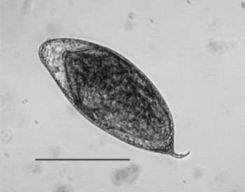
The formalin-preserved schistosome eggs collected from L. cyclotis in Dzanga Ndoki NP (CAR) measured 142–182 μm (average 164.7 μm) in length (excluding spine) by 65–80 μm (average 70.9 μm) wide with a spine length of 17–24 μm (average 19.9 μm) (fig. 1). These are comparable to measurements of fresh eggs of B. loxodontae by Vogel & Minning (Reference Vogel and Minning1940): length 145–203 μm (average 168 μm) by 56–83μm (average 71 μm) with spine length 15–37 μm (average 26 μm). Kinsella et al. (Reference Kinsella, Deem, Blake and Freeman2004) did not include measurements of schistosome eggs they obtained from forest elephants, but our eggs look similar to the one they photographed. Egg measurements of B. nairi from faecal samples from Vogel & Minning (1945) were 138–183 μm (average 161 μm) long by 70–91 μm (84 μm) with spine length 9–25 μm (average 16 μm), and 141.9–181.5 μm (average 155.6 μm) by 66–108.9 μm (average 81.04 μm) with spine length 6.6–18.2 μm (average 9.2 μm) from Sundaram et al. (Reference Sundaram, Padmanabha Iyer, Peter and Alwar1972). Measurements from eggs in utero were 80 × 30 μm from Mudaliar & Ramanujachary (Reference Mudaliar and Ramanujachary1945) and 93–121 μm (average 81 μm) by 58–69 μm from Sundaram et al. (Reference Sundaram, Padmanabha Iyer, Peter and Alwar1972).
Light microscope image of Bivitellobilharzia loxodontae egg. Scale bar = 90 μm.

In addition to the schistosome eggs, we found the following parasites, listed by year followed by type of parasite/ciliate and prevalence: in 2008, intestinal strongylid nematodes 62%, Protofasciola robusta 8%, Mammomonogamus sp. 8%, ciliates 100%; in 2009, intestinal strongylid nematodes 79%, P. robusta 14%, Mammomonogamus sp. 6%, B. loxodontae 17%, ciliates 79%; and in 2010, intestinal strongylid nematodes 68%, P. robusta 23%, B. loxodontae 20%, ciliates 71%. The above data are from the 100 faecal samples collected from 2008 to 2010, a subset of 16 of the above were used for the molecular part of this project. The number of eggs or other stages found per sample was not recorded. Specimen compositions of the faecal samples were similar to those found in L. cyclotis by Kinsella et al. (Reference Kinsella, Deem, Blake and Freeman2004).
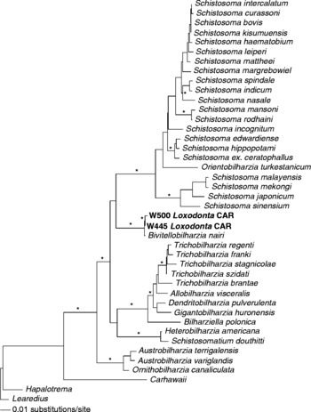
The results of the phylogenetic analysis grouped the tentatively assigned CAR schistosome B. loxodontae with B. nairi (fig. 2) at the base of the clade containing species of Schistosoma and Orientobilharzia, a result consistent with previous work (Brant et al., Reference Brant, Morgan, Mkoji, Snyder, Rajapakse and Loker2006). Based on cox1 mitochondrial DNA region, putative B. loxodontae and B. nairi were 12% different, a value similar to those of closely related species pairs: Schistosoma edwardiense–S. hippopotami 17.5%, S. spindale–S. indicum 17%, S. mansoni–S. rodhaini 12% and S. curassoni–S. bovis 5.2%. The average pairwise difference among the CAR schistosomes we recovered and species of Schistosoma was 23–25%, and for B. nairi 19–23%. Samples of B. loxodontae we examined from two different CAR elephants were 0.5% different. GenBank accession numbers for B. loxodontae 28S and cox1 are (JN579947–JN579950).
Minimum evolution (ME) estimated tree from the 28S gene. Samples in bold are those collected for this study. Node support is indicated by asterisks that indicate maximum parsimony (MP) and ME bootstrap values of >90 and Bayesian inference (BI) posterior probabilities of >98.

Discussion
The results presented here are the first genetic sequences from schistosomes from African elephants, and represent the first time these sequences have been placed in a phylogenetic context with other members of Schistosomatidae. Owing to the host from which it was recovered, the host's locality and the similarity with described egg morphology; we conclude that the species we recovered was B. loxodontae. Although the morphological differences between B. loxodontae and B. nairi are rather modest and include differences in adult body size, length of ovary and morphology of egg spine (Sundaram et al., Reference Sundaram, Padmanabha Iyer, Peter and Alwar1972), the sequence data clearly indicate that the two species are different. We affirm their status as separate species, from different host species and continents. The grouping of B. loxodontae along with B. nairi within Bivitellobilharzia to the exclusion of other species supports the validity of Bivitellobilharzia as a separate genus. This genus falls basal to the major mammalian clade that includes Schistosoma, despite low nodal support, but a result consistent with past studies (Morand & Muller-Graf, Reference Morand and Muller-Graf2000; Brant et al., Reference Brant, Morgan, Mkoji, Snyder, Rajapakse and Loker2006).
Morphological phylogenetic analyses by Carmichael (Reference Carmichael1984) and Morand & Muller-Graf (Reference Morand and Muller-Graf2000) showed that Bivitellobilharzia shared the absence of a cirrus sac, presence of dorsal tubercles on the males and a seminal vesicle that is small and globular with Schistosoma and Orientobilharzia. However, unlike Morand & Muller-Graf (Reference Morand and Muller-Graf2000), Carmichael (1984) had placed Bivitellobilharzia basal to the mammalian schistosomes that occur only in North America, Heterobilharzia and Schistosomatium. The morphological character, ‘vitelline follicles paired along both paired cecae’, is listed by Carmichael (1984) and Morand & Muller-Graf (Reference Morand and Muller-Graf2000) as the only character shared among these three genera. Based on the molecular phylogenetic results, this shared feature of Bivitellobilharzia and the North American mammalian schistosome genera is not a synapomorphy. Bivitellobilharzia shares more morphological similarities and genetic similarities with Schistosoma and Orientobilharzia. The three genera are also similar with respect to their shared lack of cercarial eyespots.
The natural snail hosts for Bivitellobilharzia species remain unknown, though Vogel & Minning (Reference Vogel and Minning1940) recovered B. nairi cercariae from experimentally exposed lab-reared snails of the Planorbidae, Biomphalaria pfeifferi originally from Africa and Planorbis sp. from Germany. The cercariae lacked eyespots and resembled those of Schistosoma, except for having a larger body. This is different from the North American mammalian schistosomes Heterobilharzia and Schistosomatium, which have cercariae with eyespots and use snails in the family Lymnaeidae (Price, Reference Price1931; Lee, Reference Lee1962). Vogel & Minning (Reference Vogel and Minning1940) also exposed lab-reared snails to B. loxodontae miracidia; interestingly, none of the African planorbids were infected but seven individuals of a lymnaeid snail, Stagnicola palustris (Muller, 1774) became infected, and each shed 1–2 cercariae (Vogel & Minning, Reference Vogel and Minning1940). They stated that the cercariae were of the same size and morphology and had the same behaviour as B. nairi cercariae. These results need to be verified; it is rare for closely related congeners to use snails in different families (but see Brant & Loker, Reference Brant and Loker2009).
A considerable effort has been made to find naturally infected snail hosts for both B. nairi (Brant et al., Reference Brant, Morgan, Mkoji, Snyder, Rajapakse and Loker2006) and B. loxodontae (Loker, unpublished), but to our knowledge they have yet to be reported. Given that the cercariae may closely resemble those of Schistosoma spp., elephant-infecting cercariae may well have been collected but assumed to be either ruminant or human schistosomes and consequently overlooked. The only large survey in the general region of central Africa was by Vercammen-Grandjean (Reference Vercammen-Grandjean1960) in the southern extreme of Lake Kivu on the border of Democratic Republic of the Congo and Rwanda. He listed S. mansoni among the cercariae collected and compared his cercarial measurements with other studies, some from the same general area, but no schistosomes were recorded from lymnaeid snails (van den Berghe, Reference van den Berghe1939; Fain, Reference Fain1953). van den Berghe (Reference van den Berghe1939) stated that Radix natalensis (Krauss, 1848) was widely distributed across then Belgian-Congo (indeed across Africa in general). If we assume that B. loxodontae cercariae are similar in size to B. nairi cercariae from Vogel & Minning (Reference Vogel and Minning1940), then there are several measurements of putative S. mansoni that overlap with those of B. nairi. It is possible that some of these cercariae could have been those of B. loxodontae, but this is supportable only if this species proves to be able to infect African Biomphalaria. Another possibility is that the relevant snails from forest elephant habitats have yet to be collected and examined. Cercarial surveys have been extensive in Asia, including areas where B. nairi exists, yet, interestingly, definitive reports of the presence of its cercariae are lacking. Once again, the cercariae may resemble those of Schistosoma sp. and could have been overlooked. Schistosoma indicum, S. nasale and S. spindale are commonly collected from the planorbid snail Indoplanorbis exustus that occurs in much of the range of Asian elephants. Similarly, both Orientobilharzia species and Schistosoma incognitum employ lymnaeid snails as hosts in Asia. Attempts have been made using B. nairi miracidia to infect experimentally several snail species, including both planorbid and lymnaeid snails, but again, no infections resulted (Brant et al., Reference Brant, Morgan, Mkoji, Snyder, Rajapakse and Loker2006). The absence of data regarding natural intermediate hosts could simply reflect a lack of concerted effort in either sampling or experimental snail infections. It could also be an indication that there is something unusual about the life cycles of elephant schistosomes. Perhaps in nature they involve amphibious or land snails that are not readily collected, or their cercariae are not shed in a conventional way, or the prevalence of infection in snails is exceedingly low, though this seems unlikely given the number of eggs passed in elephant faeces.
The distribution of B. loxodontae throughout Africa is not known. Remarkably, the few reports to date of this schistosome are all from the forest elephant, L. cyclotis. The original description of B. loxodontae was from elephants that died in captivity in an animal park in Hagenbeck, Germany (Vogel & Minning, Reference Vogel and Minning1940). These forest elephants were originally captured and probably held in the Api Elephant Domestication Centre in Api, Bas Uele, Belgian-Congo (latitude 3.711134; longitude 25.410599), now the Democratic Republic of Congo (DRC). Though the Api centre was a Belgian-run holding area to domesticate elephants (thus the exact elephant origins are unknown), all the elephants would have come from the vast expanse of surrounding forest. The similarity of the habitats of these elephants in the region in general, including our samples and those from Kinsella et al. (Reference Kinsella, Deem, Blake and Freeman2004), suggests that these schistosomes are the same species. The lack of reports of infection in the savanna elephant L. africana may simply reflect a lack of sufficient survey effort, or might be because the (unknown) snail intermediate host is restricted to environments in forested habitats not readily frequented by this mammal.
Acknowledgements
Funding to CAR was supported by the Grant Agency of the Czech Republic (206/09/0927). The University of New Mexico also supported this study, through National Institutes of Health (NIH) grants to E.S.L (RO1 AI44913 and P20 RR018754), and a National Science Foundation (NSF) grant to S.V.B. (DEB 1021427). Technical assistance at the University of New Mexico Molecular Biology Facility was supported by NIH grant 1P20RR18754 from the Institute Development Award Program of the National Center for Research Resources. We express our sincere appreciation to the Government of the Central African Republic and the World Wildlife Fund for granting permission to conduct our research in the Central African Republic. Research permits were obtained from Ministre de l'Education Nationale, de l'Alphabetisation, de l'Enseignement, Superieur et de la Recherche (037/MENAESR/D.CAB/DGESR/SCGPRS.08, 038/MENAESR/D.CAB/DGESR/SCGPRS.08, 069/MENESR/DIRCAB/DGESR/DRS/SCGPRS.09 and 056/MENESR/DIRCAB/DGESR/DRS/SCGPRS.10). This work adhered to the research protocol defined by Dzanga-Sangha Protected Areas. We are indebted to the Primate Habituation Project for logistical support in the field. We thank our BaAka trackers for their invaluable help and absolutely wonderful companionship in the field.