Patients with cyanotic CHD can become significantly desaturated at ductal closure (e.g., pulmonary atresia variants) or with inadequate antegrade flow from the ventricle into the pulmonary arteries (e.g., tetralogy of Fallot). Even after 75 years of its introduction, the Blalock–Taussig shunt, which connects the systemic and pulmonary circulations to restore sufficient pulmonary blood flow, remains an imperfect palliation, Reference Williams, Bansal and Kim1 with 30-day mortality in the region of 10% Reference Williams, Bansal and Kim1–Reference Brown, Crowe and Franklin3 and some reports are suggesting an increasing mortality in more recent years. Reference Brown, Crowe and Franklin3
Transcatheter alternatives have emerged to augment pulmonary blood supply through stenting the arterial duct or by stenting the right ventricular outflow tract. Although both were first described over 20 years ago, Reference Gibbs, Rothman, Rees, Parsons, Blackburn and Ruiz4,Reference Gibbs, Uzun, Blackburn, Parsons and Dickinson5 neither generated much early traction, and more widespread use has only recently emerged with technological improvements. Reference Gewillig, Boshoff, Dens, Mertens and Benson6–Reference Bertram, Emmel and Ewert10
Here, we document the practice and outcomes in our unit across over the last 7 years. From 2015, there was an active unit decision to move away from surgical arterial shunt for insufficient pulmonary blood supply towards transcatheter palliation. Right ventricular outflow tract stenting was chosen in preference to ductal stenting where antegrade flow was present.
Materials and methods
This is an observational, retrospective, single-centre cohort study. All infants under 3 months of age undergoing either surgical or transcatheter intervention for insufficient pulmonary blood supply at Leeds General Infirmary between January 2012 and July 2019 were included. All patients who were either duct-dependent for pulmonary bloody supply or presenting with desaturation following ductal closure considered for intervention were identified from local hospital databases and included. Outcomes were censored to August 2020. Patients for whom outcome data were unavailable were excluded. Statistical analyses were performed in R (R foundation for statistical computing, Vienna, Austria) utilising the packages ggplot2 and survminer for plot generation.
The primary outcomes for the study were 30-day survival, survival to discharge, and survival to next stage surgery (complete repair or Glenn shunt). Secondary outcomes were time to next stage surgery, weight at next stage surgery, haemoglobin level (as a surrogate of relative desaturation), and Nakata index. For the Nakata index calculations, measurements were taken on angiography or cross-sectional imaging just before the first bifurcation of each pulmonary artery. Reinterventions were defined as surgical or catheter procedures on the pulmonary circuit prior to next stage surgery.
Continuous variables are expressed as means and 95% confidence intervals. Non-continuous variables are expressed as medians and ranges. As there were three possible primary interventions (Blalock–Taussig shunt, ductal stent, and right ventricular outflow tract stent), univariable analyses of continuous variables were by analysis of variance and count data by chi-squared tests. Univariable analyses of two groups were performed with the Student’s t-tests and Fisher’s exact tests for continuous and dichotomous variables, respectively. Time-to-event data are shown by Kaplan–Meier plot and analysed by the logrank test. Although multivariable analyses had been planned, low event rates for both mortality and reintervention made these inappropriate and so they have been omitted.
Results
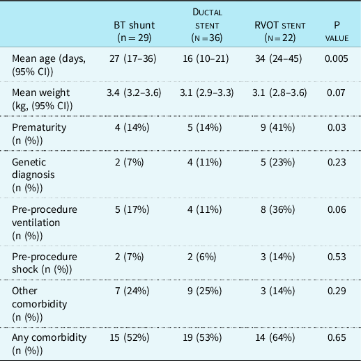
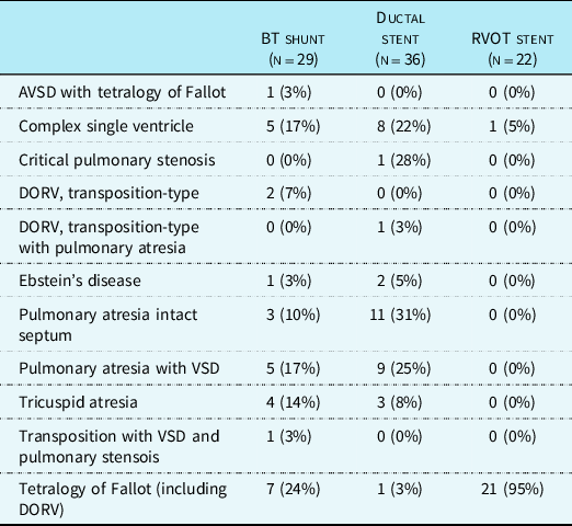
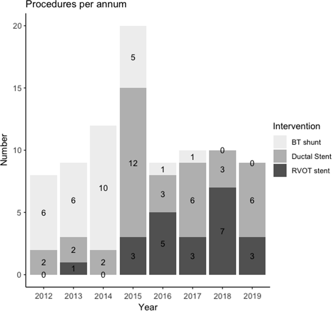
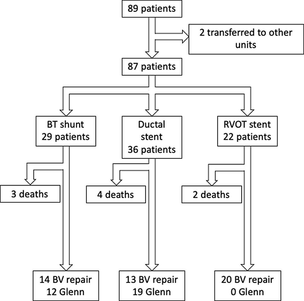
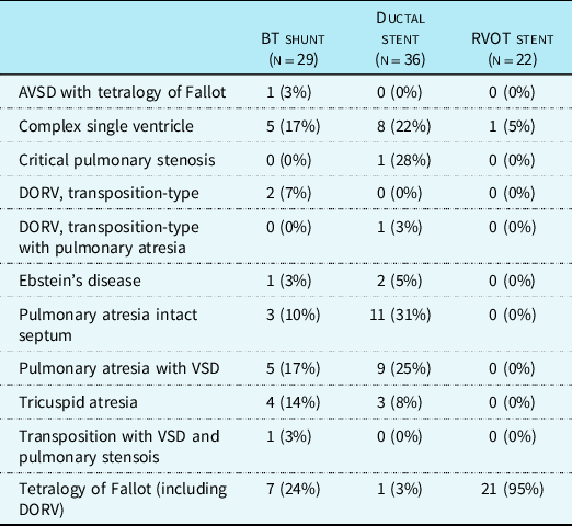
A total of 89 infants underwent palliation to secure pulmonary blood flow in our institution between January 2012 and July 2019 at under 3 months of age. Two have no follow-up data as they have moved out of region and have been excluded. Twenty-nine underwent a Blalock–Taussig shunt as a primary procedure, 36 a ductal stent, and 22 a right ventricular outflow tract stent procedure. The baseline demographics are shown in Table 1. We found the outflow tract stent group to be older at first intervention, the ductal stents the youngest, and BT shunts in between, with a statistically significant difference by analysis of variance. In addition, there was an increased incidence of prematurity in the outflow tract stent group; again the inter-group difference was significant by analysis of variance. Cardiac diagnoses are shown in Table 2, with tetralogy of Fallot patients preferentially undergoing outflow tract stenting over ductal stenting commensurate with unit policy to utilise antegrade flow wherever possible. Procedures performed per annum are shown in Figure 1 and demonstrates the time period over which the transition to percutaneous palliation occurred.
Baseline demographics

Anatomical diagnoses of palliated patients
Bar chart showing number of each procedure per annum. Note data collection ended July 2019 so the 2019 data comprises an incomplete year.

Procedural details and complications
Twenty-nine modified Blalock–Taussig shunts were performed, 25 to the right pulmonary artery, and 4 to the left. Fourteen were via median sternotomy and the remainder via left or right lateral thoracotomy. The implanted shunt size was 3 mm in 2, 3.5 mm in 13, and 4 mm in 14 patients.
Thirty-six patients underwent attempted ductal stent implant, with one technical failure where no stent was implanted. Successful approach was femoral venous in 14, femoral arterial in 11, carotid (via cut down) in 7, and axillary arterial in 3. The largest implanted stent diameter was 3 mm in 1, 3.5 mm in 25, and 4 mm in 9. One stent was implanted in 19 patients, 2 stents in 14, and 4 stents in 2 patients. The most common stent implanted was the Medtronic Integrity bare metal stent (Medtronic Inc, Minnesota, United States of America) in 25 cases (69%). All implanted stents were bare metal, with no drug-eluting stents into the ductal position during the study period.
Twenty-two patients underwent right ventricular outflow tract stenting, with approach from the internal jugular in 19 and femoral vein in 3. In one patient, the teams were unable to pass the stent across the outflow tract and so no stent was implanted. The largest implanted stent diameter was 4 mm in 1, 4.5 mm in 7, 5 mm in 10, and 6 mm in 4. The procedure ended with 1 implanted stent in 15 and 2 stents in 7. The most common implanted stent was the Cook Formula stents in 11 patients (50%). Again, all implanted stents were bare metal with no drug-eluting stents implanted during the study period.
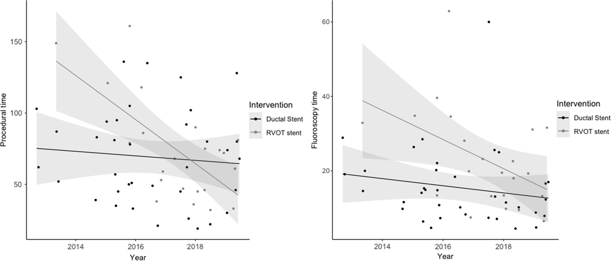
Procedural and fluoroscopy times for the catheter interventions against procedure date are shown in Figure 2. The mean procedural time was 69.2 minutes (95% confidence intervals 58–80 minutes) for ductal stenting and 73.8 minutes (57.9–89.8 minutes) for outflow tract stenting. The mean fluoroscopy time was 15.5 minutes (12–19 minutes) for ductal stenting and 22.9 minutes (17–29 minutes) for stenting the right ventricular outflow tract. There was a statistically significant fall in both procedural and fluoroscopy times for right ventricular outflow tract stenting over time (p < 0.001 and 0.03, respectively), but not for ductal stenting (p = 0.59 and 0.29, respectively), shown in Figure 2.
Scatter charts of procedural and fluoroscopy times by procedure over time with linear regression lines and 95% confidence intervals overlaid.

Post procedure, all shunts, 5 of 36 ductal stents and 9 of 22 outflow tract stents were admitted to an ICU, with all others extubated on the table and admitted to a high-dependency unit.
Outcomes
Primary outcomes are summarised in Table 3 and Figure 3. By analysis of variance, there was no difference in mortality between the groups by analysis of variance at 30 days or pre-repair (p = 0.93 and 0.97, respectively). Comparison of the shunt group with a composite transcatheter group again demonstrates no difference in mortality at either time point (p = 1 for both).
Primary outcomes

Flowchart of patient outcomes.

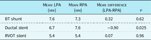
Secondary outcomes are summarised in Table 4. We found the BT shunt group to be both older and heavier at the time of next stage surgery. There was a tendency to lower Nakata index following outflow tract stenting, but this did not reach statistical significance. Branch pulmonary artery size data is shown in Table 5 with a relatively small left pulmonary artery in patients post ductal stenting reaching statistical significance.
Secondary outcomes for patients reaching next stage surgery. Reported Nakata index is prior to complete repair/Glenn shunt

Branch PA sizes

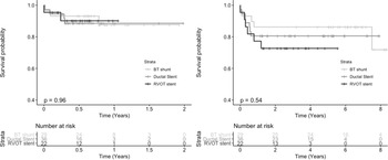
Kaplan–Meier curves to next stage surgery and overall mortality are shown in Figure 4. These demonstrate no difference in mortality by logrank test with the data censored to either complete repair or to August 2020.
Kaplan–Meier curves for mortality, with the plot on the left for mortality prior to repair and the plot on the right for mortality at any time following palliation (median follow-up = 3.9 years). Reported p values are by logrank.

Complications and reintervention prior to repair
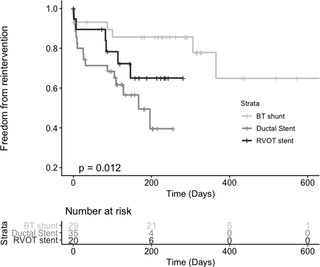
Complications and reinterventions on the pulmonary circuit are summarised in Table 6 and the Kaplan–Meier curve for reintervention is given in Figure 5. Although the groups demonstrate only a borderline difference in intervention at any time by analysis of variance, there is a statistically significant difference in reintervention by Kaplan–Meier, with the highest rate of reintervention in the ductal stent group. In the shunt group, two had serious adverse events, both comprising extra-corporeal membrane oxygenation runs. The two serious adverse events in the ductal stenting group were one perforated duct managed conservatively and one requiring intra-procedural cardiopulmonary resuscitation. For the outflow tract stent group, the four complications comprised two with perforation of the right ventricular outflow tract requiring surgical repair, one intra-procedural cardiopulmonary resuscitation and one procedural death.
Serious adverse events and reintervention prior to complete repair or Glenn shunt

Kaplan–Meier curve of reintervention prior to either complete repair or Glenn shunt. Patients are censored at death prior to complete repair or complete repair. Reported p value is by logrank.

Acute shunt reoperations in the Blalock–Taussig shunt group occurred in two (7%), with one requiring conversion to a central shunt and another requiring surgical refashioning of the shunt. Acute conversion to Blalock–Taussig shunt was required in four ductal stenting patients (11%, one following failed stent implant and the remainder following desaturation with incomplete ductal coverage) and one outflow tract stent patient (5%, stent not implanted as unable to pass stent across outflow tract). There was one further reintervention within a week for a ductal stent patient, who underwent a ductal re-stenting.
In the shunt group, four underwent reintervention on the pulmonary circuit between 7 days post-procedure and next stage surgery. These comprised one balloon dilatation of the pulmonary valve, one stenting of the shunt, one contralateral shunt implant, and bilateral pulmonary artery stenting, and one patient undergoing both a shunt stenting and a later contralateral shunt. The later pulmonary reinterventions in the ductal stent group were three balloon dilatations of the pulmonary valve, five Blalock–Taussig shunts, and four ductal restents. Late pulmonary reinterventions in the outflow tract stent group comprised three balloon dilatations of the stent (one alongside balloon pulmonary valvuloplasty), one restenting, one ductal stent (to a disconnected left pulmonary artery), and one right ventricular outflow tract patch procedure.
Discussion
We document here the experience of a single unit through a period of transition from a primary surgical to primary transcatheter approach to palliation for insufficient pulmonary blood supply. Both ductal and right ventricular outflow tract stenting have seen recently published data demonstrating equivalence in mortality Reference Glatz, Petit, Goldstein and Kelleman8,Reference Sandoval, Chaturvedi and Benson11 and some improved outcomes Reference Bentham, Zava and Harrison7,Reference Barron, Ramchandani and Murala12 in comparison with the Blalock–Taussig shunt, but there have been no previous studies reviewing all three modalities alongside one-another. Our single-centre report here provides further reassurance of equivalent outcomes with the benefits of significantly fewer intensive care admissions and the avoidance of open-chest intervention. The cost, however, is increased reintervention on the pulmonary circuit prior to next stage surgery.
Of note is the longer time to next stage surgery seen in the BT shunt group. In comparison with previous publications, length of palliation for the transcatheter intervention groups have been largely similar for both interventions, Reference Bentham, Zava and Harrison7,Reference Glatz, Petit, Goldstein and Kelleman8,Reference Barron, Ramchandani and Murala12 but time-to-next operation in the Blalock–Taussig shunt group has been significantly shorter than our cohort in recent series. Reference Bentham, Zava and Harrison7,Reference Glatz, Petit, Goldstein and Kelleman8 Although a higher mean haemoglobin in the shunt group suggests a greater degree of resting cyanosis in the surgical shunt group, this did not reach statistical significance. It is not possible from the data to ascertain whether this is truly longer palliation offered by a surgical shunt in comparison with a transcatheter palliation, or a change in unit practice towards earlier next stage surgery over the years.
Our observation of a statistically significant fall in procedural time during the course of the study for right ventricular outflow tract stenting highlights again the learning curve which is seen in a number of medical procedures. Recent large-volume data from the percutaneous aortic valve implant field has demonstrated reduced complication rates after an operator has completed 28 cases. Reference Minha, Waksman and Satler13 Studies from ductal stenting has also demonstrated fall in procedural times as well as planned procedural complexity as experience grows. Reference Santoro, Gaio and Giugno14
Adequate growth of the branch pulmonary arteries is essential for satisfactory late outcomes. Our data suggest that all three methods offer adequate growth. Although the Nakata index is smaller for the right ventricular outflow tract stent group, the majority of this group comprise patients with tetralogy of Fallot spectrum disease whom we would not routinely catheterise or further image prior to complete repair. The relatively low number of patients in this group therefore are likely those with small branch pulmonary arteries for which more information was sought prior to operation. Of note is also the difference in branch pulmonary artery sizes noted in the ductal stent group, which has also been seen in other studies. Reference Bentham, Zava and Harrison7,Reference Amoozgar, Cheriki and Borzoee15 Although we offer no new data in this regard, the finding is likely to be secondary to a degree of left pulmonary artery stenosis which can be seen at the ductal insertion in a region uncovered by the stent.
A higher rate of reintervention on the pulmonary circuit prior to next stage surgery is well recognised in patients following both ductal Reference Bentham, Zava and Harrison7,Reference Glatz, Petit, Goldstein and Kelleman8 and right ventricular outflow tract stenting. Reference Bertram, Emmel and Ewert10 Although our univariable analysis fails to reach statistical significance in this regard, the explanation is seen from the Kaplan–Meier (which does reach significance), demonstrating a number of interventions in the shunt group occurring after the last transcatheter group next stage surgery has been completed.
The importance of a multidisciplinary approach to these high-risk infants is highlighted. Two in the right ventricular outflow tract stent group required repair of injuries to the outflow tract as well as one Blalock–Taussig shunt, and 9 in the ductal stent group requiring a Blalock–Taussig shunt prior to next stage surgery. Conversely, four of the BT shunt group required transcatheter intervention prior to surgery. Although our data was analysed on an intention to treat basis, it is clear that close collaboration with the surgical team remains of paramount importance to any infant palliation programme.
The strengths of this study are the contemporaneous nature and analysis on an intention-to-treat basis. Weaknesses are the single-unit nature and the difficulties extrapolating from relatively low-count event rates. Additionally, there is an inevitable difference in method of catheter palliation related to underlying anatomic diagnosis. Due to this near dichotomous spread of some diagnoses and associated differing risk profiles of anatomical diagnoses, bias can be introduced. The overall presenting anatomic spectrum, however is unlikely to have changed significantly over the time period of the study, and analysis with a composite transcatheter group also demonstrates no difference in mortality outcomes.
In conclusion we present here a description of our unit’s experience through transition for palliation in infants with pulmonary insufficiency from a primary surgical approach with a Blalock–Taussig shunt to a primary transcatheter approach. We have demonstrated no survival disadvantage though note reintervention prior to next stage surgery is increased in the ductal stent group. We also highlight the importance of close collaboration between surgical and interventional teams.
Financial support
This research received no specific grant from any funding agency, commercial or not-for-profit sectors.
Conflicts of interest
None.
Ethical standards
The authors assert that all procedures contributing to this work comply with the ethical standards of the relevant national guidelines and with the Helsinki Declaration of 1975, as revised in 2008.