INTRODUCTION
The systematics of Actinia sea anemones is controversial, with many morphotypes of Actinia equina (Linnaeus, Reference Linnaeus1758) considered varieties by some authors (Stephenson, Reference Stephenson1935; Schmidt, Reference Schmidt1971, Reference Schmidt1972; Quicke et al., Reference Quicke, Donoghue and Brace1983, Reference Quicke, Donoghue, Keeling and Brace1985; Quicke & Brace, Reference Quicke and Brace1984; Donoghue et al., Reference Donoghue, Quicke and Brace1985) or granted specific status by others (Templeton, Reference Templeton1836; Dalyell, Reference Dalyell1848; Cocks, Reference Cocks1850; Tugwell, Reference Tugwell1856; Milne-Edwards, Reference Milne-Edwards1857; Gosse, Reference Gosse1860; Carter & Thorpe, Reference Carter and Thorpe1981; Haylor et al., Reference Haylor, Thorpe and Carter1984; Monteiro et al., Reference Monteiro, Solé-Cava and Thorpe1997; den Hartog & Ocaña, Reference den Hartog and Ocaña2003). Because of this taxonomic confusion, the binomial Actinia equina (Linnaeus, Reference Linnaeus1758) has been used for beadlet anemones found over a large geographical area, from northern Russia and the Baltic Sea to the tropical waters of West Africa and the Red Sea, South Africa, and the Far East (Stephenson, Reference Stephenson1935; Carlgren, Reference Carlgren1938; Schmidt, Reference Schmidt1972; Manuel, Reference Manuel1981; Song & Cha, Reference Song and Cha2002; Cha et al., Reference Cha, Buddemeier, Fautin and Sandhei2004).
Since 1981, studies with allozyme electrophoresis have shown that Actinia equina comprises a number of reproductively isolated and genetically highly divergent species, usually distinguished by fixed, albeit sometimes subtle, morphological, morphometric and ecological differences, like cryptic or exposed habitat and location in the intertidal zone (Carter & Thorpe, Reference Carter and Thorpe1981; Haylor et al., Reference Haylor, Thorpe and Carter1984; Solé-Cava & Thorpe, Reference Solé-Cava and Thorpe1987, Reference Solé-Cava and Thorpe1992; Monteiro et al., Reference Monteiro, Solé-Cava and Thorpe1997; Schama et al., Reference Schama, Solé-Cava and Thorpe2005).
During a genetic study of Actinia spp. by Schama et al. (Reference Schama, Solé-Cava and Thorpe2005), it became evident that anemones from South Africa previously identified as A. equina by Stephenson (Reference Stephenson1935) and Carlgren (Reference Carlgren1938) belong to a new species. The phylogenetic analyses indicated that the species from South Africa formed a sister group with Actinia tenebrosa Farquhar, Reference Farquhar1898, but were very different from the European A. equina and from any other Actinia species (Schama et al., Reference Schama, Solé-Cava and Thorpe2005). Here we describe this new species, Actinia ebhayiensis sp. nov., and compare it genetically and biometrically to Actinia equina and to the genetically close Actinia tenebrosa. Although externally very similar to A. equina (the type species of the genus), Actinia ebhayiensis can be readily distinguished from it and from all other Actinia species by molecular and morphological differences.
MATERIALS AND METHODS
Six specimens of Actinia ebhayiensis were collected from Port Elizabeth (one specimen: 33o 58′S25o40′) and Port Alfred (five specimens: 33o36′S 26o55′E), on the South African coast, in March 1998. The specimens look similar to the red column, pink pedal disc with no blue rim mid-shore morph that Quicke and collaborators examined (Quicke & Brace, Reference Quicke and Brace1984; Quicke et al., Reference Quicke, Donoghue and Brace1983, Reference Quicke, Donoghue, Keeling and Brace1985; Allcock et al., Reference Allcock, Watts and Thorpe1998). The five specimens from Port Alfred are from the same material used in the earlier genetic study of Schama et al. (Reference Schama, Solé-Cava and Thorpe2005). For comparison, specimens of A. equina were collected in Fleshwick Bay (Isle of Man, United Kingdom: 54o5′N 4o46′W) and A. tenebrosa in Half Moon Bay (Victoria, Australia: 38o08′S 144o43′E). Nematocysts of A. fragacea Tugwell, Reference Tugwell1856 from the UK (two specimens: 54o5′N 4o46′W) and A. cari Delle Chiaje, Reference Delle Chiaje1825 from Bay of Piran (North Adriatic Sea, one specimen: 45o31′N 13o34′E) were also measured. Animals were kept in aquaria (except for A. tenebrosa) for up to two weeks and then anaesthetized for four hours in a solution of 7.5% magnesium chloride diluted 1:1 with salt water. Individuals were fixed for 7 days in 4% formaldehyde diluted with salt water and then stored in 70% ethanol.
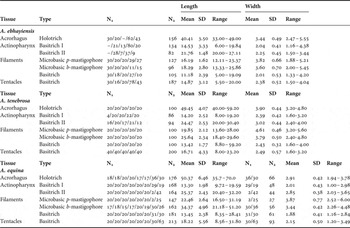
Nematocysts were analysed from fresh and fixed tissue squashes, mounted with fresh water and without stain, under a compound microscope at 1000 × magnification. The length and width of 20 undischarged capsules within haphazardly selected fields were measured from four tissues (tentacle, acrorhagi, mesenterial filaments and actinopharynx) each; on five individuals of the South African specimens, eight individuals of A. equina from the UK and five individuals of A. tenebrosa from Australia (Table 1). When two discrete size-classes of the same nematocyst type were present within a tissue, they were counted separately and labelled I and II. The classification of nematocysts follows that of England (Reference England1991). Spirocysts were excluded from the analyses as they present large size variations correlated to anemone size as shown in Francis (Reference Francis2004).
Nematocyst measurements (in micrometres) of Actinia ebhayiensis sp. nov., Actinia tenebrosa and Actinia equina. Mean, range, standard deviation (SD), sample size per individual (N1) and total sample size (N2), of measurements of length and width of capsules.

Nematocyst data were analysed using the program SYSTAT 7.0.1 (© 1997, SPSS) and the R statistical package. A normality test (Kolmogorov–Smirnov) and a homogeneity test were performed to confirm that assumptions required for parametric tests were met. An analysis of variance (ANOVA) and post hoc Tukey tests were used to infer significant differences in length, width and length/width ratio among samples.
The length/width ratio, which basically measures how rounded the nematocysts are, was used because it is less influenced by the size of the anemones (Chintiroglou & Simsiridou, Reference Chintiroglou and Simsiridou1997). Due to the large intra-specific variation observed, a hierarchical ANOVA was performed to separate the effects of intra- and inter-specific variation (Allcock et al., Reference Allcock, Watts and Thorpe1998; Williams, Reference Williams1998; Watts et al., Reference Watts, Allcock, Lynch and Thorpe2000). A linear discriminant analysis using the length of 1678 nematocysts from 16 individuals was used to summarize the differences found among the species. This type of analysis uses all measurements to discriminate among species (Klecka, Reference Klecka1980). Linear discriminant analysis has already been used in sea anemone nematocyst analysis although in a very different context. Ardelean & Fautin (Reference Ardelean and Fautin2004) used this type of analysis to differentiate nematocysts from different regions of the same specimen.
Pedal disc diameter and column height were recorded from live specimens. Photographs of the external morphology were taken using a Nikon CoolPix 5400 digital camera. General morphology was studied under a dissecting microscope. For anatomical and histological analyses samples sections were embedded in paraffin. Serial sections 5–6 µm thick were stained using haematoxylin and eosin and the Mallory triple stain method (Kiernan, Reference Kiernan1990). All A. ebhayiensis samples were deposited in the cnidarian collection of the National Museum of Rio de Janeiro (MNRJ6386–6391), Brazil and A. tenebrosa specimens were deposited in Museum Victoria (MV F112736, MV F112740–112745), Australia.
Genetic data (18 allozyme loci) from Schama et al. (Reference Schama, Solé-Cava and Thorpe2005) were re-analysed to compare the different species through a factorial correspondence analysis (FCA), using the program GENETIX 4.05 (Belkhir et al., Reference Belkhir, Borsa, Chikhi, Raufaste and Bonhomme1996–2004). This type of analysis is especially useful for estimating associations between multiple qualitative variables, where no a priori hypothesis is present (Valentin, Reference Valentin2000). FCA simplifies the analyses of complex data: the multivariate nature of correspondence analysis can reveal relationships that would otherwise not be detected in a series of pair-wise comparisons.
RESULTS
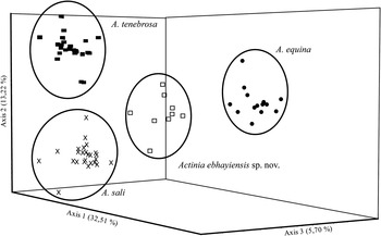
In order to have a clear view of the results on the FCA, only the morphological, genetic and geographically closer species of the genus were re-analysed. The results clearly discriminated the species (Figure 1) and more importantly, Actinia ebhayiensis could be readily distinguished in those analyses from European A. equina and also from the genetically closer Actinia tenebrosa (from Australia) and the morphologically and geographically closer (from Cape Verde) Actinia sali Monteiro, Solé-Cava & Thorpe, Reference Monteiro, Solé-Cava and Thorpe1997.
Factorial correspondence analysis based on 18 allozyme loci of Actinia species (data from Schama et al., Reference Schama, Solé-Cava and Thorpe2005). A. equina (•); A. tenebrosa (■); A. sali (X) and Actinia ebhayiensis sp. nov. (□).

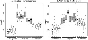
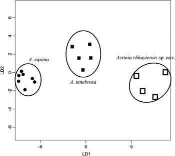
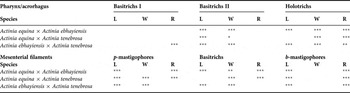
Significant differences (hierarchical ANOVA and post hoc Tukey test, after verification of normal distribution of nematocyst measurements) were observed in a number of nematocyst types from different tissues between Actinia ebhayiensis, Actinia equina sensu stricto and A. tenebrosa as follows. Except for the basitrichs of the tentacles, significant differences were found in all nematocyst types among the three species (Table 2). The lengths of the acrorhagial holotrichs were significantly larger in A. equina and A. tenebrosa when compared to A. ebhayiensis and their length/width ratio and width were significantly different among all three species (Table 2). All three species also differed significantly in the length and width of the basitrichs II of the actinopharynx, length of the microbasic b-mastigophores and p-mastigophores from the mesenterial filaments and width of the basitrichs from the filaments (Table 2). In A. ebhayiensis, the basitrichs II of the actinopharynx and the microbasic b-mastigophores and p-mastigophores of the mesenterial filaments were smaller than in A. equina and A. tenebrosa (Figure 2) and the basitrichs of the mesenterial filaments, although thinner in A. ebhayiensis than in A. tenebrosa, were wider than in A. equina (Table 1). The length/width ratio of the basitrichs I of the actinopharynx was significantly different between A. ebhayiensis and A. tenebrosa and all three species differed significantly in the length/width ratio of the microbasic b-mastigophores from the filaments, which are more elongated in A. equina and A. tenebrosa. The basitrichs from the filaments were also significantly smaller in Actinia ebhayiensis than in A. tenebrosa and A. equina. Also the width of the microbasic p-mastigophores from the filaments was significantly larger in A. tenebrosa than in A. ebhayiensis and A. equina. The three species could also be clearly distinguished in the discriminant analysis, with high posterior probabilities, in 100% of the cases (Figure 3).
Box and whiskers plot of nematocyst length of Actinia ebhayiensis sp. nov., Actinia equina and Actinia tenebrosa. (A) Microbasic b-mastigophores of the mesenterial filaments; (B) microbasic p-mastigophores from the mesenterial filaments. Horizontal line inside the box shows that the median, upper and bottom part of the box are the first and third quartiles. The whiskers show two standard deviations; points either above the third quartile or below the first quartile are plotted individually.

Discriminant analysis based on length measurements of all nematocysts types. Actinia ebhayiensis (□); Actinia equina (•) and Actinia tenebrosa (■).

Results of the post hoc pairwise Tukey test of species analysed in the analysis of variance of the nematocyst measurements. (L) Length; (W) width; (R) length/width ratio differences found. * P < 0.05; ** P < 0.01; ***P < 0.001.

Synonymy: Actinia equina: Stephenson Reference Stephenson1935; Carlgren Reference Carlgren1938; Kruger & Griffiths Reference Kruger and Griffiths1998; Acuña & Griffiths Reference Acuña and Griffiths2004; Actinia sp. 1: Schama Reference Schama2001; Schama et al., Reference Schama, Solé-Cava and Thorpe2005.
TYPE MATERIAL
Holotype: MNRJ-6386—South Africa, Port Elizabeth, 25 March 1998, Claudia Russo col. Paratypes: MNRJ-6387, MNRJ-6388, MNRJ-6389, MNRJ-6390, MNRJ-6391—South Africa, Port Alfred, 26 March 1998, Claudia Russo col.
DESCRIPTION
Oral disc smooth, circular, uniformly red, usually more transparent than column; mesenterial insertions visible. Mouth central, usually elevated in hypostome.
Tentacles smooth, red, retractile, cone shaped, one-third the length of column, arranged in five cycles, approximately 90 in number. Tentacles fully enclosed by column when animal contracted.
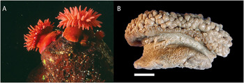
Column smooth, entirely red. Height of column 0.4–1.4 cm (N = 6; mean 0.8 cm), dome shaped in contracted specimens. Parapet and fosse well delimited. Conspicuous blue acrorhagi in fosse, simple or compound, up to 24 in single cycle. Mid-column narrower than oral and pedal disc in live specimens. Margin smooth (Figure 4).
External morphology of Actinia ebhayiensis sp. nov. (A) Live specimens, from Port Alfred (photography by Dr Toufiek Samaai); (B) preserved holotype (scale bar = 0.5 cm).

Pedal disc pink, circular, adherent, usually broader than oral disc, diameter 0.8–1.8 cm (N = 6; mean 1.1 cm).
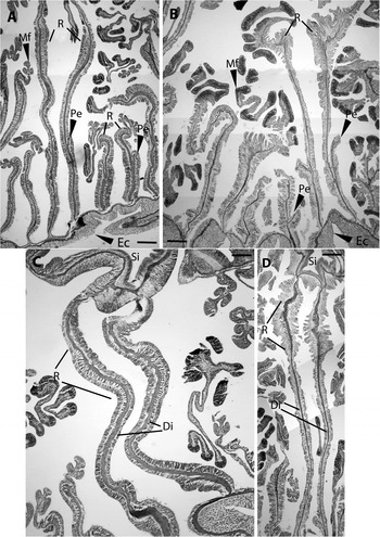
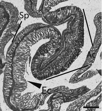
Four cycles of mesenteries arranged hexamerously: 6, 6, 12 and 24. First three cycles perfect and fourth imperfect. No gametic material observed, juveniles found in ceolenteron of some specimens. A ribbed actinopharynx extends half the length of column. Two siphonoglyphs, directive mesenteries attached (Figure 5C). Sphincter endodermal, diffuse (Figure 6). Tentacles with longitudinal ectodermal musculature. Retractor muscles diffuse (Figure 5A), long, usually ending close to parietobasilar muscles. Parietobasilar muscles diffuse, weak, with a short broad mesogleal pennon (Figure 5A).
Histology of Actinia ebhayiensis sp. nov. (A & C) and A. equina (B & D). Actinopharynx (Ph); siphonoglyph (Si); directive mesenteries (Di); retractor muscle (R); parietobasilar muscle pennon (Pe); mesenterial filaments (Mf). Scale bar = 200 µm.

Longitudinal section through the sphincter muscle (Sp) and acrorhagus (Ac), showing ectodermis (Ec) of Actinia ebhayiensis. Scale bar = 200 µm.

Cnidom: spirocysts, holotrichs, basitrichs, microbasic b-mastigophores and microbasic p-mastigophores (Figure 7). See Table 1 for size and distribution.
Representative cnidae from Actinia ebhayiensis sp. nov. (A) Holotrich; (B) basitrich; (C) microbasic b-mastigophore; (D) microbasic p-mastigohpore; (E) spirocyst. Scale bar = 20 µm.

HABITAT
Rocky shores, usually in crevices or under rocks, from high to low intertidal zone. More common in the supralittoral zone, in high energy and low suspension areas. Individuals tend to form aggregations on rocks with 2–3 cm spacing among them.
DISTRIBUTION
Port Elizabeth and Port Alfred, South Africa. It may also occur on the Indian and Atlantic coasts of South Africa, since individuals identified as Actinia equina were found on the east and west coasts of the Cape Peninsula (Carlgren, Reference Carlgren1938; Kruger & Griffiths, Reference Kruger and Griffiths1998; Acuña & Griffiths, Reference Acuña and Griffiths2004; Branch et al., Reference Branch, Griffiths, Branch and Beckley2007). Its occurrence in other parts of Africa is unknown as all occurrences of red Actinia along the African coast to date were identified as A. equina (Kruger & Griffiths, Reference Kruger and Griffiths1998; Acuña & Griffiths, Reference Acuña and Griffiths2004; Branch et al., Reference Branch, Griffiths, Branch and Beckley2007). The presence of more than one species of the genus in the same locality is well documented for a number of places and cannot be discarded, but they usually present clear morphological differences between them (Ocaña et al., Reference Ocaña, Brito and González2005; see also Perrin et al., Reference Perrin, Thorpe and Solé-Cava1999 for a review). To better understand the species distribution along the South African coast, more extensive sampling needs to be conducted. It is unlikely that A. equina occurs on the South African coast since Ocaña and collaborators extensively sampled the Macaronesia islands and never found the species, indicating that it does not disperse this far. Many studies have shown that phylogeographical breaks seem to play an important role in the distribution of benthic marine animals (Hellberg, Reference Hellberg2009) further emphasizing the importance of a continuous distribution for the dispersal of these animals.
ETYMOLOGY
The species is named after Port Elizabeth, the city where the holotype has been collected. In Xhosa, the major ethnic group in that region, the city is called Ebhayi.
DIFFERENTIAL DIAGNOSIS
Although very similar to the type species of the genus, A. equina, Actinia ebhayiensis can be distinguished from it by the presence of 9 diagnostic genetic loci (mdh-1, mdh-2, got-1, pep-1, pep-2, xod, odh, pgi-1 and pgi-2: see Schama et al., Reference Schama, Solé-Cava and Thorpe2005) and by significant nematocyst differences. Of the 18 nematocyst measurements, A. ebhayiensis differs significantly from A. equina in 12. The most striking differences are the smaller size, in A. ebhayiensis, of the acrorhagial holotrichs and the microbasic b-mastigophores and p-mastigophores from the mesenterial filaments (Figure 2). The two species also differ in muscle morphology: in A. ebhayiensis the retractor muscles are weaker and the parietobasilar muscles have a smaller, non-detached pennon than in A. equina (Figure 5A–D). Another difference between these two species is the relative size of two different types of nematocysts. The microbasic b-mastigophores of the mesenterial filaments are smaller than the basitrichs II of the actinopharynx in A. ebhayiensis, a characteristic previously observed only in A. schmidti (Schmidti, 1971, 1972; Chintiroglou & Simsiridou, Reference Chintiroglou and Simsiridou1997; Monteiro et al., Reference Monteiro, Solé-Cava and Thorpe1997), which differs from A. ebhayiensis by its much larger size, nematocyst differences (Table 3) and the apparent absence of broods (Monteiro et al., Reference Monteiro, Solé-Cava and Thorpe1997). Similar to A. tenebrosa (Ayre, Reference Ayre1984) Actinia ebhayiensis may reproduce asexually, as indicated by broods found in the coelenteron of many specimens (Carlgren, Reference Carlgren1938; Griffiths, Reference Griffiths1977; this study). In all brooding Actinia species genetically studied to date (A. equina, A. tenebrosa and Actinia bermudensis (McMurrich, Reference McMurrich1889)), the broods were produced exclusively through asexual reproduction (Black & Johnson, Reference Black and Johnson1979; Orr et al., Reference Orr, Thorpe and Carter1982; Monteiro et al., Reference Monteiro, Russo and Solé-Cava1998).
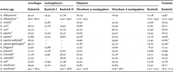
Nematocysts of Actinia spp. Length measurements in micrometres. Mean or range taken from this study (1) and from the literature: (2) Monteiro et al. (Reference Monteiro, Solé-Cava and Thorpe1997); (3) Schmidt (Reference Schmidt1972); (4) Carlgren (Reference Carlgren1950); (5) Carlgren (Reference Carlgren1938) (as A. equina); (6) Chintiroglou & Stefanidou (Reference Chintiroglou and Stefanidou1996); (7) Chintiroglou & den Hartog (Reference Chintiroglu and den Hartog1995); and (8) Allcock et al. (Reference Allcock, Watts and Thorpe1998).

*, Allcock et al. (Reference Allcock, Watts and Thorpe1998) was the only one to differentiate two size-classes, the table contains the bigger one, and the smaller one has a mean of 15.00 for the red/pink morph and 2.80 for the green/grey morph.
Although no nematocyst or histological analyses were made, in this study, on the geographically closer species A. sali from Cape Verde or A. nigropunctata den Hartog & Ocaña, Reference den Hartog and Ocaña2003 from the island of Madeira, they are clearly distinct from A. ebhayiensis both genetically (Figure 1; Schama et al., Reference Schama, Solé-Cava and Thorpe2005) and morphometrically (Table 3). Actinia sali is externally similar to A. ebhayiensis, but, among other nematocysts differences, its microbasic b-mastigophores from the mesenterial filaments are larger than the basitrichs II of the actinopharynx (Carlgren, Reference Carlgren1950; Monteiro et al., Reference Monteiro, Solé-Cava and Thorpe1997; Table 3). Actinia nigropunctata differs from A. ebhayiensis by the presence of numerous black spots on its column, the tendency towards restriction of its retractor muscles and the distinctiveness of its holotrichs (den Hartog & Ocaña, Reference den Hartog and Ocaña2003). In A. ebhayiensis the holotrichs have spirally arranged tubes, like most Actinia species, whereas in A. nigropunctata the tubes are erratically arranged (Ocaña et al., Reference Ocaña, Brito and González2005).
In a genetic analysis of some Actinia species, A. tenebrosa appears as a sister species of A. ebhayiensis, although two allozyme loci are fixed for different alleles in each species, clearly separating these species (Schama et al., Reference Schama, Solé-Cava and Thorpe2005). Morphologically these species can be distinguished by a greater number of tentacles in A. tenebrosa (121–144, against 88–100 in A. ebhayiensis) and A. tenebrosa also has weaker longitudinal muscles with no pennons (Carlgren, Reference Carlgren1924). Also in A. tenebrosa the sphincter, is well developed with a tendency to form humps (Carlgren, Reference Carlgren1924), whereas in A. ebhayiensis, the sphincter is weak and, diffuse (Figure 6). Significant nematocysts differences were also found: the two species differ significantly in 12 out of 18 measurements analysed (Table 2). Actinia ebhayiensis has significantly smaller acrorhagial holotrichs, bastitrichs II of the actinopharynx and microbasic b-mastigophores and p-mastigophores from the filaments than A. tenebrosa (Table 1).
DISCUSSION
This study describes the new species Actinia ebhayiensis sp. nov. We confirm that, though morphologically very similar to the type species of the genus, A. ebhayiensis is differentiated from A. equina on genetic and morphological grounds. The two species completely differ in nine (out of 18) allozyme loci (Schama et al., Reference Schama, Solé-Cava and Thorpe2005; see below), and significantly differ in several nematocyst measurements (Figures 2 & 3; Tables 1 & 2). Furthermore, the microbasic p-mastigophores from the mesenterial filaments and the holotrichs from the acrorhagi are much smaller in Actinia ebhayiensis sp. nov. than in other species of the genus (except A. fragacea; Table 3).
Although the use of nematocysts as characters in lower taxonomic differentiation is still under debate (Ardelean & Fautin, Reference Ardelean and Fautin2004; Francis, Reference Francis2004; Fautin, Reference Fautin2009), they have been widely used in sea anemone taxonomy (Stephenson, Reference Stephenson1935; Carlgren, Reference Carlgren1938, Reference Carlgren1950; Schmidt, Reference Schmidt1971, Reference Schmidt1972; Shick, Reference Shick1991); including differentiation of intra-specific colour morphs (Chintiroglou et al., Reference Chintiroglou, Christou and Simsiridou1997; Allcock et al., Reference Allcock, Watts and Thorpe1998).
Carlgren (Reference Carlgren1900) suggested that any description of a sea anemone should not be considered to be complete without nematocyst measurements, whereas Weill (Reference Weill1934) considered that cnidae size had little taxonomic value. It was usually the presence or absence of the different types of cnidae that were deemed important in differentiating species, since most authors argued that cnidae size may vary with nutrition state and size of the anemones (Chintiroglou, Reference Chintiroglou1996; Chintiroglou & Simsiridou, Reference Chintiroglou and Simsiridou1997) or were altogether too variable within a single individual (Ardelean & Fautin, Reference Ardelean and Fautin2004).
Nevertheless modern statistical analysis of the cnidae in different species of sea anemones has shown that random samples of cnidae follow a normal distribution and statistical tests that took into account the intraspecific variability could be used to compare different species when proper statistical sampling is conducted (Williams, Reference Williams1996, Reference Williams1998; Allcok et al., 1998; Watts et al., Reference Watts, Allcock, Lynch and Thorpe2000).
Although authors have argued that the length and width of nematocysts may vary with body size and weight, the relative proportions of cnidae size (such as length/width ratio) seem to be less influenced by anemone size (Schmidt, Reference Schmidt1971, Reference Schmidt1972; Chintiroglou & Simsiridou, Reference Chintiroglou and Simsiridou1997). In this study, we observed that between species of Actinia both length and width can be important characters when based on statistically significant differences. The different species can also be discriminated by the length/width ratio of some nematocyst types, corroborating the other statistical differences found (Table 2).
Many marine invertebrate species that were considered cosmopolitan turned out to be, under genetic scrutiny, complexes of morphologically similar but nevertheless distinct species (Knowlton, Reference Knowlton1993, Reference Knowlton2000; Thorpe & Solé-Cava, Reference Thorpe and Solé-Cava1994; Klautau et al., Reference Klautau, Russo, Lazoski, Boury-Esnault, Thorpe and Solé-Cava1999). The alleged cosmopolitanism of A. equina seems to be a typical case, and what was considered to be one single species of worldwide distribution is actually a group of at least six different species (ordered by discovery date: A. fragacea, A. prasina, A. schimidti, A. sali, A. nigropunctata and A. ebhayiensis).
Certainly, the joint use of genetic, morphology and biometrics characters is necessary for a better understanding of this complex genus. The number of allozyme diagnostic loci and the consistent biometric and morphological differences clearly separate Actinia ebhayiensis from all other species of the genus. Actinia ebhayiensis corresponds to Actinia sp. 1 in Schama (Reference Schama2001) and Schama et al. (Reference Schama, Solé-Cava and Thorpe2005), where several diagnostic gene loci (DGL) were found between Actinia from South Africa and the supposedly conspecific A. equina from the United Kingdom (DGL = 9; Nei's (Reference Nei1978) genetic distance, D = 1.48). Conspecific populations of invertebrate species usually have D < 0.18 (Thorpe & Solé-Cava, Reference Thorpe and Solé-Cava1994). In the same study, other species of Actinia were found to be highly divergent genetically from the South African samples: A. prasina (DGL = 9; D = 1.56), A. sali (DGL = 2; D = 0.25), A. bermudensis (DGL = 2; D = 0.32), A. nigropunctata (DGL = 10; D = 1.89), A. tenebrosa (DGL = 2; D = 0.28) and A. schmidti (previously A. equina mediterranea form I, sensu Schmidt; DGL = 3; D = 0.58). The levels of genetic distance were within the range usually found between distinct, congeneric species (Thorpe & Solé-Cava, Reference Thorpe and Solé-Cava1994). The genetic differences between Actinia species studied are better represented through a FCA, which clearly shows the genetic differences found between Actinia ebhayiensis and the genetically close species A. tenebrosa and the geographically closer A. sali (Figure 1).
It is evident that the binomial ‘Actinia equina’ has been incorrectly used to identify a number of distinct species from several geographical areas (A. prasina and A. fragacea in the UK; A. schmidti in the Mediterranean; A. sali, A. nigropunctata and, now, Actinia ebhayiensis in the eastern Atlantic). Pending a revision of the genus Actinia, we propose that the name Actinia equina be designated for specimens from the north-east Atlantic and for the asexually reproducing Mediterranean specimens. Specimens from other geographical locations, such as the Indo-Pacific Ocean, the Red Sea, and the Far East, should be re-evaluated using both morphological and molecular characters.
ACKNOWLEDGEMENTS
This paper is part of the PhD thesis of Renata Schama in the Genetics Program of the Federal University of Rio de Janeiro. We thank Dr Claudia Russo for the collection of South African samples, Dr Nelson Ferreira for invaluable help revising earlier versions of this manuscript, Dr Paulo Paiva for help with the statistical analyses, Dr Toufiek Samaai for photographs of live specimens, Dr Rodolpho M. Albano, Vagner Bernardo and Dr Paula B Gomes for help with the histological figures and Elizabeth Greaves for assistance in collection of live specimens. This work was supported by CNPq (Brazilian Science and Technology Ministry, RS, grant number: 141719/2001–0).