INTRODUCTION
The term mucus is broadly used for any slimy secretion from an epithelial surface produced by animals (Davis & Viney, Reference Davis and Viney1998). It is a network of proteins and polysaccharides entangled to form a weak gel containing more than 95% water (Wainwright et al., Reference Wainwright, Biggs, Currey and Gosline1976; Davies et al., Reference Davies, Jones and Hawkins1990; Smith et al., Reference Smith, Quick and Peter1999). Invertebrates use mucus as an external surface coating to reduce friction or hydrodynamic drag, and to limit water loss. In addition mucus can serve as an aid in locomotion and feeding, a mating rope, or a ‘scaffolding’ that provides anchorage and protection for eggs and a barrier against infection (Davies et al., Reference Davies, Jones and Hawkins1990). In marine invertebrates, mucus typically forms a slippery coating that prevents bacteria and debris from accumulating on the body surface (Baier et al., Reference Baier, Gucinski, Meenaghan, Wirth, Glantz, Glantz, Leach and Ericson1985) and possesses a number of defence mechanisms (Clare, Reference Clare1995; Suzuki et al., Reference Suzuki, Tasumi, Tsutsui, Okamoto and Suetake2003). These organisms require efficient mucosal defence mechanisms as they are under constant threat from a rich mixture of microorganisms in the surrounding water. Therefore, it is not surprising that these organisms have developed an impressive array of secreted compounds of the innate immune system including lytic compounds (Canicattì, Reference Canicatti1989; Canicattì & d'Ancona, Reference Canicatti and d'Ancona1990), bioactive peptides (Pomponi et al., Reference Pomponi, Willoughby, Wright, Pecorella, Sennet, Lopez, Samples, Le Gal and Halvorson1998; Ebran et al., Reference Ebran, Julien, Orange, Auperin and Molle2000), toxins, carbohydrate anti-adhesives (Guzman-Murillo & Ascenico, Reference Guzman-Murillo and Ascencio2000; Bavington et al., Reference Bavington, Lever, Mulloy, Grundy, Page, Richardson and McKenzie2004; Carsten & Schupp, Reference Carsten and Schupp2008), lectins (Iwanaga & Lee, Reference Iwanaga and Lee2005) to reduce the bacterial adhesion (McKenzie & Grigovala, Reference McKenzie and Grigovala1996). In the Annellida Polychaeta the epidermal gland cells produce a mucus that has various functions thus mucus production constitutes a key factor determining the ability of many polychaete species to survive in their environment (Bonar, Reference Bonar1972). As reported by Storch (Reference Storch, Westheide and Hermans1988) mucus intervenes in fertilization and egg protection, consolidates the tunnel wall of burrowing polychaetes and may also play a role in the absorption of metabolites. Mouneyrac et al. (Reference Mouneyrac, Mastain, Amiard, Amiard-Triquet, Beaunier, Jeantet, Smith and Rainbow2003), in their studies on Hediste diversicolor, suggested that the secretion of mucus by gland cells in the epidermis is a specific response to toxicants. In sabellid polychaetes a heterogeneous mucus is involved in different functions, such as the prevention of the proliferation of pathogenic microorganisms, protection and absorption of metabolites, as well as the building of the tube (Mastrodonato et al., Reference Mastrodonato, Lepore, Gherardi, Zizza, Sciscioli and Ferri2005). In particular, Sabellidae use mucus to cement different substances in tube building, mixing faeces, pseudofaeces and sediment (Dales, Reference Dales1961; Lewis, Reference Lewis1968). Three sets of glands add mucus to the particles used in tube-building: the ciliated glandular epidermis between the bases of the sacs, the mucous glands on the inner sides of the parallel folds, and the mucous glands on the under surface of the ventral collar folds. Because the particles were coated with mucus this churning causes them to become entangled together in a common mass with the mucus acting as a bio-adhesive in the tube building. Inspired by biomimetic applications, there has been a growing interest in the molecular mechanisms of adhesive bonding in marine organisms because of the efficiency of their adhesives in aqueous media compared with man-made materials (Santos et al., Reference Santos, Costa, Franco, Gomes-Alves, Flammang and Coelho2009). Despite the technological potential and roles in attachment, construction and obstruction, as well as in predation and defence (Graham, Reference Graham, Wnek and Bowlin2008) a considerable dearth of information remains regarding the biochemical composition of marine adhesives. Some studies have focused on the characterization of permanent adhesives characteristic of sessile organisms staying at the same place throughout their adult life. Comparatively, non-permanent adhesives, more hydrated than permanent ones and consisting of a mixture of proteins and polysaccharides, have received much less attention (Smith et al., Reference Smith, Quick and Peter1999; Flammang et al., Reference Flammang, Ribesse and Jangoux2002; Hamwood et al., Reference Hamwood, Cribb, Halliday, Kearn and Whittington2002; DeMoor et al., Reference DeMoor, Waite, Jangoux and Patrick2003; Pawlicki et al., Reference Pawlicki, Pease, Pierce, Startz, Zhang and Smith2004; Smith, Reference Smith, Smith and Callow2006; Santos et al., Reference Santos, Costa, Franco, Gomes-Alves, Flammang and Coelho2009).
In the present study we focused on the polychaete Sabella spallanzanii Gmelin, one of the best known and abundant Mediterranean sabellid. In this species a large amount of mucus is secreted when specimens are subjected to different stress conditions. As suggested in previous investigations, apart from the above described role in the tube building, the mucus seems also to be involved in the fertilization process and in defence (Canicattì et al., Reference Canicattì, Ville, Pagliara and Roch1992; Giangrande et al., Reference Giangrande, Licciano, Pagliara and Gambi2000). However, little is known about the biochemistry of S. spallanzanii mucus. In the present work we describe, over an annual cycle, some physical and chemical properties of S. spallanzanii mucus such as viscosity, osmolarity, electrical conductivity, elemental composition, the protein and carbohydrate content, the total lipids and fatty acid composition. In addition polychlorinated biphenyls (PCBs) content was evaluated in order to ascertain if it is below the maximum accepted value of contamination in pharmaceutical preparations. Implications for future investigations related to employment of S. spallanzanii mucus as a source of compounds of applied biotechnological interest are also discussed. In particular, we explored the potential of this matrix either as a good food source, on account of its nutritional value, or a source of bioactive substances with antimicrobial activity of pharmaceutical and marine technological interest.
MATERIALS AND METHODS
Animals and samples preparation
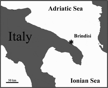
Sampling was undertaken in the harbour of Brindisi (southern Adriatic Sea, Italy) using SCUBA equipment (depth range = 5–15 m) over four periods: April 2006 (T1), July 2006 (T2), November 2006 (T3) and January 2007 (T4) (Figure 1). At each sampling period 200 adult specimens of Sabella spallanzanii were collected and transferred to the laboratory. In order to stimulate the secretion of the mucus, all the individuals have been removed from the tube where they live and kept for 30 minutes in a Petri dish. Within the secreted mucus, we checked for trapped material by microscopic observations, whilst we excluded any contamination of other excretion products by pH measurements. Secreted mucus was collected and centrifuged at 12,000g for 30 minutes at 4°C. A previous work (Canicattì et al., Reference Canicattì, Ville, Pagliara and Roch1992) showed that the protein content of the mucus varies between individuals. To avoid the introduction of this variable, in the present work for each sampling period the whole group of 200 individuals was pooled into five samples of 40 worms which were stored at −80°C until use.
Map of the Apulian coast showing the location of the sampling site.

Mucus viscosity, osmolarity and electrical conductivity
The handling procedure and storage at −80°C for up to three months did not affect the subsequent viscosity measurements of mucus samples (data not shown), a similar finding to Charman & Reid (Reference Charman and Reid1973) and Lopez-Vidriero et al. (Reference Lopez-Vidriero, Jones and Reid1980). Viscosity was measured at 200 rpm in 1-ml aliquots with a cone-plate viscometer (cone angle of 1.565°, model LVT-C/P 42, Brookfield Engineering Laboratories, USA) connected to a circulating water bath (Thermoline, Australia) set at 17±0.1°C. Due to differences in temperature and equipment used between studies, comparison of viscosity data can be difficult without reference to a common, known viscosity. Thus we documented the relative viscosity of mucus in respect to the viscosity of water, similar to Rosen & Cornford (Reference Rosen and Cornford1971) and Cone (Reference Cone, Ogra, Mestecky, Lamm, Strober, Bienenstock and McGhee1999). The viscosity of water is 1 centipoise (cP) at 20°C and only slightly dependent on temperature (Withers, Reference Withers1992).
Osmolarity was measured using a VAPRO vapour pressure osmometer (model 5520, WESCOR, UT, USA). All measurements were carried out in triplicate. The electrical conductivity was measured by using a conductimeter GLP 31(Crison).
Water content
The wet weights of mucus of 15 samples for each sample period (3 replicates for each of the five groups of 40 individuals) were measured on an analytical balance. They were then dehydrated in a SpeedVac and their dry weight was measured.
Determination of the inorganic composition
The inorganic composition and the PCBs content were determined for each sample after lyophilization of sample solution at 52°C and 0.061 mbar using a LIO 5P CINQUEPASCAL freeze-dryer.
Carbon, H and N analyses were performed using a 1106 Carlo Erba elemental analyser, while an AA-6200 Shimadzu atomic absorption flame emission spectrophotometer was used for the determination of Fe, Ca, Mg, Zn, Cu, K and Na. A P/N 206-17143 Shimadzu hydride vapour generator was coupled to the atomic absorption spectrophotometer in order to analyse the Sn and Se content. In general, each sample was mineralized to oxidize the organic fraction. To this end a weighted sample of the mucus (~10 mg) was treated with HNO3 (1 ml) and H2SO4 96% w/w (2.5 ml) at high temperature until no more fumes were released. The residue was treated again with the acids two more times. The final liquid residue was dissolved in water to give a 100 ml solution. For each element a calibration curve was obtained by using standard solutions. The quantitative analysis of phosphorous was performed using a UV-1601 Shimadzu spectrophotometer using the method reported in the literature (Kitson & Mellon, Reference Kitson and Mellon1944; Quinlan & Desesa, Reference Quinlan and Desesa1955). A 785 DMP Metrohm Titrino was used for the quantitative determination of the inorganic chloride using a pothenziometric determination.
Determination of the PCBs content
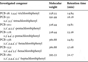
The quantitative analysis of the PCBs was performed using a gas chromatograph equipped with a capillary column (HT-5, 25 m × 0.22 mm × 0.10 µm) and connected with a mass spectrometric detector (GC-MS). The oven temperature programme was as follows: 125°C for 1 minute; 25°C min−1 to 200°C; 4°C min−1 to 260°C, 60°C for 1 minute (total run time: 20 minutes) and the injector temperature was 300°C. Helium was used as the carrier gas at a constant flow of 0.7 ml min−1. The determination was done according to the CEN 15308 method UNI EN 15308:2008 Characterization of waste–Determination of selected PCBs in solid waste by using capillary gas chromatography with electron capture or mass spectrometric detection which take into account seven congeners of PCBs (Table 1). Molecular weights and retention times of these PCB congeners were also reported.
Molecular weights and retention times of the investigated polychlorinated biphenyl (PCB) congeners.

Protein and carbohydrate concentration
The protein concentration of each mucus sample was measured using the Bradford assay (Reference Bradford1976), with bovine serum albumin (BIO-RAD) as standard.
The carbohydrate concentration of the mucus was assayed using the method described by Dubois et al. (Reference Dubois, Gilles, Hamilton, Rebers and Smith1956) and Kennedy & Pagliuca (Reference Kennedy, Pagliuca, Chapling and Kennedy1994). The assay was calibrated with known amounts of D-glucose.
Lysozyme-like activity
To detect lysozyme activity, inoculated Petri dishes were used as standard assay; 700 µl of 5 mg/ml of dried Micrococcus luteus cell walls (Sigma) were diluted in 7 ml of 0.05 M PB-agarose (1.2%) (pH 5.0) then spread on a Petri dish. Four wells of 6.3 mm diameters were sunk in the agarose gel and each filled with 30 µl of mucus. The diameter of the cleared zone of the four replicates was recorded after overnight incubation at 37°C and compared with those of reference samples represented by hen egg-white lysozyme (Merck, Darmstadt, Germany).
Microbial strains
Human and animal pathogenic bacteria were used, and these were strains of Vibrio alginolyticus, Vibrio harveyi, Vibrio anguillarum, Salmonella sp., Escherichia coli and Pseudomonas aeruginosa. Three clinical isolates of human pathogenic fungi were also used: Candida albicans, Candida famata and Candida glabrata. Strains were routinely maintained at +4°C on Marine Agar 2216 (Beckton Dickinson & Co.), Luria–Bertani (LB), nutrient, or yeast peptone dextrose (YPD) agar used for the marine bacteria, the human and animal pathogenic bacteria, and the fungi, respectively. Bacterial and yeast strains were always harvested during the exponential growth phase, i.e. after 4 hours culture at 24°C or 37°C for the pathogenic strains, under constant rotation at 100 rpm.
Antimicrobial activity
To test antimicrobial activity, 50 µl of mucus sample were added to 10 µl of bacterial or yeast suspension (108 cells ml−1). Normal growth controls consisted of bacteria or yeast incubated with sterile liquid broth. After 30 minutes at room temperature, with gentle stirring at 100 rpm, serial dilutions of these suspensions were incubated in Petri dishes between two layers of the above described specific nutritive agar to obtain imprisoned bacterial or yeast forming punctiform colonies (technique of ‘colony forming units’ (CFU)). Routinely, 10−3, 10−4 and 10−5 dilutions were plated out in triplicate. Percentages of inhibition were determined from the differences in colony numbers in controls and tests, after 24-hours incubation at 24°C for marine strains and at 37°C for the pathogenic strains. The percentage of bacteria or yeast inhibited by mucus was inferred from the difference between the number of emerging colonies in controls and in tests.
Electrophoresis
Mucus samples were analysed by SDS-polyacrylamide gel electrophoresis (SDS-PAGE). They were run on discontinuous gels, based on the method of Laemmli (Reference Laemmli1970) and the detailed protocols of Hames (Reference Hames, Hames and Rickwood1990). The gels were 10% acrylamide, and were 8×9 cm by 1.0 mm thick. The migration buffer consisted of 25 mM Tris, 192 mM glycine, pH 8.5. After migration, gels were stained using Silver Stain kit (Sigma). Kit of molecular standards (PageRuler™ Prestained Protein Ladder range10-250 kDa) was purchased from Fermentas. It is a mixture of eight recombinant, highly purified, coloured proteins with apparent molecular weights of 10 to 250 kDa.
Total lipids and fatty acid analyses
Total lipids from each mucus sample were extracted according to the method of Folch et al. (Reference Folch, Less and Stone Stanley1957). The mucus was homogenized with chloroform/methanol (2/1) to a final volume 20 times the volume of the mucus sample. After centrifugation and siphoning of the upper phase, the lower chloroform phase contained the lipids. Total lipid content was determined by the colorimetric enzymatic method (Zöllner & Kirsch, Reference Zöllner and Kirsch1962) using commercial kit (FAR—Verona, Italy).
Fatty acid composition was determined as described by Budge & Parrish (Reference Budge and Parrish2003). In this method, fatty acid methyl esters (FAME) of lipid extracts of the mucus were prepared using 14% BF3-MeOH mixture. Lipid extracts were dissolved in 0.5 ml hexane and 1.5 ml 14% BF3-MeOH mixture. The samples were flushed with nitrogen and heated for 90 minutes at 85°C. After cooling to room temperature, FAME were extracted three times with a mixture of 2 ml hexane and 2 ml water, followed by centrifugation at 2000g for 2 minutes. Hexane layers, which contained FAME were separated, combined, and evaporated under a stream of nitrogen at room temperature. Analysis of fatty acid methyl esters was performed by gas–liquid chromatography using a 6890 Hewlett Packard series gas chromatograph equipped with an Omegawax 250 capillary column (Supelco, USA). The column temperature programming was as follows: from 150 to 250°C at 4°C min−1 and maintained at 250°C. Helium was used as the carrier gas at a flow of 1 ml min−1. Methyl esters of fatty acid were identified by a FAME mix (Supelco, USA) as standard, by comparison of retention times, and the results were reported as percentages of total identified methyl esters fatty acids.
Triglycerides and total cholesterol
Triglycerides and total cholesterol were measured by the colorimetric enzymatic (Trinder, Reference Trinder1969; Bucolo & David, Reference Bucolo and David1973) using the commercial kit (SGM, Rome, Italy).
Statistical analysis
All the results were presented as the arithmetic mean of three replications ± standard deviation. Analysis of variance (ANOVA) was used in order to evaluate differences among the mean values of the measured parameters at the different sampling periods. The analyses were performed by using the Statsoft STATISTICA v. 6.0 (Statsoft, Inc., 2001).
RESULTS
Mucus viscosity, osmolarity and electrical conductivity
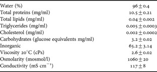
When adult specimens of Sabella spallanzanii were removed from their tubes and placed in a Petri dish, they immediately secreted a copious colourless mucous material. Statistical analysis revealed no significant differences among the viscosity values of mucus recorded in the four periods as well as osmolarity. The mean viscosity value was 2.5±0.02 cPs in respect to the 1 cPs viscosity of water measured at 20°C (Table 2). The mean osmolarity value was 1060±20 mosmol/l, showing as the worm mucus was iso-osmotic to seawater (1152±25 mosmol/l). The mean electrical conductivity of mucus was 117±8 mS cm−1 whilst the electrical conductivity of the seawater is 35 mS cm−1.
Main physical–chemical characteristics of Sabella spallanzanii mucus.

Water and inorganic content
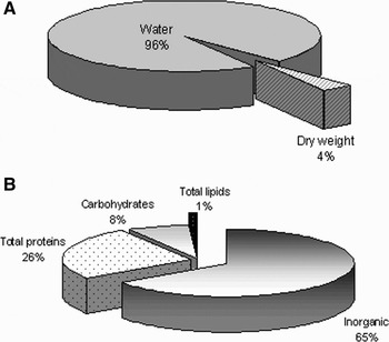
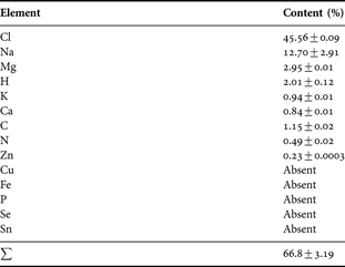
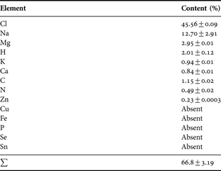
The water content of S. spallanzanii mucus was 96.2±0.3% (Figure 2A). The residual part of the dried sample is mainly represented by inorganic salts left over from evaporating seawater. Results of inorganic composition indeed showed that the 65.2±2.3% of the dry weight was composed of inorganic elements (Figure 2B). In particular Table 3 reports the mean percentage of the elements detected in the mucus. Data reveal that Cl and Na represent almost 60% of the lyophilized samples. In all samples, magnesium, calcium, and potassium each made up just 1.3–4.5% of the inorganic content.
Sabella spallanzanii mucus composition: (A) water content and dried weight; (B) organic and inorganic residuals.

Elements detected in mucus sample of Sabella spallanzanii.
The PCBs content was estimated using the CEN method 15308 and was <0.005 µg g−1.
Protein and carbohydrate concentration
Statistical analysis revealed no significant differences either among the protein concentrations of the mucus recorded in the four periods, or among carbohydrate concentrations. The mean protein content of S. spallanzanii mucus was 10.5±0.21 mg ml−1 and the carbohydrate concentration, as D-glucose equivalent, was (3.2±0.02 mg ml−1). In addition protein/glucose (PG) ratio for all replicates was 3.3 thus S. spallanzanii mucus contains three times more protein than carbohydrate (Table 2). By dry weight 26±1.2% was protein, and 8±0.21% was carbohydrate (Figure 2B). As already stated, much of the remainder of the dry weight was inorganic (65.2±3.15%). Assuming that proteins and carbohydrates were the primary organic molecules present, protein made up 76% of the organic material.
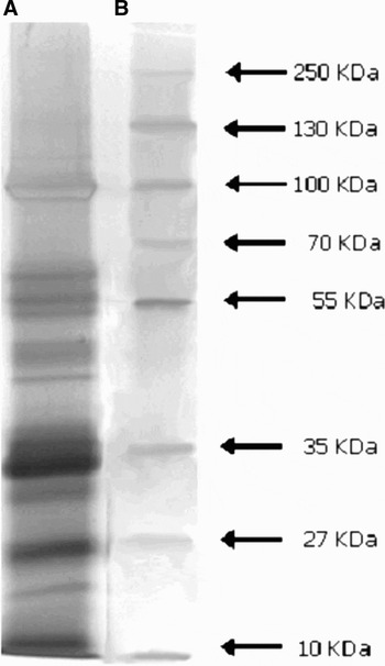
The electrophoretic analysis revealed at least ten major protein bands, with molecular weights ranging from 16 to 90 kDa, and six minor components, with molecular weights ranging from 14 to 116 kDa (Figure 3).
SDS-PAGE analysis of Sabella spallanzanii mucus. Panel A, mucus; panel B, molecular weight standards furnished by Fermentas. Molecular weights (kDa) of standard proteins are on the right.

Lysozyme-like activity
Results from the standard assay on Petri dishes showed that the mucus of S. spallanzanii exerted a natural lysozyme-like activity. By the standard assay a mean diameter of lysis of 7.9 ± 0.02 mm corresponding to 1.08 mg ml−1 of hen egg-white lysozyme was observed.
Antimicrobial activity
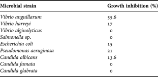
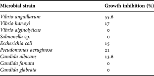
Our data enhance, in vitro, the presence in S. spallanzanii mucus of an antimicrobial activity. This activity, as shown in Table 4, was tested on Vibrio alginolyticus, Vibrio harveyi, Vibrio anguillarum, Salmonella sp., Escherichia coli, Pseudomonas aeruginosa, Candida albicans, Candida famata and Candida glabrata. By way of plate assay, we were able to quantify the degree of inhibition produced by the mucus on each strain. Vibrio anguillarum was the most inhibited (55.6% growth inhibition). A lower percentage of inhibition was instead measured on Pseudomonas aeruginosa and Vibrio harveyi. Percentages of growth inhibition were respectively 21% and 17%. Among yeast only Candida albicans was affected by the mucus treatment (13.6% growth inhibition). The other examined strains were not affected in their growth rate by the mucus.
Results of in vitro experiments showing the antimicrobial activity of Sabella spallanzanii mucus.
Total lipids and fatty acid analyses
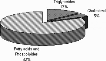
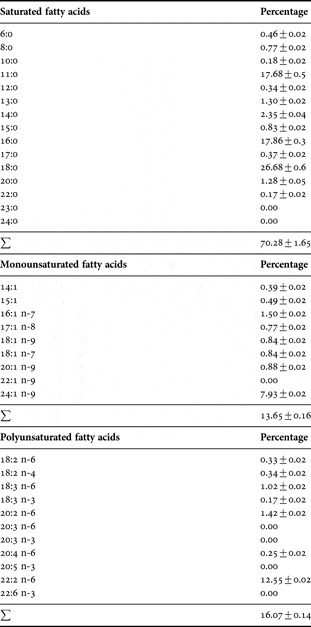
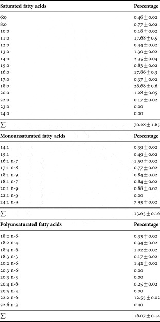
The results of the lipid analysis are presented in Figure 4. The total mean lipid content of mucus was 0.004 ± 0.0002 mg ml−1 corresponding to 0.1% of dry weight. The fatty acids profile of S. spallanzanii mucus is shown in Table 5. Stearic acid methyl (18:0) and palmitic acid methyl ester (16:0) were the predominant saturated fatty acids, while, among monounsaturated fatty acids nervonic acid methyl ester (24:1) and palmitoleic acid methyl ester (16:1) prevailed. Finally among polyunsaturated fatty acids, cis-13, 16-docosadienoic acid methyl ester (22:2, n-6) and cis-11,14-eicosadienoic acid methyl ester (20:2, n-6) were abundant. A considerable contribution of undecanoic acid methyl ester (11:0), cis-11-eicosenoic acid methyl ester (20:1) and α-linolenic acid methyl ester (18:3) to the total fatty acids was also recorded. Most of the fatty acids did not undergo statistically significant changes during the period under study. The triglycerides and the cholesterol concentrations were 0.005 ± 0.0003 mg ml−1 and 0.002 ± 0.0002 mg ml−1 respectively.
Total lipid profile of Sabella spallanzanii mucus.

Fatty acids composition of Sabella spallanzanii mucus.
DISCUSSION
Although mucus plays many different roles among marine invertebrates, relatively little is known about the structure of invertebrate mucous secretions and more in particular on the link between biochemical structure and function. The rheological properties of mucus, such as gelation, film formation, adhesiveness, and viscosity, are intimately related to the chemical composition and structure of its components and the forces which mould them into the elaborately organized three-dimensional structure of mucus. It is also reasonable to suggest that the physical–chemical properties are intimately associated with the biological function of mucus.
The present work provides a preliminary step on this topic describing some of the physical and chemical properties of Sabella spallanzanii mucus. The viscosity of mucus is dictated by its hydration state, which is dependent upon a Donnan potential between mucins and the surrounding water (Verdugo, Reference Verdugo, Nugent and O'Conner1984; Zuchelkowski et al., Reference Zuchelkowski, Pinkstaff and Hinton1985; Gupta, Reference Gupta1989; Cone, Reference Cone, Ogra, Mestecky, Lamm, Strober, Bienenstock and McGhee1999). Osmolarity may also prove useful for mucus characterization. Sabella spallanzanii mucus exhibits a low viscosity (2.5 ± 0.02 cPs) and its osmolarity is of the same order of magnitude as the seawater such that mucus achieves near ionic equilibrium with the surrounding medium. Thus on account of these results we can infer that the mucus of the studied polychaete is a fluid mucus.
As observed by different authors (Smith & Morin, Reference Smith and Morin2002) who found typical marine mucus having 96–98% water, the mucus of S. spallanzanii contains more than 90% water. The high proportion of inorganic material (about 65.2%) presumably results from dried salts left over when the seawater in a gel evaporates as already suggested by Smith et al. (Reference Smith, Quick and Peter1999) and Smith & Morin (Reference Smith and Morin2002) who observed a similar proportion of inorganic material in limpets' and periwinkles' mucus. By contrast in other marine mucous secretions such as the pedal mucus of Lottia gigantea and Collisella scabra and the mucus of two species of littorinid, Littorina littorea and Littorina obtusata, roughly less than 50% of the dry weight was constituted of inorganic residue (Connor, Reference Connor1986; Davies et al., Reference Davies, Jones and Hawkins1990).
The bulk of the organic material (about 35%) in the mucus of the studied worm is composed of proteins ranging from 14 to 116 kDa and making up 76% of the organic material (26% of the dry weight). Carbohydrates make up about 24% of the organic material and 8% of the dry weight. Similar values were recorded for limpets by Smith et al. (Reference Smith, Quick and Peter1999). Our results are also in agreement with those reported by Flammang & Walker (Reference Flammang and Walker1997) and by Flammang et al. (Reference Flammang, Van Cauwenberge, Alexandre and Jangoux1998) which provided detailed information for the mucus secreted by sea star podia composed of dry weight for 20% protein, and 8% carbohydrate, while much of the remainder was inorganic. The co-occurrence of proteins and carbohydrates also seems to be a common trait among non-permanent adhesives of marine invertebrates, being observed, besides sea stars and sea urchins, in sea cucumbers (DeMoor et al., Reference DeMoor, Waite, Jangoux and Patrick2003), snails (Smith & Morin, Reference Smith and Morin2002) and limpets (Smith, Reference Smith, Smith and Callow2006). These protein–carbohydrate complexes typically form highly hydrated adhesives with viscoelastic properties (Flammang et al., Reference Flammang, Van Cauwenberge, Alexandre and Jangoux1998; Smith, Reference Smith, Smith and Callow2006) which contrast with the rigid permanent proteinaceous adhesive cements. Finally, the lipid content of S. spallanzanii mucus is very low and accounted for 0.1% of dry weight.
Several studies attempted to estimate the nutritional values of the mucus. Criteria such as ash content, biochemical constituents, caloric content, and the relative carbon and nitrogen content are different aspects of the nutrient quality of mucus. In corals, estimates of the nutritional value of mucus have yielded variable results (Meikle, Reference Meikle1986; Meikle et al., Reference Meikle, Richards and Yellowlees1987, Reference Meikle, Richards and Yellowlees1988). Reported values for carbohydrate content range from 2.5 to 67% ash-free dry weight (AFDW), protein content from 1.6 to 72.7% AFDW, and lipid from 0 to 93% AFDW (Coffroth, Reference Coffroth1990). Thus mucus from some species of corals is considered a good food source serving as site of aggregation for microorganisms and becoming heavily fouled. Thus sediment, faecal pellets, unidentifiable debris, bacteria, ciliates, nauplii, ostracods, diatoms and filamentous algae were observed on mucus, creating a heterogeneous community (Paul et al., Reference Paul, DeFlaun and Jeffrey1986; Coffroth, Reference Coffroth1990). By contrast, S. spallanzanii mucus was low in ash and contained low lipid content as well as triglycerides, cholesterol and fatty acids suggesting that it has a low nutritional value. These features together with the high inorganic content and low organic one, suggest that it is a poor food source.
An issue of interest is the proteic pattern of the S. spallanzanii mucus: as observed from the electrophoretic analysis, it contains a complex of at least ten major and six minor proteins. This is in accordance with the general multiprotein nature of other marine invertebrate adhesives. Researchers on mussel byssus have so far identified nine proteins (for review, see Sagert et al., Reference Sagert, Sun, Waite, Smith and Callow2006), whereas barnacle cement is known to be made up of at least ten different proteins (Kamino, Reference Kamino, Smith and Callow2006). For non-permanent adhesives, much less information is available, but some multiprotein complexes have been found in the adhesives of sea cucumbers and limpets (for review, see Flammang, Reference Flammang, Smith and Callow2006; Smith, Reference Smith, Smith and Callow2006). Recently SDS-PAGE analysis of Paracentrotus lividus footprint material revealed that the soluble fraction contains about 13 protein bands with molecular masses ranging from 10 to 200 kDa (Santos et al., Reference Santos, Costa, Franco, Gomes-Alves, Flammang and Coelho2009).
The presence in S. spallanzanii mucus of a haemolytic activity (Canicattì et al., Reference Canicattì, Ville, Pagliara and Roch1992) and an antibacterial activity suggest a role of this compartment in defending the worms from microbial attack serving as medium into which the antimicrobial substances are released. In particular we observed a lysozyme-like activity with a mean diameter of lysis of 7.9 ± 0.02 mm on Petri dish assay corresponding to 1.08 mg ml−1 of hen egg-white lysozyme. Morover, by the in vitro test, we observed an antimicrobial activity towards several microorganisms including Vibrio anguillarum, Vibrio harveyi, Pseudomonas aeruginosa and Candida albicans. The substrate for lysozyme is contained not only in Gram-positive bacteria; the lysozyme spectrum of the Gram-negative bacteria has been established by Peterson & Hartsell (1955). Therefore, in our case, it is possible to hypothesize that lysozyme from S. spallanzanii mucus could be, at least in part, responsible for the observed in vitro microbial growth inhibition. However, we cannot exclude that other killing factors are responsible for these events or that both lysozyme and other defence factors act synergically in antimicrobial defence. The defensive role of mucus is important taking into account that S. spallanzanii lives in eutrophic environments such as harbours where bacteria, including pathogens to man and marine organisms, are abundant (Barg & Phillips, Reference Barg and Phillips1998). Although the worms may be aggressed by microorganisms, they are able to survive their attack on account of the presence of a powerful immune system which includes antimicrobial and haemolytic activities. Thus in the studied sabellid, the mucus not only plays an essential role in the feeding mechanisms, tube building and fertilization (Stabili et al., Reference Stabili, Schiros', Licciano and Giangrande2009), but is involved in defending the worms from microbial attack. This role has already been demonstrated for other marine invertebrates mucus (Davis & Viney, Reference Davis and Viney1998; Clare, Reference Clare1995).
The findings from this study not only contribute to the limited knowledge available on the mucus composition in invertebrates, but also have implications for future investigations related to the employment of S. spallanzanii mucus as a source of compounds with antimicrobial activity of pharmaceutical and marine technology interest. As regards pharmaceuticals we have to consider that the increasing development of bacterial resistance to traditional antibiotics has reached alarming levels, thus necessitating the strong need to develop new antimicrobial agents. Lysozyme exhibits antimicrobial activity against different microorganisms, and was chosen recently as a model protein to develop more potent bactericidal agents with broader antimicrobial specificity. Several strategies are attempting to convert lysozyme to be active in killing Gram-negative bacteria other than Gram-positive bacteria thus introducing, for the first time, a new conceptual utilization of lysozyme which would be an important contribution for medicine and modern biotechnology (Ibrahim et al., Reference Ibrahim, Aoki and Pellegrini2002). The utilization of S. spallanzanii mucus to extract bioactive substances of pharmaceutical interest is encouraged also on the basis of the results obtained on lipids as well as PCB concentrations. Notably lipids are related to bioaccumulation of lipophilic pollutants, including PCBs, in living substrates (Jones & de Voogt, Reference Jones and de Voogt1999; Quinnell et al., Reference Quinnell, Hulsman and Davie2004; Brown & Bythell, Reference Brown and Bythell2005; Ritchie, Reference Ritchie2006); the low value in lipid content within the mucus of S. spallanzanii indicated the low probability of accumulation of persistent lipophilic pollutants. In addition, the estimated PCB content is below the maximum accepted value of contamination in pharmaceutical preparations (<0.005 µg g−1).
Lastly, the antibacterial proteins of S. spallanzanii mucus could be employed to avoid the settlement of bacteria which is the primary colonizing process in the marine biofouling development. Alternative marine technologies employing biogenic compounds that function as natural anti-settlement agents are sought, taking into account that paints containing TBT and copper (Jelic-Mrcelic et al., Reference Jelic-Mrcelic, Sliskovic and Antolic2006) and organic biocides (Turley et al., Reference Turley, Fenn, Ritter and Callow2005) have been banned after 2008 (e.g. Hellio et al., Reference Hellio, Berge, Beaupoil, Gal and Bougougnon2002; Stupak et al., Reference Stupak, Garcia and Perez2003; Konstantinou & Albanis, Reference Konstantinou and Albanis2004; Ostroumov, Reference Ostroumov2008).
In conclusion Sabella spallanzanii mucus provides an accessible, renewable resource that could reward wider exploration. Further studies will be accomplished to isolate and characterize the effectors responsible for the antibacterial activity observed.
ACKNOWLEDGEMENT
Financial support was provided by the Project ACTIBIOMAR (www.actibiomar.it) granted by Apulian Region.