Introduction
Enterobius vermicularis (Nematoda), a pinworm, is a global helminthic parasite of humans that is especially common in temperate climates. Approximately 4–28% of children are infected worldwide (Bethony et al., Reference Bethony2006). In Thailand, prevalence of E. vermicularis infection among primary schoolchildren ranges from 0.19% to 38.82% (Kaewkes et al., Reference Kaewkes1983; Jongsuksantigul et al., Reference Jongsuksantigul, Chaeychomsri and Techamontrikul1992; Nithikathkul et al., Reference Nithikathkul2001; Polseela et al., Reference Polseela2004; Saksirisampant et al., Reference Saksirisampant2006). Prevalence is generally related to local levels of public health support, education and personal hygiene (Li et al., Reference Li2015). Human infection is closely associated with high population density, socio-economic status and the habit of thumb sucking (Kim et al., Reference Kim2013). Enterobiasis reinfection occurs commonly by the faecal–oral route. Enterobiasis is one of the main factors causing weight loss and leads to debilitating loss of sleep among children (Kucik et al., Reference Kucik, Martin and Sortor2004). Adult worms live in the human large intestine. Female worms emerge at night to lay eggs in the perianal region. Resulting symptoms are perianal itching and irritability (Cerva et al., Reference Cerva, Schrottenbaum and Kliment1991). Eggs become infective within a few hours of being laid and can survive several days on clothing or bed linens. This can facilitate person-to-person transmission (Beaver et al., Reference Beaver, Jung, Cupp, Beaver, Jung and Cupp1984). Transmission occurs through direct contact, and therefore this infection is common among children in kindergartens and primary schools (Park et al., Reference Park2005). Self reinfection by the anal–oral route is also very common. In chronic infections, this worm can slow cognitive development in children (Li et al., Reference Li2015). In some cases, E. vermicularis has invaded the kidneys and fallopian tubes, causing severe outcomes and even death (Pampiglione and Rivasi, Reference Pampiglione and Rivasi2009; Serpytis and Seinin, Reference Serpytis and Seinin2012). It can also cause clinical manifestations such as acute appendicitis (Cerva et al., Reference Cerva, Schrottenbaum and Kliment1991).
Enterobius vermicularis DNA has been successfully amplified and sequenced using material from various sources. For example, parasite DNA in pre-Columbian human coprolites as old as 6110 years was sequenced and found to be very similar to that of modern E. vermicularis (Iniguez et al., Reference Iniguez2006). DNA extracted from eggs in modern faecal samples has been sequenced to explore the genetic diversity, phylogeography and host specificity of this cosmopolitan parasite. Nakano et al. (Reference Nakano2006) obtained adult worms from captive chimpanzees in five zoos and other institutions in Japan, and eggs from humans in Japan and sequenced a portion of the cytochrome c oxidase subunit 1 (cox1) gene. Phylogenetic analysis of DNA sequences identified three clusters, each differing in amino-acid sequence (types A, B and C). Sequences from chimpanzees were represented in each cluster, and humans only in cluster A. Piperaki et al. (Reference Piperaki2011) sequenced a portion of the cox1 gene from E. vermicularis eggs from adhesive-tape swabs from Greek children. All sequences were grouped in type B. Additional E. vermicularis cox1 sequences from humans in Denmark, Germany, Greece, Poland and Iran also fell into type B (Ferrero et al., Reference Ferrero2013; Hagh et al., Reference Hagh2014; Kubiak et al., Reference Kubiak, Dzika and Paukszto2017). No sequences from worms in humans have been placed in type C to date, and sequences in type A seem to be restricted to eastern Asia. To date, there have been no studies on genetic variation of Enterobius species infecting human populations in Thailand, a country in South-East Asia. Here, we used a partial sequence of the internal transcribed spacer 2 (ITS2) region of ribosomal DNA to confirm the identity of the recovered Enterobius species. An alignment of the cox1 sequences was used to evaluate the genetic diversity of E. vermicularis from Thailand. The aim is to explore molecular diversity of Enterobius eggs recovered from schoolchildren.
Materials and methods
Survey areas and study populations
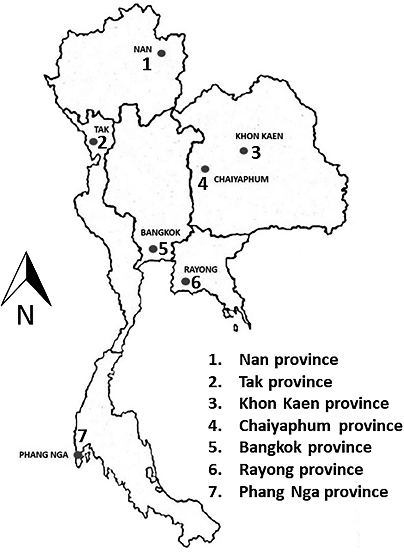
The survey was conducted between May 2015 and December 2016 at six primary schools and three orphanages in northern, north-eastern, central, eastern and southern Thailand (fig. 1). Four schools were in rural areas (> 70 km from urban areas; two in Tak Province and two in Nan Province). Two study sites were in urban areas (Bangkok and Khon Kaen). The three orphanages were in Bangkok, Khon Kaen and Rayong.
The locations of the sample collection sites in Thailand.

Sample collection
With the help of the teachers, the adhesive tape perianal swab technique (1–2 slides for each child) was used as the standard examination method (Beaver et al., Reference Beaver, Jung, Cupp, Beaver, Jung and Cupp1984). Prior informed consent was obtained from the parents or guardians of each participant. The examinations were conducted before the child washed or went to the toilet in the morning, to prevent any pinworm eggs being washed from the perianal region. The adhesive tape perianal swab specimens were examined microscopically for pinworm eggs or adult worms by parasitologists and experienced medical technologists within 2–3 hours in the field. All positive samples were stored at −20°C, with the positions of eggs marked. Descriptive statistical analysis was used to analyse the prevalence rate (SPSS version 16.0, SPSS Inc., Chicago, USA).
DNA extraction and molecular analysis
Perianal swabs (n = 19) from each infected child, each holding c. 30–50 Enterobius eggs, were individually crushed, and DNA was extracted using the Nucleospin tissue kit (Macherey-Nagel GmbH & Co, Duren, Germany), according to the manufacturer's instructions. The DNA was eluted in 50 μl of elution buffer, 5 μl of which was used for amplification. Newly designed primers specific for a portion of the cox1 sequence (EU281143) were EVCOXF (5′-TGGTTTTTTGTGCATCCTGAGGTTTA-3′) and EVCOXR (5′-AGAAAGAACGTAATGAAAATGAGCAAC-3′), amplifying a region of 441 bp. For the partial ITS2 (HQ646164), primers used were EVITS2F (5′-GTACAACTGTTAGCGGTGGATCAC-3′) and EVITS2R (5′-ATTCAGCGGGTAATCACGCC-3′), amplifying a region of 623 bp. All primers were designed using the tool for finding specific primers (Primer-BLAST; NCBI, Bethesda, USA).
DNA amplification
Polymerase chain reaction (PCR) was carried out in a 25 μl reaction volume containing 0.625 U of Taq polymerase (Roche, Mannheim, Germany), 2.5 mm dNTP mixture, 1.8 mm MgCl2 and 0.2 μm of each primer. All cycling conditions were as follows: initial denaturation at 94°C for 2 minutes, with the first 10 cycles at 94°C for 1 minute, 40°C for 1 minute and 68°C for 2 minutes. This was followed by 30 cycles of 94°C for 1 minute, 45°C for 2 minutes and 68°C for 2 minutes, and a final elongation at 68°C for 7 minutes. Positive (E. vermicularis adult DNA) and negative (H2O) controls were included in all runs. The amplicons were electrophoresed in 1% agarose gel, and then gels were stained with ethidium bromide and observed under a UV transluminator (BioDoc-It™ Imaging System, UVP, Cambridge, UK).
Sequence and population genetics analysis
DNA sequencing was conducted by First BASE Laboratories Sdn Bhd (Selangor, Malaysia). Cox1 sequences were obtained from 19 swabs, and ITS2 sequences from 18 of the same swabs. We placed our data in a global context using sequences of Enterobius vermicularis publicly available in the GenBank database. A multiple alignment of all available E. vermicularis cox1 and ITS2 sequences was done using ClustalW (Thompson et al., Reference Thompson, Higgins and Gibson1994) implemented in BioEdit Sequence Alignment Editor (Hall, Reference Hall1999). A phylogenetic tree was inferred from the cox1 alignment using the maximum-likelihood (ML) method implemented in MEGA version 6.0 (Tamura et al., Reference Tamura2013). The best-fit substitution model for the cox1 alignment was the Hasegawa–Kishino–Yano model (HKY + G) (Nei and Kumar, Reference Nei and Kumar2000). Enterobius anthropopitheci (accession no. AB254450) was used as the outgroup.
The haplotype diversity was calculated using DnaSP version 5.0 (Librado and Rozas, Reference Librado and Rozas2009). Ninety-five cox1 sequences (19 sequences from this study, the remainder from Korea, Japan, Czech Republic, Greece, Iran and Denmark, obtained from GenBank) derived from human and chimpanzee hosts were used in the analysis. DnaSP v5 was used to prepare an input file for drawing a median-joining network in Network software version 5.0.0.0 (Fluxus Technology Ltd., www.fluxus-engineering.com).
Results
Prevalence of enterobiasis
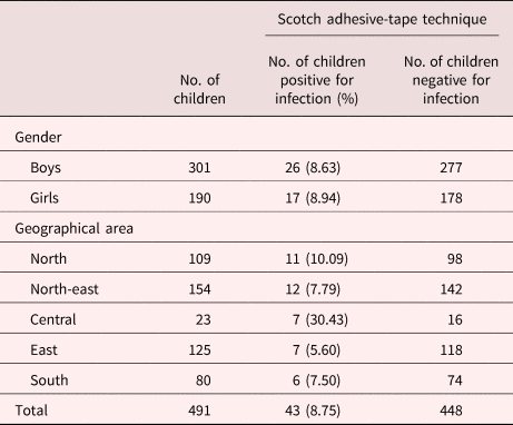
In total, 608 primary schoolchildren were included in the study but complete data were available for only 491 of these. The children ranged in age from 3 to 8 years old. Among the 491 enrolled children, there were no significant differences in infection rate by age or gender. The overall prevalence was 8.75% (43/491). The prevalence in males was 8.63% (26/301), slightly lower than in females (8.94%; 17/190) (P = 0.483). The highest prevalence was found in central Thailand (7/23; 30.48%), specifically in the crowded slum areas of Bangkok (table 1). The infection rates were 30.43%, 10.1%, 7.8%, 5.6% and 7.5% in central, northern, north-eastern, eastern and southern parts of Thailand, respectively.
Results of the adhesive tape perianal swab technique used to investigate the prevalence of infection with Enterobius vermicularis eggs among schoolchildren in five regions of Thailand.

Pinworm genetic variation
The bands produced by PCR amplification of the cox1 gene (441 bp) and ITS2 region (623 bp) are demonstrated in supplementary fig. S1. Database searches showed that all Thai sequenced egg samples were from E. vermicularis. Representative sequences of the cox1 gene (n = 17) and ITS2 region (n = 5) have been deposited in GenBank with the accession numbers MH208464–MH208480 and MH203023–MH203027, respectively. The 18 partial ITS2 sequences came from various regions in Thailand and were identical, with the exception of two sequences from Bangkok (CI1 and CI2 - MH203023), which exhibited substitutions at one position. When aligned with shorter sequences of ITS2 types (Nakano et al., Reference Nakano2006), i.e. X (AB222054), Y-1 (AB222055), Y-2 (AB222056), Y-3 (AB222057) and Z (AB222058), all ITS2 from Thailand were most similar to Y-2 (AB222056), differing only at a single nucleotide.
Phylogenetic analysis based on 95 cox1 sequences (19 from this study and 76 from GenBank) placed all sequences from Thailand into types A or B of Nakano et al. (Reference Nakano2006) and Hasegawa et al. (Reference Hasegawa, Sato and Torii2012) (supplementary fig. S2). Twenty-seven variable sites were seen in an alignment of Thai sequences. Nine sequences from Thailand—one from central Thailand (CC3; MH208466), three from eastern Thailand (EC1, EC2 and EC3; MH208467–MH208469), three from northern Thailand (NC1, NC2 and NC3; MH208470–MH208472) and two from north-eastern Thailand (NEC3 and NEC5; MH208475 and MH208476)—fell into type A (supplementary table S1). Also in type A were reference sequences from Japan (AB221470–AB221472) and Korea (EU281143). Sequences from remaining Thai samples were placed in type B with reference sequences from Europe (Denmark, Greece, Czech Republic and Germany), Africa (Sudan) and Asia (Japan and Iran), as demonstrated in supplementary fig. S2.
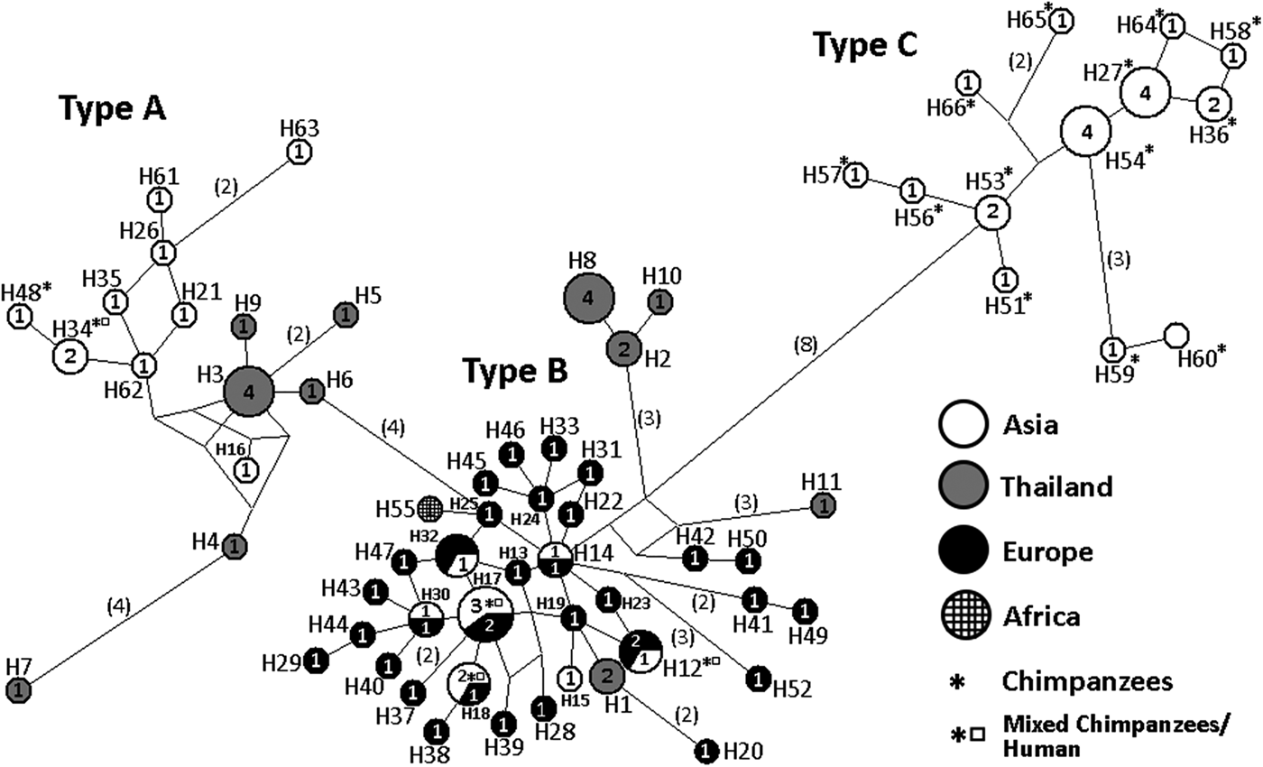
The median-joining network identified 66 haplotypes among the 95 available sequences. The haplotype diversity (Hd) was 0.9888. The 19 sequences from Thailand were divided into 11 different haplotypes. In agreement with the phylogenetic tree, nine sequences fell into type A and represented six haplotypes (fig. 2; supplementary table S1). The remaining ten sequences from Thailand represented five haplotypes in type B (fig. 2; supplementary table S1). No sequences from Thailand fell into type C. The very high haplotype diversity within Thailand was mainly due to synonymous substitutions: within each type the amino acid sequence did not vary (data not presented).
The median-joining network of Enterobius vermicularis based on sequences of the cox1 gene. Ninety-five sequences were distributed among 66 haplotypes (19 sequences from this study and 76 sequences from other studies lodged in the NCBI database). The sequences from Thailand fell into two types (A and B). The size of each circle is proportional to the number of sequences belonging to that haplotype. The colours and cross-hatching correspond to different locations of sample collection. Sequences originating from captive chimpanzees in Japan are indicated by an asterisk. An asterisk accompanied by a square symbol indicates sequences from chimpanzees (in Japan) and humans. All remaining sequences were from humans only. Numbers in parentheses along each branch indicate the number (when > 1) of mutations occurring along that branch.

Discussion
This study demonstrated that E. vermicularis infection is currently a public health problem in Thailand. The prevalence of E. vermicularis infection among children indicates that prevention and control measures should be implemented in schools and at the community level. The prevalence found in this study (8.75%) differs from that in previous reports concerning Thai schoolchildren in Phisanulok Province, northern Thailand (5.87%) (Polseela et al., Reference Polseela2004) and in Samut Prakarn Province, central Thailand (38.82%) (Nithikathkul et al., Reference Nithikathkul2001). This suggests that prevalence of enterobiasis, in tandem with risk factors (i.e. low socio-economic and sanitary conditions), has not changed in recent years. We also noted the behaviours of our subjects and found that finger sucking and anal scratching were still risk factors for E. vermicularis infection (P < 0.01), as previously reported elsewhere (Kim and Yu, Reference Kim and Yu2014).
Amplification of the cox1 and/or ITS2 sequences was possible for only 19 of 43 adhesive tape swabs. This could be due to the low concentration of DNA from the swabs, or the presence of some inhibitors in the tape, or possibly to DNA degradation during extraction. Our 18 partial ITS2 sequences of E. vermicularis from different regions of Thailand showed high similarity: only two sequences varied from the common sequence, and then only at a single position. Our sequences are similar or identical to sequences of samples from Germany (HQ646164; Zelck et al., Reference Zelck, Bialek and Weiss2011).
The mitochondrial cox1 gene revealed considerable genetic diversity in phylogenetic analysis (fig. 2). This region has been proposed as being appropriate for recognition of cryptic species (Blouin, Reference Blouin2002; Kang et al., Reference Kang2009). Mitochondrial DNA sequence data have been widely used as a rich source of genetic markers for phylogenetic analysis and study of genetic variability in many helminth groups (Le et al., Reference Le, Blair and McManus2000). Such sequences can provide a more comprehensive picture of E. vermicularis evolution than ITS2, as has been demonstrated by a study on E. vermicularis from captive chimpanzees and humans (Nakano et al., Reference Nakano2006).
The phylogenetic tree and the network, both inferred using cox1 gene sequences, demonstrated that all Thai samples fell into types A and B of Nakano et al. (Reference Nakano2006). Type A includes pinworms from humans and is known only from eastern Asia (Nakano et al., Reference Nakano2006; Kang et al., Reference Kang2009). Type B sequences have come from E. vermicularis in humans in Denmark, Germany Greece and Poland (Europe), Iran (Middle East Asia) and Japan (eastern Asia) as well as from captive chimpanzees in Japan (Nakano et al., Reference Nakano2006; Piperaki et al., Reference Piperaki2011; Ferrero et al., Reference Ferrero2013; Kubiak et al., Reference Kubiak, Dzika and Paukszto2017). The presence of cluster B sequences in worms from captive chimpanzees in Japan and from humans in Europe and Asia suggests possible zoonotic transmission of some original chimpanzee worms from humans (supplementary fig. S2). Zoonotic transmission from primates to humans in Asia is an additional possibility.
Nakano et al. (Reference Nakano2006) identified a third type of Enterobius cox1 sequence, type C. All known representatives of this type have been found in captive chimpanzees in Japan. None of our sequences from Thailand fell into type C. The geographical and host origins of E. vermicularis haplotypes in type C are unknown. To date, cox1 sequences of E. vermicularis have been published from only a limited number of countries. In eastern/South-East Asia, data were formerly available only from Japan, and now also from Thailand. Type A and B sequences have been found in both countries, but the situation in most countries remains unknown. Much more extensive geographical sampling is required before general patterns can be suggested.
In conclusion, infection caused by E. vermicularis is still a health problem for schoolchildren in Thailand. To our knowledge, this study is the first molecular identification and genetic diversity analysis of E. vermicularis.
Author ORCIDs
Pewpan M Intapan https://orcid.org/0000-0002-2138-8783
Wanchai Maleewong https://orcid.org/0000-0002-6726-3759
Supplementary material
To view supplementary material for this article, please visit https://doi.org/10.1017/S0022149X18000962
Acknowledgements
The authors would like to thank Dr David Blair for valuable suggestions and assistance with the presentation of this paper through Khon Kaen University Publication Clinic.
Financial support
This study was supported by a TRF Senior Research Scholar Grant, Thailand Research Fund grant number RTA5880001. KT was supported in part by the Research and Diagnostic Center for Emerging Infectious Diseases, Khon Kaen University. OS was supported by a scholarship under the Post-Doctoral Training Program from Research Affairs and Graduate School, Khon Kaen University (grant no. 58101).
Conflict of interest
None.
Ethical standards
All procedures performed in studies involving human participants were in accordance with the ethical standards of the Khon Kaen University Ethics Committee for Human Research (HE571064) and with the 1964 Helsinki Declaration and its later amendments or comparable ethical standards.