Introduction
Ceruminal glands are modified sweat glands located in the skin of the external auditory canal. They may give rise to rare malignant tumours such as ceruminous mucoepidermoid carcinoma.Reference Thompson, Nelson and Barnes1 In this paper, we report the first case in the English literature of ceruminous mucoepidermoid carcinoma of the external auditory canal, and highlight the impact of concurrent chemoradiotherapy on the outcomes of this disease. This report is of particular importance because it is unlikely that a prospective trial will be performed in this patient population.
Case report
In late 2007, a 47-year-old Hispanic female presented with a 2-month history of otalgia, dizziness, pulsatile tinnitus and a right-sided mass filling the external auditory canal. Biopsy revealed high grade ceruminous mucoepidermoid carcinoma. The patient underwent excision of the right external auditory canal and temporal bone, with superficial parotidectomy, excision of the intra-parotid lymph nodes and the malleus of the right middle ear, and dissection of the right side of the upper neck. Pathology showed a completely excised high grade ceruminous mucoepidermoid carcinoma in situ of the temporal bone and external auditory canal. In addition, all surgical margins, as well as 39 lymph nodes, were negative for cancer. Pathological analysis of tumour–node–metastasis (TNM) revealed a T2 N0 (stage II) tumour. All margins were negative with no lymphovascular or perineural invasion. Due to the favourable surgical findings, the patient was offered a close follow up rather than proceeding with immediate treatment.
One year later, the patient developed pain in the surgical region. A computed tomography (CT) scan of the temporal bone showed tumour recurrence within the flap reconstruction. A biopsy of the new lesion revealed recurrent ceruminous mucoepidermoid carcinoma.
Pre-operative positron emission tomography (PET) and CT showed a mass in the surgical region (with a standardised uptake value of 9). The imaging revealed a bulky, enhancing soft tissue mass replacing the right parotid gland, with extension into the right external auditory canal and mastoidectomy bed that was inseparable from the adjacent cervical erector muscles. In addition, a right-sided, 7-mm level I lymph node, and multiple right-sided level II–IV lymph nodes (with a standardised uptake value of 2), were observed.
Management
The patient was discussed at our institutional multidisciplinary head and neck tumour conference. The consensus was to proceed with surgery followed by salvage post-operative radiotherapy (RT) with chemotherapy as a radiosensitiser.
Surgery
The patient underwent right auriculectomy, total parotidectomy with facial nerve sacrifice, right lateral temporal bone resection, mastoidectomy, labyrinthectomy, and right neck dissection at levels II–IV, with reconstruction.
Pathology
Pathological analysis revealed right external ear ceruminous mucoepidermoid carcinoma grade 3 of ceruminous gland origin. Gross examination showed the white, firm mass was 4 × 3.7 × 2.5 cm in size. It involved the cavum conchae, tragus and the subcutaneous tissue, completely obliterating the external auditory canal. Microscopic examination revealed ceruminous mucoepidermoid carcinoma of the right external auditory canal harbouring intermediate cells, frequent epidermoid areas with focal squamous pearl formation, occasional mucinous cells (with both intraluminal and intracytoplasmic mucin production), and a lack of myoepithelial cells. The tumour included areas of ceruminous mucoepidermoid carcinoma with focal apocrine (ceruminous) differentiation, which showed luminal apocrine snouts and dark pink, mildly granular cytoplasm. The tumour was diffusely invading nearby structures, including the dermis of the external ear, cartilage of the pinna, subcutaneous soft tissues and temporal bone. There was perineural invasion, with lymphatic spread. In addition, pathological examination of the surgically removed lymph nodes revealed that 2 of the lymph nodes (out of a total of 25) had extracapsular extension of the malignant cells. The tumour showed a solid growth pattern, markedly pleomorphic nuclei and a prominent desmoplastic reaction that lacked an inflammatory component. The immunohistochemical investigation revealed luminal carcinoembryonic antigen and cytokeratin-7 labelling in the neoplastic cells which were of high molecular weight; those of focally low molecular weight were cytokeratin positive. The Ki-67 proliferation (labelling) index was approximately 30 per cent. All final surgical margins were negative and the pathological analysis revealed a T3 N1 M0 (recurrent stage IV) tumour.
Radiation therapy
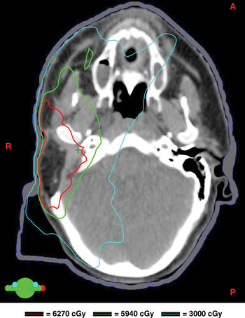
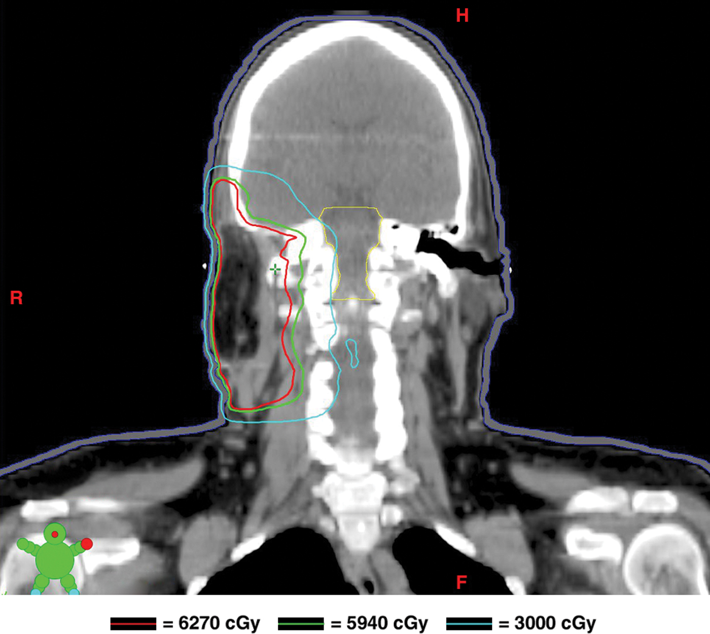
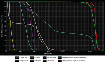
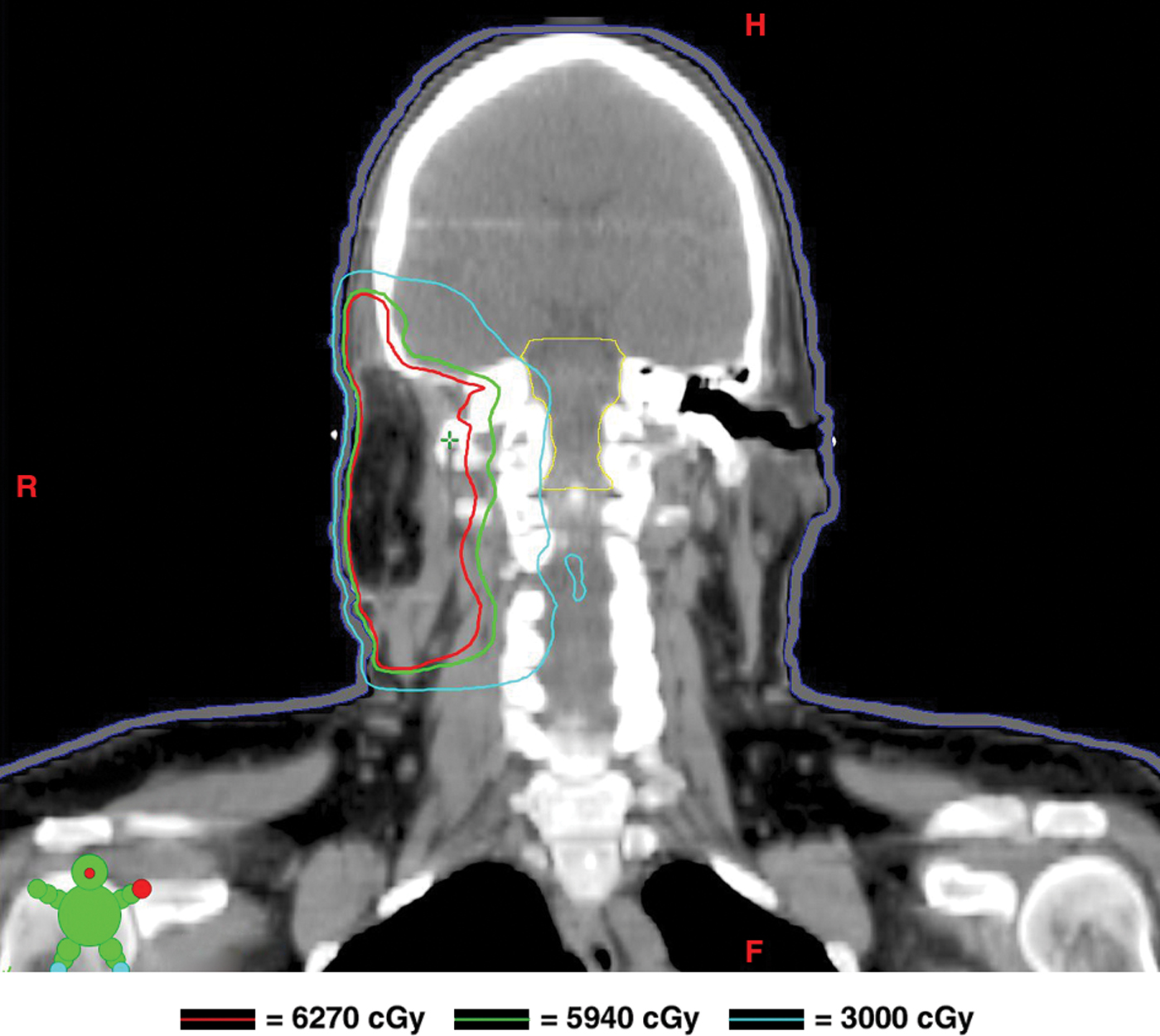
Five weeks post-surgery, the patient underwent CT simulation and intensity-modulated radiation therapy based treatment. The planning target volume included the tumour region and parotid fossa (the latter of which comprised the intra- and infra-parotid lymph nodes), ipsilateral preauricular and retroauricular lymph nodes, and ipsilateral neck (levels II–IV) lymph nodes. Intensity-modulated radiation therapy with seven beams was chosen in order to preferentially spare the left parotid, and minimise the radiation to the nearby critical structures (Figures 1–3). Chemotherapy was administered weekly with carboplatin as a radiosensitiser. The patient received a total dose of 6270 cGy (190 cGy × 33 fractions) to the tumour region in the right ear canal and right upper neck, with daily 5 mm bolus to deliver full dose to the scar and the skin. She also received 5940 cGy (180 cGy × 33 fractions) to the ipsilateral high risk neck region and to the lymphatics in transit. The treatment was delivered via a linear accelerator (six megavoltage photons) and was completed in the autumn of 2008. The patient was able to tolerate the treatment without breaks, although she did experience the expected acute toxicity related to concurrent chemoradiotherapy in the form of grades I–II xerostomia, dysgeusia, mucositis, dysphagia and dermatitis.
Axial computed tomography image of the head and neck showing the radiation doses delivered to each target volume. A = anterior; R = right; P = posterior

Coronal computed tomography image of the head and neck showing the radiation doses delivered to each target volume. H = head; R = right; F = feet
Dose–volume histogram of planning target volumes and critical structures. R = right

Follow up
Forty months following the date of salvage surgery, and 37 months after the date of concurrent chemoradiotherapy completion, a comprehensive physical examination and PET and CT scans confirmed the absence of any loco-regional recurrence or distant metastasis.
Discussion
In general, malignant tumours of the external auditory canal are extremely rare, with approximately 250 cases per year reported in the USA. Although the true incidence of ceruminous gland tumours is difficult to determine, the incidence of malignant external auditory canal tumours has been reported as follows: 86 per cent squamous, 6 per cent basal, 6 per cent adenoid cystic carcinoma, 2 per cent adenocarcinoma, 1 per cent melanoma, and less than 1 per cent mucoepidermoid, acinic cell and Merkel cell carcinomas.Reference Wenig2 Few cases of ceruminous mucoepidermoid carcinoma involving the external auditory canal have been reported in the English literature.Reference Pulec3–Reference Crain, Nelson, Barnes and Thompson9 Only five cases were reported with adequate detail. The median age of these 5 patients was 49 years (28–62 years), and comprised 2 males and 3 females.Reference Pulec3–Reference Crain, Nelson, Barnes and Thompson9 No staging system currently exists for the classification of primary glandular tumours arising in the external auditory canal. Instead, for many years, a system for external auditory canal tumour staging has been used.Reference Arriaga, Curtin, Takahashi, Hirsch and Kamerer10
A review of the literature indicated that ceruminous mucoepidermoid carcinoma is associated with a poor outcome (mean follow up of 1.5 years). Three out of five patients died of the disease. One died of the disease but no details were reported, another died after 3 years due to local recurrence and liver metastasis (as revealed by pathology), and the other patient had recurrent disease at 18 months and subsequently died.Reference Pulec3, Reference Kinney and Wood4 The remaining 2 patients (for whom the follow up was limited to 2.2 years) had no evidence of disease at 24 and 29 months.Reference Magliulo, Fusconi and Pulice6, Reference Bared, Dave, Garcia and Angeli8
• Ceruminous mucoepidermoid carcinoma of the external auditory canal is rare
• A 47-year-old female underwent right external auditory canal excision and upper neck dissection for this condition
• The carcinoma recurred one year later and was managed with salvage resection followed by concurrent chemoradiotherapy
• The patient had no evidence of disease at 37 months' follow up
• The trimodality approach, using surgery plus concurrent chemoradiotherapy, provided reasonable loco-regional control with tolerable toxicity
Due to the rarity of ceruminous mucoepidermoid carcinoma of the external auditory canal, there is no universal consensus on the management of the disease, but surgery has been utilised in all reported cases.Reference Crain, Nelson, Barnes and Thompson9 There is no data to support the routine usage of RT with or without chemotherapy. We think that post-operative (adjuvant or salvage) RT with or without chemotherapy would be wise in cases with adverse prognostic features (such as locally advanced disease, close or positive margins, multiple lymph nodes, extra capsular extension, perineural invasion, and invasion of the lymphovascular space). In the case reported here, it seems that concurrent chemoradiotherapy offered better locoregional control, with acceptable long-term comorbidities of grade I trismus and xerostomia (the initial tumour had recurred within one year despite aggressive surgery and favourable pathology). Our report highlights the efficacious effect of the trimodality salvage approach involving surgery followed by concurrent chemoradiotherapy to control an aggressive recurrent disease.
Conclusion
The trimodality approach appears to be a reasonable and successful approach for a rare disease such as ceruminous mucoepidermoid carcinoma. It provided three years locoregional control, with acceptable comorbidities. Further detailed case reports are warranted in order to optimise the management of this rare malignancy.