Introduction
The bark beetle genus Dendroctonus Erichson (Coleoptera: Curculionidae: Scolytinae) has been recognised as being very difficult taxonomically because few reliable characters have been identified for distinguishing the species (Hopkins Reference Hopkins1909; Wood Reference Wood1963, Reference Wood1982). Species in the Dendroctonus frontalis complex (D. adjunctus LeConte, D. approximatus Dietz, D. brevicomis LeConte, D. frontalis Zimmermann, D. mesoamericanus Armendáriz-Toledano and Sullivan, D. mexicanus Hopkins, and Dendroctonus vitei Wood), have proven to be particularly difficult due to their morphological similarity, the broad variation of their external morphological characters (Vité et al. Reference Vité, Islas, Renwich, Hughes and Kliefoth1974, Reference Vité, Hughes and Renwich1975; Wood Reference Wood1974, Reference Wood1982; Lanier et al. Reference Lanier, Hendrichs and Flores1988; Armendáriz-Toledano et al. Reference Armendáriz-Toledano, Niño, Sullivan, Macías-Sámano, Victor, Clarke and Zúñiga2014b; Víctor and Zúñiga Reference Víctor and Zúñiga2016), and the frequency with which populations coexist in space and time in the same trees (Zúñiga et al. Reference Zúñiga, Cisneros and Salinas-Moreno1995, Reference Zúñiga, Mendoza-Correa, Cisneros and Salinas-Moreno1999; Salinas-Moreno et al. Reference Salinas-Moreno, Mendoza, Barrios, Cisneros, Macías-Sámano and Zúñiga2004, Reference Salinas-Moreno, Vargas, Zúñiga, Víctor, Ager and Hayes2010; Moser et al. Reference Moser, Fitzgibbon and Klepzig2005; Armendáriz-Toledano et al. Reference Armendáriz-Toledano, Niño, Sullivan, Kirkendall and Zúñiga.2015). These circumstances have made practical identification of specimens difficult and have raised doubts concerning the taxonomic status of these species, despite the abundant information that has been published on the natural history and management of the several economically important pest species in this group.
Dendroctonus vitei was described from Patzun, Guatemala based on ecological, reproductive, morphological, and biochemical data (Wood Reference Wood1974; Vité et al. Reference Vité, Hughes and Renwich1975), but it has remained one of the least studied species in the genus Dendroctonus (Six and Bracewell Reference Six and Bracewell2015). This species went unnoticed for many years, and has been collected only sporadically from portions of its range including the Mexican states of Nuevo León, Veracruz, Oaxaca, and Chiapas (Atkinson and Equihua-Martínez Reference Atkinson and Equihua-Martínez1985; Lanier et al. Reference Lanier, Hendrichs and Flores1988; Wood and Bright Reference Wood and Bright1992). Recently, numerous specimens of D. vitei were identified (using characters of the male seminal rod) from within the Sierra Madre Oriental, the east Trans-Mexican Volcanic Belt, the Sierra Madre de Chiapas in Mexico; as well as the Cordillera Central of Guatemala (Armendáriz-Toledano et al. Reference Armendáriz-Toledano, Niño, Macías-Sámano and Zúñiga2014a).
The taxonomic identification of D. vitei has been extremely problematic in part because its taxonomic description was based entirely on specimens from the type locality (Wood Reference Wood1974); hence geographic variation of diagnostic characters was never evaluated. In addition, most of the diagnostic morphological characters were not sufficiently illustrated (Vité et al. Reference Vité, Hughes and Renwich1975; Wood Reference Wood1982).
Several studies have proposed that D. vitei can be distinguished from other species of the D. frontalis complex by its relative size, colour, punctation of the pronotum, vestiture of the elytral declivity, sculpturing of the frons, and seminal rod shape (Vité et al. Reference Vité, Islas, Renwich, Hughes and Kliefoth1974; Wood Reference Wood1974; Lanier et al. Reference Lanier, Hendrichs and Flores1988). However, with the exception of seminal rod shape and to a lesser extent body colour, these characters are not geographically consistent and they overlap among species within the D. frontalis complex, which makes their use unreliable (Armendáriz-Toledano et al. Reference Armendáriz-Toledano, Niño, Sullivan, Kirkendall and Zúñiga.2015).
Nonetheless, seminal rod shape is geographically consistent and a reliable attribute for identifying D. vitei males (Lanier et al. Reference Lanier, Hendrichs and Flores1988). By using seminal rod shape as the diagnostic character for the species, this study aimed to find additional morphological characters useful for identification of D. vitei from the antenna, stridulatory apparatus, male and female genitalia, and external morphology. In addition, we present a re-description of this species, information on its natural history, karyology, and chemical ecology derived from new and published data.
Material and methods
Specimens
More than 1000 specimens of the D. frontalis complex collected from different locations from Mexico and Guatemala were reviewed exhaustively. The identification of D. vitei males was carried out by means of the seminal rod, which has a ventral process conspicuous longer than the dorsal process, with the two processes joined by a broad, concave arch (Armendáriz-Toledano et al. Reference Armendáriz-Toledano, Niño, Macías-Sámano and Zúñiga2014a). To identify putative females of this species, we used the body colour (dark black), and the pronotum width (1.56±0.18 mm standard deviation) as in Lanier et al. (Reference Lanier, Hendrichs and Flores1988); these were collected in the same sections of the tree than males and at the same locations.
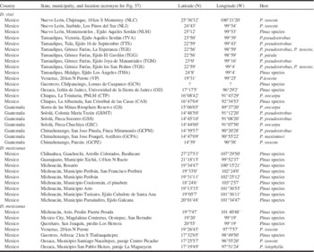
A total of 150 adult specimens of D. vitei from 22 localities were used to find new characters and redescribe this species (Table 1). The male specimens of D. vitei reported by Armendáriz-Toledano et al. (Reference Armendáriz-Toledano, Niño, Macías-Sámano and Zúñiga2014a) were included in this study (Table 1). A comparison of the variation of D. vitei characters was performed with respect to D. mexicanus (Table 1), because it is its sibling species (Víctor and Zúñiga Reference Víctor and Zúñiga2016) and frequently both species are confused. In addition, both species can coexist in sympatry and syntopy in several localities from Mexico and Guatemala. The sex of specimens was determined by the presence of frontal tubercles on the males and the morphology of the seventh abdominal tergite (Lyon Reference Lyon1958; Mendoza-Correa and Zúñiga Reference Mendoza-Correa and Zúñiga1991).
Country, locality, reference acronym, coordinates, and host of Dendroctonus mexicanus and Dendroctonus vitei specimens examined in this study.

PNLM, Parque Nacional Lagunas de Montebello.
Adults of both species were examined and photographed following the protocols reported by Armendáriz-Toledano et al. (Reference Armendáriz-Toledano, Niño, Sullivan, Kirkendall and Zúñiga.2015). The removal and mounting of the morphological structures (antenna, elytra, seventh tergite, and genitalia) was carried out following the protocols reported by López et al. (Reference López, Armendáriz-Toledano, Macías Sámano, Shibayama-Salas and Zúñiga2014) and Armendáriz-Toledano et al. (Reference Armendáriz-Toledano, Niño, Sullivan, Kirkendall and Zúñiga.2015). Details of morphological structures were photographed with an environmental scanning electron microscope (ESEM-Evo® 40 VP; Zeiss, Ontario, California, United States of America). The elements of elytral sculpture (punctures, granules, pubescence, etc.) were measured and quantified directly in the slides with an ocular micrometer under a phase contrast microscope (400×).
A total 33 attributes, 28 from the external morphology (head, antennae, pronotum, elytra, abdomen) and five from male and female genitalia were examined. Ten of them were coded as double state (DS) or binary, seven as meristic quantitative (M), and 16 as continuous quantitative (C) characters (Tables 2–4). The number of individuals analysed (=number of counts or measurements) per character was variable (Tables 2–4). Lastly, normality of the distribution of continuous characters was tested independently by a Shapiro–Wilkinson test and by examination of distribution histograms, and the homogeneity of variances was determined using Cochran’s test. Contrasts of each character between D. vitei and D. mexicanus were evaluated by a Student’s t-test for continuous characters and a Mann–Whitney test for discrete characters (Zar Reference Zar2010).
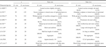
Frequencies of double-state or binary characters used to compare Dendroctonus vitei and Dendroctonus mexicanus.

An asterisk indicates a significant difference between the species (Mann–Whitney test, *P⩽0.001; **P⩽0.05).
EEP, elevation of lateral sections of epistomal process; FS, frons sculpture; DSA, distribution of sensillae on the anterior surface of the antennal club; EPC, elevation of pronotal callus; SS, setae of the elytral disc and declivity; AP, acute projections on the posterior edge of squamiform plates in the median proximal area of the eighth tergite; CS, cornu size; DSt, density of striae in the nodulus; PSS, proportion of the spermatheca covered by striae; AA, seminal rod anchor anatomy.
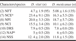
Means values±SD of meristic characters used to compare Dendroctonus vitei and Dendroctonus mexicanus.
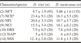
An asterisk indicates a significant difference between the species (Mann–Whitney test, *P⩽0.001).
NFT, number of frontal tubercles; NCE, number of crenulations on the elytral disc; NPI, number of punctures in elytral declivity interspaces; NCI, number of crenulations in elytral declivity interspaces; DRS, number of ridges in the transversely sulcate area (file) of the stridulatory apparatus; NAP, number of acute projections on squamiform plates; NSS, number of striae in the spermatheca.
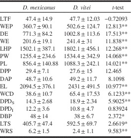
Mean, standard deviation, and results of Student’s t-test applied to contrasts of continuous morphological characters between Dendroctonus vitei and Dendroctonus mexicanus.

*P⩽0.05; **P⩽0.001.
LFT, frontal tubercles length; WEP, width of epistomal process; DE, distance between the eyes; WE, width of left eye; LHP, length of the head-pronotum; PW, width of the pronotum at its posterior margin; PL, pronotum length; DPP, diameter of pronotal punctures; DAP, distance among pronotal punctures; EL, elytra length; WCD, width of crenulations in interspace two of elytral declivity; DPDS, diameter of the punctures in stria 2 of elytral declivity; DPDI, diameter of punctures in interstria 2 of elytral declivity; DBP, distance between the punctures in stria 2 of elytral declivity; LTS, length of transverse sulcate area of stridulatory apparatus; WRS, width of ridges of sulcate area of stridulatory apparatus.
Characters analysed
(1) Elevation of the lateral sections of the epistomal process (EEP)
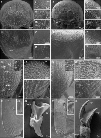
The epistomal process is a structure present on the head of all members of the genus. It consists of a pair of lateral elevations (=sections or arms) in the epistomal area at the inferior-median region of the frons. A median section that is flat and usually shiny separates both elevations (Figs. 1, 8, 12). Wood (Reference Wood1974, Reference Wood1982) differentiated D. vitei from D. mexicanus by the former having the lateral sections of epistomal process more strongly elevated, and the sculpturing of the median section almost as coarse as the lateral elevations (Table 2). This is a double-state character.
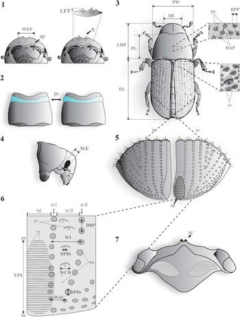
General anatomy of Dendroctonus adult. 1. Ventral view of head. 2. Dorsal view of pronotum. 3. Insect dorsal view. 4. Lateral view head and pronotum. 5. Internal surface of elytral declivity. 6. Sculpture of elytral declivity. 7. Seventh tergite of males (propygidium). Length of frontal tubercles (LFT), width of epistomal process (WEP), distance between the eyes (DE), width of left eye (WE), length of head and pronotum (LHP), width of pronotum at posterior margin (PW), pronotum length (PL), diameter of pronotal punctures (DPP), distance among pronotal punctures (DAP), elytra length (EL), width of crenulations in interspace two of elytral declivity (WCD), diameter of the punctures in the stria two of elytral declivity (DPDS), diameter of the punctures in interstria 2 of elytral declivity (DPDI), distance between the punctures in stria 2 of elytral declivity (DBP), length of transverse sulcate area of stridulatory apparatus (LTS). cr, crenulations; ep, epistomal process; in I, interspace 1; in II, interspace 2; le, left elytron; pu, punctures; re, right elytron; rs, ridges of sulcate area; sa, sulcate area of stridulatory apparatus; sc, scraper of stridulatory apparatus.

Dendroctonus mexicanus (left) and Dendroctonus vitei (right) adults, respectively. 8, 12. Male head. 9, 13. Male epicranial surface. 10, 14. Surface of male frons. 11, 15. Surface of male epistomal process. 16, 19. Lateral view of male pronotum. 17, 20. Lateral posterior surface of male pronotum. 18, 21. Preepisternal and episternal area of male. 22, 26. Female right elytron. 23, 27. Punctures and crenulations on anterior region of elytral disc. 24, 28. Elytral disc. 25, 29. Female elytral declivity. 30, 33. Sulcate area of male stridulatory apparatus. 31, 34. Ridges of sulcate area of male stridulatory apparatus. 32, 35. Seminal rod. cr, crenulations; dp, dorsal process; ep, epistomal process; ft, frontal tubercles; gr, granules; in I, interspace 1; in II, interspace 2; in III, interspace 3; pu, punctures; st I, stria 1; st II, stria 2; st III, stria 3; vp, ventral process.

(2) Number of frontal tubercles (NFT)
The males of species in the D. frontalis complex, except D. adjunctus, have numerous and prominent granular tubercles on the lateral areas of the frons. Wood (Reference Wood1974) remarked that the frontal tubercles in D. vitei are less numerous than in D. mexicanus (Fig. 1; Table 3). This is a meristic quantitative character.
(3) Frons sculpture (FS)
Frontal sculpture consists of granules, punctures, and fused granules resembling small crenulations. Wood (Reference Wood1974, Reference Wood1982) distinguished D. vitei from D. mexicanus by the former having a more finely sculptured frons (Figs. 8–15; Table 2). This is a double-state character.
(4) Distribution of sensillae on the anterior surface of the antennal club (DSA)
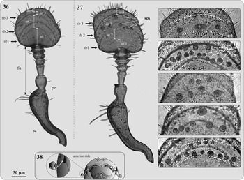
Different types of sensillae are located on the four antenomeres of the antennal club of Dendroctonus species. Their position in the sensorial bands and distribution on the antennal club are apparently not similar among species, which suggests that these differences might be useful taxonomic characters for these beetles (Figs. 36–38; Table 2). This is a double-state character.
Anterior side of right antennae. 36. Dendroctonus mexicanus without groups of sensillae arranged in circular clusters. 37. Dendroctonus vitei with groups of sensillae arranged in circular clusters on the third antennomere and on third sensorial band. 38. Frontal view of head. an1, first antennomere; an2, second antennomere; an3, third antennomere; an4, fourth antennomere, fu, funiculus; pe, pedicel; sb1, sensorial band 1; sb2, sensorial band 2; sb3, sensorial band 3, sc, scape; scs, sensillae clusters.

(5) Elevation of pronotal callus (EPC)
Females of the D. frontalis complex have a pronotal callus, which can be dorsally or transversally elevated. Due to the presence of this callus, the lateral surfaces of the anterior pronotum look bulging and emarginate in dorsal view. Wood (Reference Wood1974) indicated that the pronotal callus in D. vitei is more poorly developed than in D. mexicanus (Fig. 2; Table 2). This is a double-state character.
(6) Setae of the elytral disc and declivity (SS)
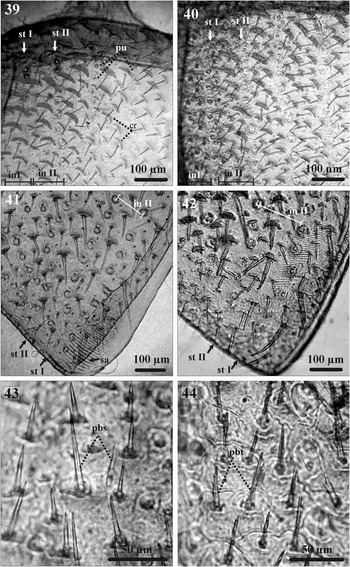
The surface of the elytra is covered by abundant setae of different sizes; these are categorised as short, medium, and long. The setae on the surface of the elytral interspaces usually differ among Dendroctonus species (Figs. 43–44; Table 2). This is a double-state character.
(7) Number of crenulations on the elytral disc (NCE)
The elytral surface is covered by flat, cuticular elevations with convex and concave margins (crenulations), which are less abundant towards the elytral declivity. Wood (Reference Wood1974, Reference Wood1982) indicated that D. vitei have more crenulations in interstriae of the elytral disc than D. mexicanus. The crenulations in this study were counted in the first anterior third of interstriae II–III of elytral disc of right elytron (Figs. 3, 39–40; Table 3). This is a meristic quantitative character.
(8) Number of punctures in elytral declivity interspaces (NPI)
Interspaces of the elytral declivity are covered by small punctures varying in size and number among species (Wood Reference Wood1982). Punctures in interspace II of the right elytron in this study were counted in the area corresponding to the last 10 distal punctures of striae II and III (Figs. 6, 41, 42; Table 3). This is a meristic quantitative character.
(9) Number of crenulations in elytral declivity interspaces (NCI)
Interspaces of the elytral declivity are covered by crenulations that vary in size and number between the species. Wood (Reference Wood1974, Reference Wood1982) distinguished D. vitei from D. mexicanus by the less numerous and smaller crenulations of the elytral declivity of the former. In this study the crenulations were counted in the anterior third of interstriae II–III of the elytral disc of the right elytron (Fig. 6; Table 3). This is a meristic quantitative character.
(10) Number of ridges in the transversely sulcate area (file) of the stridulatory apparatus (DRS)
Dendroctonus males produce sound with a stridulatory apparatus composed of a bifid process (“stridulating process” sensu Hopkins Reference Hopkins1909) located on the posterior margin of the propygidum (seventh abdominal tergite) and a transversely sulcate area (file) on the ventral surface of the elytral declivity straddling the elytral suture at the apex of each elytron. The number of ridges in this structure is variable among species. The ridges per 50 µm on the distal region of the file on the right elytron were counted (Figs. 5, 7, 30–31, 33–34; Table 3). This is a meristic quantitative character.
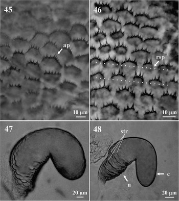
(11) Acute projections on the posterior edge of squamiform plates in the median proximal area of the eighth tergite (AP)
The eighth tergite has transverse, parallel rows of squamiform plates. These plates display several acute projections on their distal edge. The relative size of these projections is variable among species and has been demonstrated to be a useful taxonomic character within the genus (Armendáriz-Toledano et al. Reference Armendáriz-Toledano, Niño, Sullivan, Macías-Sámano, Victor, Clarke and Zúñiga2014b). In the present study this character was evaluated on the median anterior region of the tergite (Figs. 45–46; Table 2). This is a double-state character.
Elytra of Dendroctonus mexicanus (left) and Dendroctonus vitei (right). 39–40. Elytral disc. 41–42. Elytral declivity. 43–44. Elytral declivity pubescence. in II, interspace 2; sa, sulcate area of stridulatory apparatus; st I, stria 1; st II, stria 2; pbs, pubescence with smooth surface; pbt, pubescence with saw-toothed outer surface.

Eighth tergite and spermathecae (female terminalia) of Dendroctonus mexicanus (left) and Dendroctonus vitei (right). 45–46. Squamiform plates in the median proximal area of the eighth tergite. 47–48. Spermathecae. ap, acute projections; rsp, ridges of squamiform plates str, striations. strations; n, nodulus; c, cornu.

(12) Number of acute projections on squamiform plates (NAP)
The number of acute projections was counted on 10 squamiform plates per specimen on the median anterior region of the tergite (Figs. 45–46; Table 3). This is a meristic quantitative character.
(13) Cornu size (CS)
The female spermatheca is reniform and divided into a nodulus and cornu (Fig. 48). The cornu is the distal portion of the spermatheca beyond the middle constriction (Armendáriz-Toledano et al. Reference Armendáriz-Toledano, Niño, Sullivan, Macías-Sámano, Victor, Clarke and Zúñiga2014b), whereas the nodulus is the proximal portion. Variation in cornu size was shown to be a useful taxonomic character for separating some species in the D. frontalis complex (Figs. 47–48) (Table 2). This is a double-state character.
(14) Number of striae in the spermatheca (NSS)
The surface of the spermatheca has transverse striae that partially or completely encircle it and are distributed predominantly in the nodulus (Armendáriz-Toledano et al. Reference Armendáriz-Toledano, Niño, Sullivan, Macías-Sámano, Victor, Clarke and Zúñiga2014b). The striae number varies between certain Dendroctonus species (Figs. 47–48; Table 3). This is a meristic quantitative character.
(15) Density of striae in the nodulus (DSt)
In the proximal region of the nodulus the striations of spermathecae are more closely spaced than in the distal region. The striation density in this region differs between Dendroctonus species (Armendáriz-Toledano et al. Reference Armendáriz-Toledano, Niño, Sullivan, Macías-Sámano, Victor, Clarke and Zúñiga2014b), hence it can be of taxonomic value (Figs. 47–48; Table 2). This is a double-state character.
(16) Proportion of the spermatheca covered by striae (PSS)
The striations in the spermatheca can cover different proportions of nodulus. The area covered by the striae is different between species of D. frontalis complex (Armendáriz-Toledano et al. Reference Armendáriz-Toledano, Niño, Sullivan, Macías-Sámano, Victor, Clarke and Zúñiga2014b) (Figs. 47–48; Table 2). This is a double-state character.
(17) Seminal rod anchor anatomy (AA)
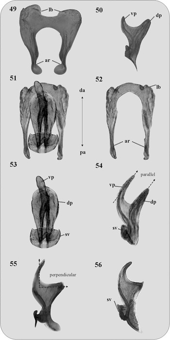
The seminal rod anchor is a sclerotised structure that is positioned dorsally to the seminal rod. Viewed dorsally, this structure has two proximally extending, parallel arms joined by a distal arch. The distal area of arch possesses two ear-like lobes extending distally and laterally from each side (Figs. 49–52; Table 2). This is a double-state character.
Accessory apparatus of male genitalia. 49. Dendroctonus mexicanus: seminal rod anchor in ventral view. 50. Seminal rod in lateral view. 51. Dendroctonus vitei: seminal rod anchor coupled with seminal rod in ventral view. 52. Anchor seminal rod in ventral view. 53. Seminal rod in ventral view. 54–56. Seminal rod in lateral view. da, distal area; dp, dorsal process; lb, lobe; pa, proximal area; sv, seminal valve; vp, ventral process.

(18–33) Continuous quantitative characters
These were: length of the frontal tubercles (LFT), width of the epistomal process (WEP), distance between the eyes (DE), width of the left eye (WE), length of the head and pronotum (LHP), width of the pronotum at its posterior margin (PW), pronotum length (PL), diameter of the pronotal punctures (DPP), distance among pronotal punctures (DAP), elytra length (EL), width of the crenulations in interspace 2 of the elytral declivity (WCD), diameter of the punctures in stria 2 of the elytral declivity (DPDS), diameter of the punctures in interstria 2 of the elytral declivity (DPDI), distance between the punctures in striae 2 of the elytral declivity (DBP), length of transversal sulcate area or file of the stridulatory apparatus (LTS), width of the ridges of the sulcate area of the stridulatory apparatus (WRS) (Figs. 1, 3–4, 6). The characters DPP, DAP, WCD, DPDS, DPDI, and DBP were measured in the areas of each structure showed in Figure 1. In each species, the average value of these characters was obtained from a total 10 specimens (5♀, 5♂); 10 cuticular elements (punctures, crenulations, etc.) were measured in each individual.
Results
The characters EEP, FS, DSA, EPC, SS, AP, CS, and AA showed differences between D. vitei and D. mexicanus (Table 2), five of them showed exclusive character states of species: the FS (Figs. 8, 15); the DSA (Figs. 36–37); the SS (Figs. 43–44); CS (Figs. 47–48); and AA (Figs. 49, 51–52).
Dendroctonus vitei specimens have sparse sculpture on the frons (Figs. 12–15), sensillae on the third and fourth antennomere of the anterior face of the antennal club clustered in circular concavities resembling pit-craters (Fig. 37), setae in the elytral disc and declivity that possess a saw-toothed tip (Figs. 42, 44), the cornu similar in length to the nodulus (Fig. 48), and the distal lobes of the seminal rod anchor poorly emarginate and with thin arms (Fig. 51). Contrariwise, D. mexicanus specimens have abundant sculpture on the frons (Figs. 8–11), sensillae on the antennal club not clustered in concavities (Fig. 36), setae on the elytral disc and declivity with a sharp tip and a smooth outer surface (Figs. 41, 43), a cornu that is half the length of the nodulus (Fig. 47), and the distal lobes of the seminal rod anchor strongly emarginate and prominent with thick arms (Fig. 49). Characters EEP, EPC, and AP showed strong overlap between species (Table 2).
Among the seven meristic characters (NFT, NCE, NPI, NCI, DRS, NAP, NSS), only the NCE and the number of acute projections on the posterior edge of squamiform plates in the median proximal area of the eighth tergite (NAP), showed differences between species. Dendroctonus vitei had from four to five acute projections per plate on the eighth tergite
 $\left( {{\rm \bar{X}{\equals}5}\,\pm\,{\rm 0}{\rm .5}} \right)$
and from 8–29 crenulations on the elytral disc
 $$\left( {{\rm \bar{X}}{\equals}23.4\,\pm\,9.1} \right)$$
. Dendroctonus mexicanus had from 5–6
 $$\left( {{\rm \bar{X}}{\equals}6\,\pm\,0.5} \right)$$
acute projections, and from 12–23 crenulations
 $$\left( {{\rm \bar{X}}{\equals}16.3\,\pm\,5.5} \right)$$
. The five remaining meristic characters (NFT, NPI, NCI, DRS, NSS) had similar averages in both species (Table 3).
Lastly, a Student’s t-test supported statistically significant differences in 12 of the 16 continuous characters analysed (WEP, DE, WE, LHP, PW, PL, EL, WCD, DPDS, DBP, LTS, WRS). In general, D. vitei possessed higher mean values in these characters than D. mexicanus, except for DBP, DPDI, and WRS (Table 4).
Discussion
The evaluation of 10 double-state characters indicates that five of them (FS, DSA, SS, CS, and AA) are useful to differentiate D. vitei and D. mexicanus. Among these, DSA and AA, together with seminal rod shape, can differentiate this species from other D. frontalis complex species in addition to being diagnostic characters for D. vitei (F.A.T. and G.Z., unpublished data).
The presence of sensillae clustered into pit-crater-like cavities in the third and fourth antenomeres of D. vitei antennae is unique, and nothing similar has been documented in other species of Dendroctonus (Payne et al. Reference Payne, Moeck, Willson, Coulson and Humphreys1973; Dickens and Payne Reference Dickens and Payne1977; Chen et al. Reference Chen, Zhang, Wang and Kong2010; López et al. Reference López, Armendáriz-Toledano, Macías Sámano, Shibayama-Salas and Zúñiga2014). In addition, their occurrence in both sexes allows reliable differentiation of both male and female D. vitei from other species of the D. frontalis complex with which they can coexist in syntopy or sympatry. Further studies should be carried to describe in detail the organisation, ultrastructure, and function of these unique, presumably sensory-related, structures on the antennae of D. vitei.
Our results show that the structure of the seminal rod anchor of D. vitei easily discriminates D. mexicanus and D. vitei males. This character has only been documented in D. ponderosae Hopkins (as D. monticolae Hopkins), and its use for taxonomic purposes has not been evaluated until now (Cerezke Reference Cerezke1964). Nevertheless, the analysis of this character in Dendroctonus species present in Mexico shows that it is different in some of them (F.A.T. and G.Z., unpublished data). Examination of the seminal rod anchor in the remaining Dendroctonus species would establish whether it might be a character of indisputable taxonomic value. With respect to the male seminal rod, several authors have demonstrated its great usefulness in the identification of D. frontalis complex species (Vité et al. Reference Vité, Hughes and Renwich1975; Lanier et al. Reference Lanier, Hendrichs and Flores1988; Armendáriz-Toledano et al. Reference Armendáriz-Toledano, Niño, Sullivan, Macías-Sámano, Victor, Clarke and Zúñiga2014b).
Other characters useful for identifying D. vitei and D. mexicanus are FS, CS, and SS. Although these characters are different in these species, their taxonomic use for other members of the genus should be evaluated more deeply. For example, the cornu size in D. vitei is distinctive for this species, but cornu size in D. mexicanus is similar to that of D. frontalis and D. mesoamericanus (see figures 3–4 in Rios-Reyes et al. Reference Rios-Reyes, Valdez-Carrasco, Equihua-Martínez and Moya-Raygoza2008 and figure 3C–D in Armendáriz-Toledano et al. Reference Armendáriz-Toledano, Niño, Sullivan, Kirkendall and Zúñiga.2015).
Overlapping characters
Whereas 14 body measurements, including characters that give a general measurement of the body size (LHP, PW, PL, EL) (Table 4), five characters on the head (EEP, WEP, DE, WE), one on the pronotum (EPC), and five describing the elytral sculpture (NCE, DPDS, WCD, DBP, LTS), show statistically significant differences between D. vitei and D. mexicanus (Tables 2–4), the distribution of these character states strongly overlap. Wood (Reference Wood1974, Reference Wood1982) and Lanier et al. (Reference Lanier, Hendrichs and Flores1988) used some of them (e.g., WEP, PW, DPI, DPDs, WCD) for the identification of these species; however, because they did not include samples of many more locations of D. vitei, it was not possible to analyse and compare the geographic variation of these attributes. Consequently, the overlapping makes these characters unreliable for the separation of both taxa.
Lastly, the differences observed in the morphology of the stridulatory apparatus of D. mexicanus and D. vitei suggest that they could be useful in the taxonomy of the entire genus (Table 4). In three D. frontalis complex species (D. approximatus, D. brevicomis, and D. frontalis) differences have been found in the length of the sulcate area as well as in the number and density of ridges in the strulatory file (Michael and Rudinsky Reference Michael and Rudinsky1972; Yturralde and Hofstetter Reference Yturralde and Hofstetter2015). Detailed analysis of intraspecific and interspecific variation of these characters should be performed to determinate the morphological specificity of this structure.
New records
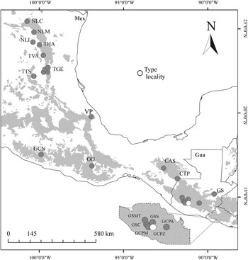
Based on diagnostic characters reported in this study, our collections confirm the presence of D. vitei in Ixtlán, Oaxaca (Lanier et al. Reference Lanier, Hendrichs and Flores1988) and validate records reported by Armendáriz-Toledano et al. (Reference Armendáriz-Toledano, Niño, Macías-Sámano and Zúñiga2014a). In addition, it provides new records from Mexico and Guatemala (Table 1, Fig. 57). MEXICO: Nuevo León, Iturbide municipality, Los Pinos del Sur, Pinus teocote Schlechtendal and Chamisso (Pinaceae); Montemorelos municipality, ejido Aquiles Serdán. Tamaulipas, Gómez Farías municipality, ejido La Esperanza, P. pseudostrobus Lindley and P. teocote; Gómez Farías municipality, ejido El Gavilán, P. patula Schlechtendal and Chamisso; ejido Joya de Manantiales, P. pseudostrobus; ejido los San Pedros, P. pseudostrobus and P. teocote; Hidalgo municipality, ejido Los Ángeles. Guerrero, Chilpancingo municipality, Nixtatipa Lomas de Cuapanco. GUATEMALA: Chimaltenango, San José Pinula, finca Miramundo, P. pseudostrobus; San José Poaquil, Astillero, P. maximinoi Moore. Specimens representative of these location were deposited in the Museo de History Natural de la Ciudad y Cultura Ambiental and Colección Nacional de Insectos del Instituto de Biología, Universidad Nacional Autonoma de México (CNIN), Mexico City, Mexico.
Dendroctonus vitei distribution. Acronyms for locations are defined in Table 1. Mex, Mexico; Gua, Guatemala.

Redescription of Dendroctonus vitei Wood
Diagnosis
Figs. 12–15, 19–21, 26–29, 33–35, 37, 40, 42, 44, 46, 48, 51–56, 58–59.
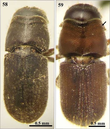
Dorsal view of both male and female adults of Dendroctonus vitei. 58. ♂ from Mexico: Oaxaca: Ixtlán de Juárez: Universidad de la Sierra de Juárez. 59. ♀ from Mexico: Tamaulipas: Gómez Farías: Ejido Joya de Manantiales. Arrow indicates pronotal callus.

Dendroctonus vitei is distinguished from D. mexicanus by means of five diagnostic characters: in both sexes (1) presence of sensillae clustered into cavities resembling pit-craters on the anterior side of the third and fourth antennomeres of the antennal club (Fig. 37); (2) FS with punctures less impressed and crenulations less prominent, less abundant in lateral areas of epistomal process (Fig. 12); (3) setae on elytral declivity with a toothed tip and a saw-toothed outer surface (Fig. 44); (4) male seminal rod anchor with thin arms and lateral edges of arch slightly emarginate, forming two poorly developed lobes (Figs. 51, 52); and (5) seminal rod divided into dorsal and ventral processes. The ventral process consisting of a long, curved, spine-shaped projection larger than dorsal projection in lateral view (Figs. 54–56); ventral process flattened, paddle-like, and with shape resembling a beaver tail in ventral view (Figs. 51, 53). The dorsal process broadly triangular in lateral view (Figs. 54–56), and widely oval and shorter and wider than ventral process in dorsal view (Fig. 53). Both processes parallel (Fig. 54) or perpendicular to each other (Figs. 55–56).
Description of adult male
Total length 3.4–5.2 mm; 2.7 times longer than wide; head and pronotum of same colour or darker than elytra; head black; pronotum and elytra medium-brown to black (Figs. 58, 59); chromosome formula=5AA+Xyp.
Head
Length 0.28–1.05 mm
 $\left( {{\rm \bar{X}}{\equals}0.7\,{\rm mm}} \right)$
, surface of epicranium and vertex covered with small punctures that are deep and variable in diameter (Figs. 12–15), vertex and frons completely convex in lateral view. Frons armed with a pair of transverse elevations surrounded by four to six prominent tubercles and small granules just below upper level of eyes (Figs. 12, 14), length of highest frontal tubercles 0.01–0.075 mm
 $\left( {{\rm \bar{X}}{\equals}0.047\,{\rm mm}} \right)$
; frontal tubercles separated by a weak median groove extending from just above epistomal process to upper level of eyes (Fig. 12); surface of median groove shiny and smooth, some specimens with small punctures on lateral areas of the groove; surface between eyes surrounded by punctures and abundant, weakly crenulate granules (i.e., truncate, weak protuberances) that form a series of nearly concentric, semicircular ridges around the median area of the frons, weakly crenulate granules less impressed and less abundant towards the epistomal region in upper and lateral areas of epistomal process (Fig. 12); area immediately above epistomal process flat or slightly concave, shiny, and with sparse, small punctures. Width of epistomal process 0.37–0.82 mm
 $\left( {{\rm \bar{X}}{\equals}0.56\,{\rm mm}} \right)$
, 0.5 (0.37–0.80) times the distance between eyes
 $\left( {{\rm \bar{X}}{\equals}1.03\,{\rm mm}} \right)$
, arms of epistomal process slightly elevated and oblique ~24° from horizontal (Fig. 12), surface smooth and shiny, some specimens with a few small punctures, underside with a dense brush of yellow setae. Pubescence of head yellow and of three different sizes that are sparsely distributed around frons and less abundant on area immediately above epistomal process (Figs. 12, 15): largest setae distributed on vertex, those of medium size between frons and lateral areas of eyes, shortest setae surrounding epistomal process.
Antenna
Composed of a scape, funicle, and club (Dickens and Payne Reference Dickens and Payne1977). Antennal club articulate with fifth funicular antennomere and composed of four fused antennomere and divided by three bands of sensillae (Fig. 37). On the anterior side of club, 5–18 groups of sensillae are arranged in clusters resembling pit-craters; these are distributed transversely in two, often poorly organised, uneven lines with respect to the proximal-distal axis of the antenna. The proximal line of sensillae clusters located on the third antennomere and the distal line on the third sensorial band between the third and fourth antennomeres (Fig. 37). Clusters of sensillae more numerous (3–13 groups,
 $${\rm \bar{X}{\equals}8}$$
) in the proximal than the distal line (1–6;
 $${\rm \bar{X}{\equals}3}$$
). In both lines, clusters of sensillae similar in diameter, 10.0–27.5 µm
 $\left( {{\rm \bar{X}}{\equals}15.2\,{\rm \mu m}} \right)$
.
Pronotum
Length 0.80–1.87 mm
 $\left( {{\rm \bar{X}}{\equals}1.09\,{\rm mm}} \right)$
, width 1.27–1.90 mm
 $\left( {{\rm \bar{X}}{\equals}1.54\,{\rm mm}} \right)$
, widest on posterior third, anterior region scarcely constricted; dorsal surface with rather coarse, deep, close punctures, and interspaces smooth and shiny (Fig. 19); some dorsal punctures with emarginate edge, which is strongly elevated in punctures located on lateral margins of pronotum; punctures on lateral median areas less defined and give the appearance of smooth and shiny areas; lateral punctures (on preepisternal and episternal areas) shallower, less abundant, and reduced in diameter (Figs. 20–21), in some specimens dull and with fine ridges; pubescence yellow; longer and more dense on dorsal anterior area, pubescence of preepisternal and episternal areas shorter and less abundant than dorsal area (Fig. 19).
Elytra
Length 1.75–2.97 mm
 $\left( {{\rm \bar{X}}{\equals}2.4\,{\rm mm}} \right)$
, ≈ 1.5 times longer than wide, 2.2 times longer than pronotum, sides straight on basal two-thirds, rather broadly rounded behind; declivity convex with striae weakly impressed (Fig. 26); punctures of striations with a diameter ranging from 19–27 µm
 $\left( {{\rm \bar{X}}{\equals}23\,{\rm \mu m}} \right)$
, surface between punctures smooth with a distance between punctures from 12–45 µm
 $\left( {{\rm \bar{X}}{\equals}28.7\,{\rm \mu m}} \right)$
; in ~75% of specimens the diameter of punctures was 1.2 times the size of interspaces between punctures (Figs. 26, 42); declivital interstria II wider
 $\left( {{\rm \bar{X}}{\equals}131\,{\rm \mu m}} \right)$
) than I
 $\left( {{\rm \bar{X}}{\equals}98\,{\rm \mu m}} \right)$
and III
 $\left( {{\rm \bar{X}}{\equals}114\,{\rm \mu m}} \right)$
; declivital interstria II not constricted apically; surface of interstriae with weakly marked rugae, small punctures and crenulations. Punctures on declivital interstriae well defined and smaller than those of striae (Figs. 29, 42); average diameter of interstrial punctures 0.62 times diameter of punctures on striations; crenulations in interstriae of variable size, more defined and abundant in anterior area of elytral disc and scarce or absent towards posterior elytral disc (Figs. 26, 29, 42); width of the crenulations on anterior area of elytral disc from 29–88 µm
 $\left( {{\rm \bar{X}}{\equals}65.7\,{\rm \mu m}} \right)$
≈0.49 times width of interspaces. Width of crenulations on anterior area of elytral declivity from 34–53 µm
 $\left( {{\rm \bar{X}}{\equals}41.0\,{\rm \mu m}} \right)$
, 0.3 times width of interspaces. Setae in interstriae I–III of elytral declivity originating from centre of punctures with more than one size class, shortest about one-third length of longest, and longer punctures showing a gradual progression of sizes; colour yellow to amber in mounted specimens (Fig. 59); most setae (all sizes) on elytral disc and declivity with saw-toothed tip and surface (Figs. 42, 44). Ventral surface of both elytral declivities with a transversely sulcate area covering the suture and apex of each elytron (Figs. 33–34); when the elytra are closed this forms a continuous file-like surface, which makes contact with the stridulating scraper (a bifid process) of the seventh abdominal tergite (propygidium); average length of transversely sulcate area of 592.5 µm, average width of ridges of sulcate area 6.2 µm, total of ~84 ridges; ridge width decreasing from distal to proximal region of sulcate area (Fig. 33).
Genitalia
Composed of four sclerotised structures: tegmen, spicule, penis, and accessory apparatus (seminal rod and seminal rod anchor). General anatomy of tegmen, spicule, and penis similar to that described for Dendroctonus ponderosae Hopkins (as D. monticolae Hopkins) (Cerezke Reference Cerezke1964). Sclerotised plate (seminal rod anchor) positioned dorsally of the seminal rod (Figs. 51–52). This structure displays two roughly parallel, lateral arms that thicken distally and are then joined by an arch. The dorsal process of the seminal rod is enclosed within this U-shaped structure (seminal rod anchor, Fig. 51). Outside, lateral margins of the arch are slightly emarginate forming two poorly developed, ear-like lobes (Figs. 51–52). The seminal rod, consisting of a sclerotised structure connected to the ejaculatory duct, is situated within the seminal rod anchor. Proximal region of seminal rod consisting of a seminal valve, with the distal portion divided into dorsal and ventral processes (Figs. 51–56). Ventral process consisting of a long curved, spine-shaped (in lateral view) projection larger than dorsal one (Figs. 54–56); ventral process flattened, paddle-like, and with shape resembling a beaver tail (in ventral view) (Figs. 51, 53), width of ventral process ~0.4 times the width of dorsal process, dorsal process broadly triangular in lateral view (Figs. 54–56), widely oval and shorter and wider than ventral process (in dorsal view) (Fig. 53); dorsal process from perpendicular (Fig. 55–56) to parallel respect to ventral one (Fig. 54).
Description of adult female
Total length 1.7–5.2 mm
 $\left( {{\rm \bar{X}}{\equals}4.1} \right)$
; 2.7 times longer than wide; body colour similar to male; chromosome formula=5AA+XX.
Head
Length 0.7 mm; surface of epicranium and vertex covered with well-defined punctures, deep and variable in diameter; less abundant and smaller than in males; shape of vertex and frons in lateral view similar to male. Frons with slight, transverse elevations covered with small granules and without prominent tubercles of male; transverse elevations separated by a median groove as in males; surface of median groove shiny or with scarce, small punctures; punctures and granules distributed around the median groove less abundant and smaller than in male; distribution of punctures and granules between eyes similar to males; area immediately above epistomal process slightly concave (some specimens flat), shiny and with more sparse and smaller punctures than in males; width of epistomal process (0.28–0.75 mm (
 $\left {{\rm \bar{X}}{\equals}0.4\,{\rm mm}} \right$
)) narrower than in males, 0.44 times the distance between eyes (
 ${\rm \bar{X}}{\equals}0.7\,{\rm mm}$
; 0.48–0.97 mm); arms of epistomal process slightly elevated or almost flat, oblique and about 24° from horizontal, surface smooth and shiny with punctures less abundant and smaller than in male; underside with a less dense brush of setae than in male, pubescence of head slightly more abundant than male, size and distribution of setae similar to male.
Pronotum
Length from 0.7–1.3 mm
 $\left( {{\rm \bar{X}}{\equals}1.1} \right)$
, width from 1.2–1.8 mm
 $\left( {{\rm \bar{X}}{\equals}1.5} \right)$
, widest on posterior third, anterior region slightly constricted; pronotal constriction with a transverse callus that is slightly bulging both laterally and dorsally in most specimens (90%, n=40) but evidently bulging in others (10%, n=40); vestiture and sculpture of pronotum similar to males.
Elytra
Length from 2.0–2.9 mm
 $\left( {{\rm \bar{X}{\equals}2}{\rm .5}} \right)$
, 1.6 times longer than wide, 2.3 times longer than pronotum; sides parallel on basal two-thirds, rather broadly rounded behind; declivity convex with striae weakly impressed; vestiture and sculpture of elytral declivity similar to males.
Eighth tergite
Segment characterised by an oval distal edge and two oval proximal lateral edges converging on an anterior median line. Distal surface of tergite is covered by abundant setae of different sizes. Eighth tergite displays parallel lines of squamiform plates of sclerotised cuticle. Lines better defined in anterior area and fade in posterior area. Squamiform plates possess from three to seven acute distal projections
 $\left( {{\rm \bar{X}{\equals}4}{\rm .7}} \right)$
of variable size (Fig. 46).
Spermatheca
Reniform, divided into a proximal nodulus and distal cornu. The surface of nodulus transversely covered by striations, becoming confused in distal region of nodulus; striations in the proximal region of nodulus not aggregate; collectively, striations cover almost entire nodulus; cornu displaying an oval shape in lateral view, as long as nodulus or longer than it (Fig. 48).
Distribution
This species is present in the departments of Chimaltenango and Sololá (Cordillera Central) in Guatemala, and in the states of Chiapas (Sierra Madre de Chiapas), Oaxaca and Guerrero (Sierra Madre del Sur), Veracruz (Eje Volcánico Transmexicano), and Tamaulipas and Nuevo León (Sierra Madre Oriental) in Mexico (Fig. 57).
Natural history
Dendroctonus vitei has been recorded in Mexico at elevations lower than 2500 m, typically between 1000–1500 m; in pines from the section Trifoliae: P. maximinoi, P. michoacana Martínez, P. montezumae Lambert, P. oocarpa Schiede ex Schlechtendal, P. pseudostrobus, and P. teocote (Vité et al. Reference Vité, Hughes and Renwich1975; Atkinson and Equihua-Martínez Reference Atkinson and Equihua-Martínez1985; Armendáriz-Toledano et al. Reference Armendáriz-Toledano, Niño, Macías-Sámano and Zúñiga2014a).
The status of D. vitei as a forest pest has not been evaluated, however it has commonly been recorded in infestations occurring in Central America; for example, those in Chimaltenango, Guatemala in 1973 and 1974 that were attributed to D. adjunctus (Vité et al. Reference Vité, Hughes and Renwich1975). Our observations indicate that D. vitei can attack and kill healthy trees. We have found this species in Oaxaca and Nuevo León states colonising one or two trees, but not forming multi-tree infestations.
Dendroctonus vitei can coexist in sympatry or syntopy with other species of the genus: D. adjunctus in Guatemala (Vité et al. Reference Vité, Hughes and Renwich1975), D. frontalis (Lanier et al. Reference Lanier, Hendrichs and Flores1988) in Oaxaca, and D. mexicanus in Veracruz, Guerrero, and Nuevo León (Armendáriz-Toledano et al. Reference Armendáriz-Toledano, Niño, Sullivan, Kirkendall and Zúñiga.2015). In syntopy with D. mexicanus, D. vitei has been collected at the base of the stem, 1–2 m from the ground, however the brood galleries of both species show spatial overlap.
Dendroctonus vitei galleries have an S-shape; they cross each other frequently or may be parallel when in close proximity, with typical width from 2.0–2.5 mm and length from 30–100 cm (Vité et al. Reference Vité, Islas, Renwich, Hughes and Kliefoth1974; F.A.T., unpublished data). Most galleries extend in a downward direction from the entrance hole, and changes in gallery direction often coincide with the presence of thin bark or the crossing of another gallery (Vité et al. Reference Vité, Islas, Renwich, Hughes and Kliefoth1974; F.A.T., unpublished data). In Ixtlán, Oaxaca most of the D. vitei galleries we observed ran on the longitudinal axis of the bole.
The host may be colonised from the ground up to the lower crown; the attack density seems to be highest at 2–3 m above the ground (Vité et al. Reference Vité, Islas, Renwich, Hughes and Kliefoth1974). In Oaxaca, Veracruz, Nuevo Leon, and Guatemala it has been observed that D. vitei prefers large trees, although it has been documented in pines as small as 4.4 cm diameter at breast height (Vité et al. Reference Vité, Islas, Renwich, Hughes and Kliefoth1974; F.A.-T., personal observation).
Vité et al. (Reference Vité, Islas, Renwich, Hughes and Kliefoth1974) reported that development of one generation required 60 days under laboratory conditions. Females can deposit up to 198 eggs with a sex ratio of 1.4♂:1.0♀ (n=63). The egg incubation period is from 7–8 days, larval development requires 33 days, pupal stage seven days, and adult maturation from 12–14 days. Four instars have been identified.
Renwick et al. (Reference Renwick, Hughes and Vité1975) extracted hindgut volatiles from D. vitei in Patzun, Guatemala and analysed them using gas chromatography and mass spectrometry. They found that emerged and attacking females produced cis and trans-verbenol, and attacking females additionally produced frontalin. Emerged males produced myrtenol and large quantities of 1-phenylethanol. Attacking males produced myrtenol but traces 1-phenylethanol, 1-heptanol, 2-heptanol, and linalool were detected from attacking individuals of both sexes. To corroborate these results, we extracted hindguts of emergent male D. vitei collected in P. pseudostrobus near San José Poaquil, Chimaltenango, Guatemala, and analysed the extracts by capillary-column gas chromatography/mass spectrometry (see Supplementary Material). We duplicated the finding of Renwick et al. (Reference Renwick, Hughes and Vité1975) that emergent male beetles contained very large quantities of 1-phenylethanol (from 0.41 to 6.3 µg/beetle) and lesser amounts of myrtenol (~1/60 the concentration of 1-phenylethanol), but no frontalin or brevicomin. In addition, we detected traces of verbenone, alpha-terpineol, linalool, terpinen-4-ol, and acetophenone. The preponderance of 1-phenylethanol in emergent males has not been reported from any other studied species of Dendroctonus (Symonds and Elgar Reference Symonds and Elgar2004; Sullivan et al. Reference Sullivan, Niño, Moreno, Brownie, Macías-Sámano and Clarke2012), and may be a diagnostic, biochemical character for D. vitei. Renwick et al. (Reference Renwick, Hughes and Vité1975) additionally reported that D. vitei specimens were attracted to sticky traps baited with frontalin and attached to mechanically injured pines, suggesting frontalin can be responsible for the aggregation of this species. Synthetic frontalin attracted predominantly males, and 1-phenylethanol and the heptanols apparently had some regulatory effect on the response to frontalin. Myrtenol and trans-verbenol had no apparent effect on the response to frontalin. These results should be viewed cautiously as release rates were not reported and secondary attractants could potentially have been produced from natural beetle attacks on the trap-trees.
We propose the common name Guatemalan pine beetle for D. vitei, as the species was originally described from Guatemala.
Acknowledgements
The authors are grateful to Miguel Ángel Barrios for his comments and suggestions on an earlier version of this manuscript. They are also grateful to two anonymous reviewers. The authors thank Ariana Guadalupe Robles and Jessica B. Valencia (Servicio Nacional de Sanidad Inocuidad y Calidad Agroalimentaria) and Marco Ramírez (Carl Zeiss, Mexico) for technical assistance in obtaining environmental scanning electron microscope photographs. Many thanks to Julio César Concha (Universidad de la Sierra de Juárez) and Eduardo Jiménez Quiroz (Laboratorio de Análisis de Referencia en Sanidad Forestal de la Secretaria de Medio Ambiente) for their assistance in the field and laboratory and for providing specimens. This study was funded by the Secretaria de Investigación y Posgrado Instituto Politécnico Nacional (20161790). J.G.R. and F.A.T. were Consejo Nacional de Ciencia y Tecnología (CONACyT) fellows (608539, 267436); and F.A.T. was a member of the Programa Institucional de Formación de Investigadores of Instituto Politécnico Nacional (BEIFI-IPN).
Supplementary Material
To view supplementary materials for this article, please visit https://doi.org/10.4039/tce.2017.10