Introduction
During preimplantation development, the bovine embryo passes through proliferation and differentiation stages in order to assure implantation and fetation. Among those stages, compaction and blastocoels formation leading to the blastocyst at day 7 after fertilization and elongation are essential. During this period, the first decisions about cell fate occur, with the formation of the inner cell mass (ICM) and the trophoblast (TB), (Viebahn, Reference Viebahn1999; Degrelle et al., Reference Degrelle, Campion, Cabau, Piumi, Reinaud, Richard, Renard and Hue2005). The ICM mostly gives rise to the embryonic tissue, whereas the TB gives extraembryonic tissues. Later the embryo elongates prior to implantation. This peri-implantation stage is characterised by an active proliferation of trophoblastic cells leading to the morphological transition of the embryo from a sphere to a filamentous shape (Degrelle et al., Reference Degrelle, Campion, Cabau, Piumi, Reinaud, Richard, Renard and Hue2005). In bovine embryos, trophoblast elongation starts around day 12 after fertilization and the filamentous stage is complete around day 18 (Bazer et al., Reference Bazer1992; Guillomot et al., Reference Guillomot, Turbe, Hue and Renard2004) at which the conceptus size increases more than 1000 fold (Maddox-Hyttel et al., Reference Maddox-Hyttel, Alexopoulos, Vajta, Lewis, Rogers, Cann, Callesen, Tveden-Nyborg and Trounson2003) and the increase of cell number is accompanied by increased protein synthesis (Thompson et al., Reference Thompson, Sherman, Allen, McGowan and Tervit1998; Degrelle et al., Reference Degrelle, Campion, Cabau, Piumi, Reinaud, Richard, Renard and Hue2005).
These dramatic and rapidly occurring changes are tightly regulated at the gene expression level. Of special relevance is the expression of genes that code for pluripotency markers and that maintain the undifferentiated stage of the cells. OCT4, SOX2 and NANOG are expressed mainly in the inner cell mass of day-7 blastocysts and in the embryonic disc (ED) of elongated bovine embryos and are associated with the pluripotency of the embryo (Degrelle et al., Reference Degrelle, Campion, Cabau, Piumi, Reinaud, Richard, Renard and Hue2005). Conversely IFN-τ and TKDP, and to a lesser extent HAND1, PAGs and MASH2, are recognized as markers of trophoblast development (Hernandez-Ledezma et al., Reference Hernandez-Ledezma, Sikes, Murphy, Watson, Schultz and Roberts1992; Wrenzycki et al., Reference Wrenzycki, Wells, Herrmann, Miller, Oliver, Tervit and Niemann2001; MacLean et al., Reference MacLean, Chakrabarty, Xie, Bixby, Roberts and Green2003). In spite of the augmented expression documented for these trophoblastic genes during elongation, their transcription starts before, at blastocyst stage (Arnold et al., Reference Arnold, Bordignon, Lefebvre, Murphy and Smith2006).
The expression level of specific genes changes from blastogenesis to implantation Ushizawa et al., (Reference Ushizawa, Herath, Kaneyama, Shiojima, Hirasawa, Takahashi, Imai, Ochiai, Tokunaga, Tsunoda, Tsujimoto and Hashizume2004) reported in bovine embryos more than 500 genes are up-regulated between day 7 versus day 14, whereas only 26 genes were down-regulated in the same time period. Likewise, differential gene expression was shown in bovine embryos during elongation (Degrelle et al., Reference Degrelle, Campion, Cabau, Piumi, Reinaud, Richard, Renard and Hue2005; Rodríguez-Alvarez et al., Reference Rodriguez-Alvarez, Cox, Navarrete, Valdés, Zamorano, Einspanier and Castro2009). Recent research using somatic cell nucleus transfer (SCNT) technology suggested that aberrant expression of genes during the earliest stages of development could be the cause of embryonic losses (Arnold et al., Reference Arnold, Fortier, Lefebvre, Miglino, Pfarrer and Smith2008). Aberrant gene expression might be responsible for the observed phenotypes in embryos produced by SCNT, not only during early embryonic development, but also during gestation and parturition leading in many cases to the so called abnormal offspring syndrome (AOS) (Garry et al., Reference Garry, Adams, McCann and Odde1996; Walker et al., Reference Walker, Hartwich and Seamark1996; Ranilla et al., Reference Ranilla, Gebbie, King, Carolan, Sinclair, Watt, Dolman, Beckers and Robinson1998; Zhan et al., Reference Zhan, Kubota, Yang, Zhang, Page and O'Neill2004). Incomplete reprogramming of the donor cell could be the cause of aberrant patterns of gene expression first manifested in bovine blastocyst, but lasting throughout embryonic development (Latham & Sapienza, Reference Latham and Sapienza2004; Piedrahita et al., Reference Piedrahita, Mir, Dindot and Walker2004; Arnold et al., Reference Arnold, Fortier, Lefebvre, Miglino, Pfarrer and Smith2008). Aberrant gene reprogramming after nucleus transfer has been documented also for miRNA (Castro et al., 2009). This finding implies that de-regulated gene expression is a common phenomenon in cattle cloning.
Comparative data of gene expression for candidate genes at day 7 (blastocyst) versus day 17 (filamentous elongated) bovine embryos are not available, especially for nucleus transfer embryos, and few studies have addressed the switch of gene expression from blastogenesis to elongation in cattle. This scarcity is probably related to difficulties and costs associated with the production of elongated embryos in vitro or via SCNT. Recently we and others have developed systems for elongation of ruminant cloned embryos using heterologous recipients as intermediate hosts (Rodríguez-Alvarez et al., Reference Rodriguez-Alvarez, Cox, Navarrete, Valdés, Zamorano, Einspanier and Castro2009; Rodríguez-Alvarez et al., submitted). This approach can be an attractive model to study changes in gene expression during elongation, however the contribution of a heterologous uterine environment to correct embryonic gene expression requires still further research. We hypothesize that expression of pluripotency markers is down-regulated as development proceeds toward elongation. To test this hypothesis, the goal of this study was to study the expression of several developmentally crucial genes at the blastocyst stage, and to compare it with the expression of the same genes at day 17 of development, during filamentous stage using bovine cloned and in vitro produced embryos. For this, a real-time quantitative PCR approach was used. We found changes in the expression of candidate genes during the analysed period. This finding can have implications for understanding the switch from blastogenesis to elongation at the gene level in cattle.
Materials and Methods
Oocyte collection and in vitro maturation (IVM)
All reagents and media except otherwise stated were from the Sigma Chemica Co.
Oocyte collection and maturation were perfomed as described earlier (Rodriguez et al., 2008). In brief, cumulus–oocyte complexes (COCs) were aspirated from medium-sized (3 to 8 mm diameter) follicles from bovine ovaries. The COCs were in vitro matured in TCM199 Earle salts supplemented with follicle stimulating hormone (FSH) and luteinizing hormone (LH) (0.01 U/ml each), 17β-oestradiol (1 μg/ml), epidermal growth factor (10 ng/ml) and 10% of fetal bovine serum (FBS) at 39°C in a 5% CO2 in air atmosphere. Oocytes were matured for 21 h or 23 h for nuclear transfer (NT) procedure or IVF respectively.
Generation of NT embryos
Nuclear transfer was accomplished via handmade cloning (HMC) (Peura & Vajta, Reference Peura and Vajta2003) with minor modifications made in our laboratory (Rodríguez et al., Reference Rodríguez, Navarrete, Tovar, Cox and Castro2008). As karyoplasts we used a cell line from skin fibroblasts of adult Wagyu cattle (CWA). The cell line had yielded previously live cloned calves (Rodríguez et al., unpublished). Cells in passages three to five were grown to full confluence and left undisturbed for at least 3 days without serum starvation. For experiments involving quantification of OCT4 gene expression we used additionally a fetal cell line from a 50-day bovine fetus (LCF). This cell line was used in the same passages and conditions as CWA cells.
After 21 h of maturation, oocytes were denuded by vortexing briefly in TCM199 + 0.3 mg/ml hyaluronidase. Guided enucleation was conducted and oocytes were incubated for 2 h in TCM199 + 2.5 μg/ml demecolcine, to induce the protrusion of the meiotic cones. After this process, the zona pellucida was removed by brief pronase treatment (2 mg/ml for less than 1 min). Each oocyte was bisected under stereoscopic observation with a microblade (Ultra Sharp Splitting Blades; AB Technology) and the enucleated halves were used as cytoplasts receptors. For embryo reconstruction two demi-oocytes were attached to a single somatic cell by very brief incubation with phytohemagglutinin (1 mg/ml). The triplet was fused with a single direct current pulse of 2 kV/cm for 9 μs in manitol (0.3 M) using an electrofusion device (BLS). After fusion, nuclear reprogramming was allowed for 2 h in TCM199 supplemented with 10% FBS. Activation was induced by 7% ethanol in TCM199-HEPES for 5 min, followed by 5 h incubation in TCM199 without HEPES plus cycloheximide (10 μg/ml) and cytochalasin B (5 μg/μl) at 39°C in a 5% CO2 in air atmosphere (Rodriguez et al., 2008).
Embryo culture was carried out in synthetic oviduct fluid (SOFaci) supplemented with 0.37 mM tri-sodium citrate, 2.77 mM myo-inositol, essential and non-essential amino acids (final concentration 1×), gentamycin (50 μg/ml), 3 mg/ml essential fatty acid-free bovine serum albumin (BSA) fraction V, 0.2% FBS and 10 ng/ml epidermal growth factor (EGF). Cloned embryos without zona pellucida were cultured for 7 days by the ‘well-on-the-well’ system (WOW) (Vajta et al., Reference Vajta, Peura, Holm, Páldi, Greve, Trounson and Callesen2000) of a 4-well dish (Nalge Nunc International) in a balanced gas atmosphere consisting of 5% CO2, 5.5% O2 and 89.5% N2, 100% humidity at 39°C.
Generation of in vitro fertilized (IVF) embryos
After 23 h of maturation oocytes were in vitro fertilized with thawed semen from a single bull of proven fertility. The motile fraction of the semen was obtained using swim-up procedure in Tyrode's albumin lactate pyruvate (TALP)-HEPES (containing 2 mM sodium pyruvate, 50 μg/ml gentamycin and 3 mg/ml of BSA Fraction V) for 1h at 37°C. Approximately, 1 × 10⁶ motile sperms/ml were added to fertilization drop and incubated with 25 COCs for 24 h at 39°C in 5% CO2 in air atmosphere. The fertilization was carried out in TALP medium supplemented with 2 mM sodium pyruvate, 50 μg/ml gentamycin, 0.01 mg/ml heparin sodium salt, 5 mM glucose and 6 mg/ml of BSA fraction V (essential fatty acid free). Oocytes were denuded by brief incubation in TCM199-HEPES with 0.3 mg/ml of hyaluronidase and gentle swirling. Embryos were cultured in a 4-well dish (Nalge Nunc International) under the same conditions described for NT embryos.
Embryo transfer and recovery of day-17 elongated embryos
Day-7 IVF or HMC blastocysts produced with adult cells were transferred non-surgically to synchronic recipient heifers according to conventional procedures published elsewhere (De Armas et al., Reference De Armas, Solano, Riego, Pupo, Aguilar, Ramos, Aguirre, de la Fuente and Castro1994). Oestrus synchronization was performed using a controlled intravaginal drug release (CIDR) device (EAZI-Breed™) for 7 days and injection of PGF-2α the day that the device was removed.
A total of 30 expanded blastocysts produced by HMC were transfered to six recipients (five embryos per recipient) whereas 11 IVF embryos to two recipients (five and six embryos per recipient). Ten days after embryo transfer (day-17 of development) heifers were slaughtered, their uteri collected and flushed twice with 50 ml ice-cold Dulbecco PBS supplemented with 6% BSA fraction V. Six elongated embryos from each treatment (HMC and IVF) with visible embryonic disc were used for gene expression analysis. Two groups (treatments) of embryos were created: HMC (embryos produced by hand made cloning) and IVF (embryos produced by in vitro fertilization).
Single or pooled embryos treated in this way were left overnight at 4°C, in a 1.5 ml Eppendorf tube in RNALater solution (Ambion Inc.) and placed at –80°C until further use.
Gene expression analysis
mRNA isolation
Pre-implantation: Day-7 blastocysts derived from CWA cells were pooled in four groups of ten embryos each for mRNA extraction, cloned blastocysts derived from the fetal cell lines were pooled in two groups of ten each. IVF blastocysts were pooled in two groups of 10 each. Total RNA was isolated using a NucleoSpin RNA XS Kit (NucleoSpin RNA XS) according to instructions in the kit. Elution was done in 20 μl of diethylpyrocarbonate (DEPC)-treated water and the eluted product was stored at –80°C until use.
Peri-implantation: Day-17 elongated embryos with embryonic disc were used. Total RNA from elongated embryos was extracted using mirVana™ miRNA Isolation Kit, (Ambion Inc.). Total RNA was quantified using NanoDrop technology and the quality was assessed with Agilent 2100 Bioanalyzer (RNA Nanochip; Agilent). Six elongated bovine cloned and six elongated bovine IVF embryos were analysed.
Donor cells: Three confluent P60 dishes from each cell line were used. The culture medium was removed from the dishes. Cells were washed three times with cold PBS, scrapped in 1 ml ice-cold PBS, spinned down on a cold centrifuge, and the pellet used for total RNA extraction as described for the elongated embryos.
Reverse transcription
For cDNA conversion, 8 μl from blastocysts or 1 μg of total RNA form elongated embryos and donor cells were treated with 1 U of RNase-free DNase I (Boehringer) in a 10 μl reaction for 30 minutes at 37°C. The enzyme was heat-inactivated (65°C for 10 min) in the presence of 25 mM de EDTA (1 μl). Reverse transcription (RT) was performed as described earlier (Rodríguez et al., Reference Rodríguez, Navarrete, Tovar, Cox and Castro2008). In brief, the total volume of the DNase I treatment (11 μl) was used in a 20-μl final reaction containing 30 ng/μl random primers, 10 mM each dNTP, 4 μl first-strand buffer (5×), 200 U/ml M-MuLV (Fermentas GmbH) and DEPC-treated water. Cycling parameters were: 25°C for 10 min, 42°C for 60 min and 70°C for 10 min. The produced cDNAs were kept frozen at –20°C until use in PCR experiments.
PCR
Five genes important for embryonic development (OCT4, NANOG, IFN-τ, EOMES and FGF4) were studied by real-time quantitative PCR (qPCR) in both blastocysts and elongated embryos. ACTB was used as internal control and GDF9 as negative control.
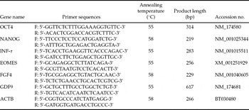
The composition of primers and PCR conditions for each assayed gene are provided in Table 1. Real-time PCR reactions were run in a StepOnePlus™ device (Applied Biosystems) and the product were detected with SYBRGreen. The qPCR reaction contained 5 μl of 2× SensiMix DNA Kit and 0.2 μl of 50× SYBRGreen (Quantace Ltd.) in a final volume of 10 μl. Standard curves for each qPCR were prepared using PCR products excised and eluted from agarose gels using Invisorb Spin DNA Extraction Kit (Invitek GmbH) and quantified by NanoDrop ND-1000. Serial dilutions of PCR products were prepared and 2 μl were used in duplicates for each point of the standard curve. Samples were also loaded as duplicates for the qPCR. The Crossing Point (CP) was calculated by the built-in software.
Oligonucleotide composition and PCR conditions for the analysed genes

Sequencing of OCT4 PCR product
PCR product obtained with OCT4 primers from cloned and IVF blastocysts and donor cells, was extracted from agarose gel using a Invisorb Spin DNA Extraction Kit (Invitek GmbH) and PCR-sequenced using a BigDye® Terminator v1.1/3.1 kit (Applied Biosystems).
Immunohistochemistry
Cells were grown on pre-coated gelatinized glass slides submerged in DMEM:F12 medium supplemented with 10% FCS and antibiotics/antimycotics. After filling the slide totally, the cells were permeabilized for 30 min in PBS containing 1% Triton X-100 and 0.25% BSA and subsequently washed carefully three times in PBS alone (3 min each time). Endogenous peroxidase was blocked with 3% H2O2 in PBS for 30 min, followed by three more washes in PBS. For de-masking OCT4 epitopes, we incubated the slides with cells in citrate buffer pH 6.0, three times 5 min each at 65°C with 20 min of cooling to RT between each incubation. Further, blocking of the slide was performed (10 min, using DAKO protein blocking kit; Gene Express). A primary anti-OCT4 polyclonal antibody (Santa Cruz) was diluted 1:250 in PBS containing 0.1% triton X + 0.25% BSA and left overnight at 4°C over a small circular section of the slide drawn with repellent marker. The slide was placed on a humid chamber. Secondary anti-human IgG-antibody conjugated with peroxidase (Santa Cruz) was used at 1:500 dilutions at 37°C for 2 h. The reaction was developed and stopped using DAKO kit. Slides were mounted and viewed under microscope at different magnifications (see Fig. 2 for details).
Statistical analysis
The mRNA expression level data was analysed by Kruskal–Wallis test. Statistical analysis was conducted using InfoStat. Differences were considered significant at p < 0.05.
Results
Embryo production
For the experiments described here a total of 17 cloning sessions were made. As a resume, 81 blastocysts (day 7) for the fetal and 155 for the adult cell line were produced; the blastocyst rate was 38.2 and 65.76% respectively. Only part of these embryos (n = 40; CWA and n = 20 fetal cell line) were used in qPCRs. IVF embryos resulted from four replicates that yielded 31 day-7 blastocysts (32%), of these 20 were used in the qPCRs. Additionally 30 expanded blastocysts produced by HMC from the CWA cell line were bulk-transferred to six recipients (five/recipient) as well as 11 IVF embryos to two recipients (five and six embryos per recipient). After flushing at day 17, elongated embryos were recovered [n = 19 (63%) for HMC and n = 6 (54.5% for IVF)]. Only six elongated filamentous embryos with embryonic disc were used from each group (HMC-CWA and IVF; no embryos from the fetal cell line were transferred).
Gene expression in preimplantation bovine cloned and IVF embryos
Forty excellent and expanded cloned day-7 blastocysts produced with CWA and 20 IVF embryos of the same quality in pools of 10 embryos were used for gene expression analysis of OCT4, NANOG, EOMES, FGF4 and IFT-τ, plus one internal control (ACTB) and a negative control (GDF9). As a result in conventional PCR, it was detected amplification for all the studied genes but EOMES (data not shown). Interestingly in the conventional PCR assays, OCT4 was detected in the adult cell line used for nucleus transfer, as well as in the fetal (LCF) cell line used as control. Transcripts of NANOG, FGF4, EOMES and IFN-τ were not readily detectable in donor fibroblasts cell cDNA.
The OCT4 PCR product from the blastocysts (both cloned and IVF) and from the cell line was sequenced. The sequence obtained displayed 99% homology with the reported bovine OCT4 sequence (NCBI number: AY490804.1; NM_174580.1). The alignment of sequence from cells (CWA) and embryos (clones and IVF) showed 99% of identity among them. NANOG and IFN-τ PCR products were also sequenced and also showed more than 99% homology with the corresponding bovine genes.
The relative presence of the transcripts was quantified using quantitative real time PCR (qPCR). In this assay, OCT4 was expressed at higher levels (p < 0.05) in the cloned embryos when compared with IVF (Fig. 1a) whereas no significant differences in gene expression at the blastocysts stage, was found for any of the other four genes studied. For comparison, the expression level of OCT4 was quantified in the adult cell line used for cloning as well as in a control fetal cell line (Fig. 1b).
Results of the quantitative PCR in: (a) day 7 or day 17 cloned and IVF bovine embryos, for FGF4, IFN-τ, OCT4, EOMES and NANOG genes. (b) The somatic cells used for nucleus transfer: CWA: adult cell line; FCL: fetal cell line (only OCT4 quantification is showed for the cells). Values on the Y axis are expressed as fg of target/fg of ATCB.

Gene expression in peri-implantation (elongated) cloned and IVF bovine embryos
Using conventional PCR (not shown) expression was demonstrated in day-17 elongated bovine cloned and IVF embryos of all the five genes assayed. However, the main goal of this research was to compare and to quantify the expression levels of these genes in the blastocysts and in elongated embryos, thus a quantitative gene expression analysis was conducted with day-17 elongated cloned and IVF embryos. Elongated embryos were efficiently generated from an adult cell line of bovine fibroblasts as well as from IVF. In separate experiments, this adult cell line yielded approximately 10% live-born cloned cattle (calculated from the number of embryos transferred to recipients; Rodríguez-Alvarez et al., submitted).
Six cloned and six IVF elongated embryos with visible embryonic disc were selected for this experiment. All the quantification on individual samples was performed three times. All genes were expressed across the samples, this included EOMES, which was not detected earlier in blastocysts. Quantification of gene expression in elongated embryos showed statistically significant differences for EOMES, NANOG and FGF4. In all three cases, the expression was higher in the IVF embryos (p < 0.05). IFN-τ and OCT4 were expressed at similar levels among the elongated embryos in spite of their origin (cloned or IVF).
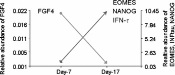
Quantified levels of expression for all genes were compared for blastocysts and elongated embryos. As a result, there were changes in the expression levels for all transcripts between the two stages. Expression of NANOG, IFN-τ and EOMES was increased in the elongated embryos (p < 0.05). Expression of FGF4 conversely dropped significantly from blastocysts to elongation, while expression of OCT4 dropped drastically in the cloned elongated embryos, but not in the IVF (Figure 2). ACTB expression (not shown) was kept constant.
General trend of the changes observed in expression of studied markers in day 7 versus day 17 bovine embryos (IVF and cloned). Values for the relative abundance of EOMES, NANOG and IFN-τ are combined and merged in the same arrow line, while the values for FGF4, expression are depicted separately. OCT4 was not included since its expression differed among cloned and IVF embryos. For this figure, the relative values for each transcript were pooled between IVF and cloned embryos for a given time point (day 7 or day 17). Values on the Y axis are expressed as fg of target/fg of ATCB.

Expression of OCT4 gene in donor cells
OCT4 gene expression did not follow a common pattern between cloned and IVF embryos. In order to ascertain whether this could be related to the observed expression of OCT4 gene in the donor cells, it was tested its expression in the cell line CWA at the protein level using polyclonal antibodies against human OCT4. Immunohistochemistry data showed unequivocally that CWA cells did express OCT4 (Figure 3). Sequencing and protein data corroborated that OCT4 expression was real and not from a pseudogene. Interestingly another cell line, of fetal origin also showed OCT4 expression at the qPCR level, although at much lower levels than the adult CWA cell line.
Immunohistochemical staining of bovine adult cells (CWA) used for nucleus transfer. Primary antibody: goat polyclonal antibody against human OCT4, secondary antibody, anti-goat IgG conjugated with peroxidase. Working dilutions are indicated in the materials and methods section. Left panel: cells expressing OCT4. Right panel: CWA cells incubated with a non-related primary antibody and revealed with the same secondary antibody. Amplification ×100.

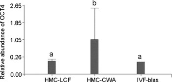
We inferred that the higher OCT4 expression detected in the cloned embryos derived from adult somatic cells could be related with OCT4 gene expression in these cells. In order to ascertain this, 20 cloned blastocysts from the fetal cell line, which expressed much lower levels of OCT4, were analysed in qPCR experiments. The blastocyst rate obtained from these cells upon nucleus transfer was also much lower (38.2%; 81/out of 212) than in the CWA cells (65.7%; 155/out of 236) (p < 0.05). In qPCR experiments, using 20, day 7 blastocysts from the fetal cell line, we found lower expression of OCT4 (p < 0.05) when compared with CWA-derived blastocysts (Figure 4).
Quantification of the expression levels of OCT4 in bovine nucleus transfer expanded (day 7) blastocyst generated from a fetal cell line (HMC-LCF) or an adult cell line (HMC-CWA) and in IVF bovine control blastocysts (IVF-blast). Values on the Y axis are expressed as fg of target/fg of ATCB. Different letters in individual columns imply statistical significant differences (p < 0.05).

Discussion
Differences in expression pattern between bovine cloned and IVF expanded day-7 blastocysts
This study was aimed at determining the potential differences in gene expression of developmentally important genes related to early embryonic development and pluripotency in SCNT and IVF embryos. The transcription of five genes was studied (OCT4, FGF4, NANOG, EOMES and IFN-τ) in bovine preimplantation and elongated embryos produced by IVF and handmade cloning. In the IVF blastocysts, all transcripts except EOMES were detected, therefore their expression in the SCNT embryos can be indicative of genomic activation and adequate nuclear reprogramming of the donor cell, especially considering that none of these transcripts (with exception of OCT4) was detected in the donor cells.
In these experiments, it was found that only OCT4 expression was significantly higher in cloned blastocysts when compared with IVF. No similar reports have been published for OCT4 and bovine cloned blastocysts. Daniels et al. (Reference Daniels, Hall and Trounson2000), using bovine granulose cells as donors, found dissimilar results: OCT4 expression had a similar pattern in both IVF and SCNT blastocysts. Similar levels of OCT4 expression in bovine cloned and IVF blastocysts have been reported by others (Hall et al., Reference Hall, Ruddock and French2005; Amarnath et al., Reference Amarnath, Li, Kato and Tsunoda2007; Long et al., Reference Long, Cai and He2007; Zhou et al., Reference Zhou, Xiang, Walker, Farrar, Hwang, Findeisen, Sadeghieh, Arenivas, Abruzzese and Polejaeva2008). Likewise reduction of OCT4 expression in bovine cloned blastocysts has been published (Misica-Turner et al., Reference Misica-Turner, Oback, Eichenlaub, Well and Oback2006; Beyhan et al., Reference Beyhan, Forsberg, Eilertsen, Kent-First and First2007). Variation in methylation level of OCT4 was also reported for cloned bovine embryos (Kremenskoy et al., Reference Kremenskoy, Kremenska, Suzuki, Imai, Takahashi, Hashizume, Yagi and Shiota2006) but so far no increase of OCT4 expression have been documented for bovine cloned blastocysts. In mice, it is generally accepted that OCT4 is down-regulated in cloned preimplantation embryos (Boiani et al., Reference Boiani, Eckardt, Scholer and McLaughlin2002, Reference Boiani, Eckardt, Leu, Scholer and McLaughlin2003, Reference Boiani, Gentile, Gambles, Cavaleri, Redi and Scholer2005; Bortvin et al., Reference Bortvin, Eggan, Skaletsky, Akutsu, Berry, Yanagimachi, Page and Jaenisch2003). The differences in OCT4 expression between the two species might be consequence of cellular distribution of its transcripts.
In bovine embryos, OCT4 is expressed both in the embryo proper inner cell mass as in the trophoblast during early elongation, this can be related with the intense trophoblastic proliferation that occurs during elongation (Keefer et al., Reference Keefer, Pant, Blomberg and Talbot2007). OCT4 gene expression is silenced at the end of elongation (Degrelle et al., Reference Degrelle, Campion, Cabau, Piumi, Reinaud, Richard, Renard and Hue2005). One possible explanation for this elevated expression of OCT4 in the cloned embryos produced from the adult cell line described here might be the expression of the gene in the donor cells. Probably the regulatory mechanisms of the growing blastocysts are unable to compensate for the constitutive levels of OCT4 present in the cells. This also indicates an inadequate nuclear reprogramming at least for this gene. Interestingly, the bovine cloned blastocyst created from a fetal cell line that expressed very low levels of OCT4, showed a similar pattern of expression like IVF embryos. Also remarkably, the blastocysts rate with the fetal cell line was almost half of the rate obtained with the adult cell line, which expressed high levels of OCT4. We speculate that the high levels of cellular OCT4 are not completely reprogrammed upon nucleus transfer and also are not detrimental for early embryonic development in cloned bovine embryos. Similar to these results Bortvin et al. (Reference Bortvin, Eggan, Skaletsky, Akutsu, Berry, Yanagimachi, Page and Jaenisch2003) showed that mouse blastocysts produced by nucleus transfer from ES cells which expressed OCT4 were able to rescue OCT4 expression in 100% of the cases, while somatic cell clones from cumulus cells, could only partially reprogram OCT4 expression.
Expression of EOMES was not detected here in bovine blastocysts, whereas Hall et al., (Reference Hall, Ruddock and French2005) found it by RT-PCR in IVF bovine blastocysts; however this may be due to culture conditions or the in vitro embryo production system rather than natural expression. Interestingly, these authors found EOMES expression in only one blastocyst. In mice, EOMES is expressed at the blastocyst stage by the polar trophoblast (Kimber et al., Reference Kimber, Sneddon, Bloor, El-Bareg, Hawkhead, Metcalfe, Houghton, Leese, Rutherford, Lieberman and Brison2008). In cattle, as in most mammals, the polar trophoblast disappears shortly after blastocysts expansion (Degrelle et al., Reference Degrelle, Campion, Cabau, Piumi, Reinaud, Richard, Renard and Hue2005) and thus its detection with the current analytical tools might be precluded by the limited availability of specific cells and their quick elimination.
We did not detect significant changes in the expression levels of FGF4, NANOG or IFN-τ between cloned and IVF embryos. For FGF4 this is in discordance with reported down-regulation in bovine cloned embryos (Daniels et al., Reference Daniels, Hall, French, Korfiatis and Trounson2001; Amarnath et al., Reference Amarnath, Li, Kato and Tsunoda2007). For IFN-τ and NANOG this is agreement with previous reports (Long et al., Reference Long, Cai and He2007).
Differences in expression pattern between bovine cloned and IVF elongated day-17 embryos
In the qPCR expression experiments with bovine elongated cloned and IVF embryos described here, it was found a similar pattern of expression for OCT4 and IFN-τ in the elongated embryos, a modest but statistically significant increase in expression of FGF4 in the IVF embryos, and a dramatic (approximately four- and 10-fold increase) in the expression of NANOG and EOMES respectively in the IVF elongated embryos.
In literature there are no data on quantified expression of OCT4, NANOG, EOMES and FGF4 in elongated bovine embryos, thus these results are difficult to discuss. Nevertheless the data presented here are indicative of an incomplete reprogramming of some important pluripotency associated gene markers upon nucleus transfer. Similar to our results, others have found equal gene expression of IFN-τ in elongated bovine embryos (Ushizawa et al., Reference Ushizawa, Herath, Kaneyama, Shiojima, Hirasawa, Takahashi, Imai, Ochiai, Tokunaga, Tsunoda, Tsujimoto and Hashizume2004; Arnold et al., Reference Arnold, Bordignon, Lefebvre, Murphy and Smith2006). The rationale for such intense overexpression of these three genes in the IVF embryos could be related to the intense proliferation of the trophoblast. In any case, a reduction in the activities (not necessarily in the gene expression levels) of some pluripotency-related genes like OCT4 and FGF4 observed in our studies, may be sufficient to permit pluripotent embryonic cells to differentiate into the trophoblast cell lineage.
Differences in the temporal expression of selected developmentally important genes in the bovine blastocysts (day-7) and elongated (day-17) embryos
It was interesting to investigate what the changes that occur in the expression pattern are after the transition from blastulation to elongation. From the presented data, we concluded that there is a variation in the expression of the studied genes in bovine embryos at the transition from blastogenesis to elongation. In general bovine cloned and IVF embryos showed a similar trend of expression. NANOG, EOMES and IFN-τ augmented their expression, FGF4 expression dropped, while OCT4 showed a particular pattern; its expression was fairly constant in the IVF embryos, but dropped drastically in the cloned embryos.
Ushisawa et al. (Reference Ushizawa, Herath, Kaneyama, Shiojima, Hirasawa, Takahashi, Imai, Ochiai, Tokunaga, Tsunoda, Tsujimoto and Hashizume2004) using a cDNA microarray approach found differential expression between bovine blastocysts at day 7 and at early elongation (day 14). More than 500 genes were up-regulated as in this study, and only 26 genes were down-regulated. Most of these up-regulated genes continued to be expressed at later stages around implantation. The simplest explanation for this finding is the increasing complexity of processes being faced by the growing elongated embryos, as well as the increase in cell number (Degrelle et al., Reference Degrelle, Campion, Cabau, Piumi, Reinaud, Richard, Renard and Hue2005). For instances, the expression of IFN-τ gene and protein and conceptus size related to proliferation are positively correlated (Hernández-Ledesma et al., Reference Hernandez-Ledezma, Sikes, Murphy, Watson, Schultz and Roberts1992; Robinson et al. Reference Robinson, Fray, Wathes, Lamming and Mann2006).
As discussed earlier, the contribution of cellular OCT4 to the embryonic levels of this transcript in the expanded cloned blastocyst at day 7 can not be excluded. Interestingly, the relative expression levels of OCT4 dropped to the same level as found in IVF embryos at elongation. This finding can be related to at least two factors: (1) OCT4 gene expression is no longer needed at high levels for elongation and proliferation of trophoblastic cells, and this role is played by other genes such as FGF4, NANOG or EOMES (Degrelle et al., Reference Degrelle, Campion, Cabau, Piumi, Reinaud, Richard, Renard and Hue2005; Arnold et al., Reference Arnold, Bordignon, Lefebvre, Murphy and Smith2006); or (2) there are compensatory mechanisms in the elongated embryos to epigenetically regulate OCT4 expression. It has been suggested that NANOG and OCT4 are involved in epigenetic regulation of trophoblast cell proliferation, differentiation and invasion in mice and humans (Hattori et al., Reference Hattori, Nishino, Ko, Hattori, Ohgane, Tanaka and Shiota2004; Tomikawa et al. Reference Tomikawa, Fukatsu, Tanaka and Shiota2006). However, there are only limited evidences of epigenetic modifications specifically in bovine trophoblastic genes (Kremenskoy et al., Reference Kremenskoy, Kremenska, Suzuki, Imai, Takahashi, Hashizume, Yagi and Shiota2006). In cloned cattle on the contrary there are numerous reports of epigenetic modifications leading to aberrant placentation, which are indirect indications that trophoblast cell differentiation is under the control of epigenetic regulation too (Daniels et al., Reference Daniels, Hall and Trounson2000; Hall et al., Reference Hall, Ruddock and French2005; Arnold et al., Reference Arnold, Bordignon, Lefebvre, Murphy and Smith2006).
OCT4 gene expression in the somatic cells
We found OCT4 gene and protein expression in the adult cell line used for nucleus transfer (Rodríguez-Alvarez et al., Reference Rodriguez-Alvarez, Cox, Navarrete, Valdés, Zamorano, Einspanier and Castro2009 and this paper). Expression of OCT4 has been reported in adult stem cells and in differentiated cells (Pochampally et al., Reference Pochampally, Smith, Ylostalo and Prockop2004; Tai et al., Reference Tai, Chang, Kiupel, Webster, Olson and Trosko2005; Tondreau et al., Reference Tondreau, Meuleman, Delforge, Dejeneffe, Leroy, Massy, Mortier, Bron and Lagneaux2005; Zangrossi et al., Reference Zangrossi, Marabese, Broggini, Giordano, D'Erasmo, Montelatici, Intini, Neri, Pesce, Rebulla and Lazzari2007; Misuno and Kosaka, Reference Mizuno and Kosaka2008; Rodríguez-Alvarez et al., Reference Rodriguez-Alvarez, Cox, Navarrete, Valdés, Zamorano, Einspanier and Castro2009). However, controversy has arisen about this issue, due to the existence of alternative splice variants of OCT4, (Takeda et al., Reference Takeda, Seino and Bell1992) and of several pseudogenes in the human and mouse genome (Liedtke et al., Reference Liedtke, Enczmann, Waclawczyk, Wernet and Kögler2007), whose expression must be ruled out if true OCT4 expression is investigated. In cattle there is one duplicated pseudo gene that has not been found expressed in embryos or other tissues (Yadav et al., Reference Yadav, Kues, Herrmann, Carnwath and Niemann2005).
Immunohistochemistry performed on cell cultures and sequencing of the PCR product for OCT4 unequivocally showed the presence of the protein and gene respectively, which indicated that the mRNA found in the RT-PCR was not due to transcription of a pseudogene. The adult cells used in cloning experiments described here were obtained from ear punches of adult cattle, where no stem cell niches had been described earlier. Nevertheless in other species, such as mice (Rim et al., 2005), and more recently in rats (Sarta et al., Reference Sarta, Schneider and Agathos2009), the presence of mesenchymal stem cells derived from ear biopsies has been described, although no data about OCT4 expression in those cells were given.
In summary we showed that there exists important differences in the expression of pluripotency markers in bovine cloned or IVF embryos during the transition from blastogenesis to elongation, further it was demonstrated that at least changes in the expression of OCT4 might be related to the source of donor cells used in nucleus transfer and that these changes might have consequences on the expression of other developmentally important genes. We also demonstrated kinetic changes in the pattern of expression of other pluripotency markers such as EOMES, NANOG and FGF4. The findings presented here may be important in the design of screening strategies to predict embryo viability and success of implantation.
Acknowledgements
Authors are indebted to Carnes Ñuble, Chillán for providing ovaries and Dr Patricio Bustamante for invaluable support. Technical help from Felipe Navarrete is also acknowledged. This work was partially funded by grant from Ministry of Agriculture of Chile, FIA PIC-2005–1-P-097 and by a fellowship from the German Academic Exchange Agency (DAAD, for R.LL.).