Introduction
Micro- and nanoplastics (MNPs) (MNPs) have emerged as pervasive environmental pollutants with potential consequences for human health. Reference Dümichen1,Reference Rotchell2 These synthetic particles, typically ranging in size from a few nanometers to 5 millimeters, originate from the breakdown of larger plastic debris, industrial processes, and intentional inclusion in consumer products. MNPs are generally categorized based on size: nanoplastics are smaller than 1 µm (<1 µm), while microplastics range from 1 µm to 5 millimeters. This size distinction is critical, as nanoplastics are more likely to cross biological barriers and penetrate cellular structures, posing unique toxicological and detection challenges compared to their larger microplastic counterparts.. Due to their minute size, MNPs can infiltrate biological systems through ingestion, inhalation, and dermal absorption, leading to concerns about their accumulation in human tissues. Recent studies have detected MNPs in human blood, lung tissues, and even the placenta, raising alarms about their impact on maternal and fetal health. Reference Hu3,Reference Xiong4
Pregnancy represents a critical window of vulnerability, where environmental exposures can have long-lasting implications for fetal development and growth. The placenta, once a relatively effective barrier against foreign substances, has been found to allow the passage of MNPs and their associated chemical contaminants, such as endocrine disruptors and heavy metals. These pollutants may interfere with key developmental processes, potentially leading to adverse birth outcomes, neurodevelopmental disorders, metabolic imbalances, and immune dysfunctions in offspring.
Despite increasing awareness and detection of MNPs in maternal and fetal compartments, the mechanisms by which these particles influence intrauterine development remain poorly understood. Current research is limited in scope, with few studies addressing the biological pathways involved, dose-response relationships, or long-term consequences of prenatal exposure. Moreover, methodological challenges in detecting and characterizing MNPs in biological tissues further constrain our ability to assess their true impact. There is a pressing need for mechanistic studies that elucidate how MNPs interact with placental and fetal systems, as well as longitudinal investigations that link early-life exposure to later health outcomes. Therefore, despite the growing recognition of MNP contamination in maternal-fetal environments, research on its specific effects on intrauterine development remains limited. Reference Ragusa5–Reference Xue7 This review aims to provide a comprehensive analysis of existing literature on prenatal exposure to MNPs, with a focus on detection methodologies and associated health risks. By synthesizing current findings, we seek to highlight critical knowledge gaps and propose future research directions to advance understanding of MNP-induced effects during pregnancy. Understanding these impacts is essential for informing public health policies, guiding maternal safety guidelines, and shaping regulatory frameworks to protect vulnerable populations, including pregnant women and developing fetuses.
Methodology
This review was involved a systematic literature search and critical analysis of peer-reviewed studies focusing on MNPs exposure during pregnancy and its impact on intrauterine development. The methodology followed a structured approach comprising literature selection, data extraction, and thematic synthesis. A comprehensive search was performed across major scientific databases, including PubMed, Scopus, Web of Science, and Google Scholar. The search terms included combinations of the following keywords: “microplastics,” “nanoplastics,” “prenatal exposure,” “placental transfer,” “fetal development,” “intrauterine growth,” “toxicity,” and “maternal health.” Additional sources, such as government reports, WHO guidelines, and regulatory documents, were consulted to provide a broader perspective on MNP exposure risks.
This systematic review was conducted in accordance with the Preferred Reporting Items for Systematic Reviews and Meta-Analyses (PRISMA) guidelines to ensure methodological rigor and transparency. A comprehensive literature search was carried out across three major electronic databases—PubMed, Scopus, and Web of Science—to identify relevant studies published between January 2010 and March 2025. The search strategy employed combinations of the following keywords: “microplastics” OR “nanoplastics”; “prenatal” OR “maternal exposure” OR “pregnancy”; and “offspring health” OR “fetal development” OR “placenta.” To supplement the electronic search, we manually screened the reference lists of relevant articles to identify any additional eligible studies.
To determine eligibility, studies had to meet the following inclusion criteria: they had to be original, peer-reviewed research articles (including in vitro, animal, or human studies); they needed to focus specifically on maternal exposure to micro- or nanoplastics and their potential health effects on offspring; and they had to be published in English. Exclusion criteria included studies that did not address prenatal exposure, review articles, conference abstracts, editorials, opinion pieces, and any non-English publications. The initial database search yielded 1,136 articles. After removing 312 duplicates, 824 unique records were screened by two independent reviewers based on titles and abstracts. Of these, 123 articles were selected for full-text review. Following detailed assessment, 47 studies met all inclusion criteria and were included in the final synthesis. Any disagreements during the selection process were resolved through discussion and consensus, with input from a third reviewer when necessary.
Data were extracted systematically from the included studies, capturing key elements such as study design, species or cell type used, exposure methods and doses, measured endpoints, and observed outcomes related to offspring health. Given the heterogeneity in study methodologies and outcome measures, a qualitative synthesis was conducted. Where feasible, findings were categorized into four primary health domains: placental function, immune modulation, neurodevelopment, and metabolic effects.
Primary inclusion criteria encompassed (i) research articles investigating MNP detection in biological samples, particularly placental and fetal tissues, (ii) experimental studies evaluating the toxicological effects of MNPs in in vivo and in vitro models, (iii) epidemiological studies assessing maternal-fetal health outcomes linked to MNP exposure, and (iv) reviews discussing MNP sources, characterization, and potential risks. Studies with insufficient methodological details, non-peer-reviewed articles, and those lacking relevance to prenatal exposure were excluded. Relevant information was systematically extracted from selected studies, including details on MNP characterization methods (e.g., spectroscopy and microscopy techniques), exposure routes, reported health effects, and proposed biological mechanisms. The extracted data were synthesized thematically to identify trends in current research, major findings, and existing gaps.The review critically evaluates mainstream and emerging techniques for detecting MNPs in biological and environmental samples. Special attention is given to spectroscopy-based methods (e.g., Raman and Fourier-transform infrared spectroscopy), electron microscopy, and novel approaches such as hyperspectral imaging and nano-tracking analysis. The strengths, limitations, and applicability of each method in assessing prenatal exposure were compared. Analysis of Health Impacts
Studies reporting potential health effects of prenatal MNP exposure were analyzed based on their findings related to oxidative stress, inflammatory responses, endocrine disruption, and developmental toxicity. Comparative assessments between animal models and human studies were conducted to draw relevant conclusions regarding potential risks. Based on the literature synthesis, significant gaps in current research were identified, including the need for standardized methodologies in MNP detection, long-term epidemiological studies on maternal-fetal health outcomes, and mechanistic investigations into placental transfer processes. Recommendations for future research and policy interventions are proposed.
Micro-nanoplastics and human health
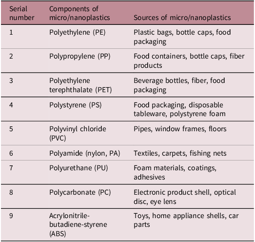
Micro/nanoplastics (MNPs) are plastic particles smaller than 5 mm in diameter, classified into microplastics (1 μm–5 mm) and nanoplastics (<1 μm). These particles originate from diverse sources, including the degradation of larger plastic waste due to environmental factors such as UV radiation, mechanical abrasion, and chemical weathering. Additionally, MNPs are intentionally manufactured for use in various products, such as plastic microbeads in personal care items, synthetic fibers from textiles, and additives in industrial processes. The most common types of MNPs include polyethylene (PE), polystyrene (PS), polycarbonate, polypropylene (PP), and polyethylene terephthalate (PET), materials widely used in packaging, consumer goods, and industrial applications. Due to their small size and persistence, these particles are found in air, water, soil, and food products, raising concerns about their accumulation in ecosystems and potential health risks to humans (Table 1). Reference Lin8
Common chemicals found in micro/nanoplastics

The global proliferation of plastic products and their improper disposal have led to the widespread contamination of ecosystems by MNPs, making them a pressing environmental concern. During their lifecycle—manufacturing, usage, and disposal—plastics release vast quantities of microscopic particles through physical abrasion, chemical degradation, and environmental weathering. Due to their highly stable chemical composition, these plastic fragments resist natural decomposition, persisting in the environment for extended periods. Their minute size enables them to be easily transported by air and water currents, facilitating their dispersal across diverse ecosystems. As a result, MNPs have been detected in remote regions, including deep-sea sediments, polar ice, freshwater bodies, agricultural soils, and even atmospheric fallout, underscoring their extensive environmental reach. 9 In the ocean, micro/nanoplastics have even been found in deep-sea sediments, Reference Peng, Chen, Chen, Dasgupta and Bai10 demonstrating the extent and depth of their spread and pollution.
The presence of MNPs poses a serious threat not only to the ecological environment but also to human health, as they can enter the food chain and water supply system through various pathways, leading to the absorption and bioaccumulation of MNPs in the human body, ultimately impacting human health. For example, marine organisms may ingest microplastics, introducing them into the food chain. Humans may ingest MNPs by consuming contaminated seafood, aquatic products, and drinking water. Once inside the human body, MNPs can potentially cause tissue damage and functional disorders in multiple systems (such as the digestive, respiratory, cardiovascular, immune, reproductive, and endocrine systems) through mechanisms such as cellular damage, inflammatory responses, metabolic disorders, genotoxicity, and immune and endocrine disruption, thereby posing potential health risks. Reference Zhao11
Additionally, MNPs can act as carriers, carrying and adsorbing numerous harmful substances such as chemical additives, environmental pollutants, and pathogenic microorganisms. These attached harmful substances can enter the human body through the food chain along with the MNPs, increasing health risks and potentially affecting future generations(Figure 1). Reference Blackburn and Green12
Micro/nanoplastics pollution sources, entry routes and effects on intrauterine development and growth.

Microplastics detection technologies
Recently, scientific research on microplastics has been booming, and the precise and rapid identification of microplastics is crucial for research in this field. Depending on the type, size, color, abundance, and weight of microplastics, the main detection methods include but are not limited to optical microscopy, scanning electron microscopy (SEM), Fourier-transform infrared spectroscopy (FTIR), Raman spectroscopy, pyrolysis-gas chromatography-mass spectrometry (Py-GC-MS), thermal extraction-desorption gas chromatography-mass spectrometry (TED-GC-MS), and thermogravimetric analysis-differential scanning calorimetry (TGA-DSC). Reference Lee and Chae13,Reference Ahmed14
Microscopy techniques
Ordinary optical microscopes can identify microplastics larger than several hundred micrometers in diameter on the basis of their shape, size, and color, offering a simple, fast, and economical method. However, this method is highly subjective and prone to high error rates. Reference He15 Ordinary optical microscopes are challenging to use for identification of microplastics particles smaller than 100 μm in diameter.
SEM uses electron microscopes for analysis and identification, providing higher magnification and clearer images than ordinary optical microscopes. However, SEM has higher detection requirements, such as the sample needing to be solid, non-toxic, and non-radioactive, and the laboratory environment must be kept under vacuum conditions. Reference Ompala16
Spectroscopic analysis methods
FTIR obtains the interferogram of microplastics, which is then processed through Fourier transformation to determine the infrared spectrum of the microplastic particles. This non-destructive detection technology is widely used for the qualitative detection and component analysis of microplastics as a result of its simple pretreatment, short analysis time, and ease of maintenance. Reference Rotchell2,Reference Song17,Reference Li18 The disadvantage of FTIR is its limited resolution of only 20 μm. Reference Käppler19 After the degradation of microplastics, the infrared spectrum changes, leading to a decrease in detection accuracy. With the continuous development of microplastics detection technology, micro-Fourier transform infrared spectroscopy (μFTIR) has gradually been applied to detect smaller microplastics samples (> 10 μm).
Raman spectroscopy utilizes the different scattering vibrations corresponding to different atomic and molecular structures. By obtaining the scattering spectrum of the sample and comparing it with reference values in the spectral database, the composition of the microplastic sample can be determined. Reference Araujo, Nolasco, Ribeiro and Ribeiro-Claro20 Compared to FTIR, Raman spectroscopy has better resolution (down to 1 μm), Reference Sobhani, Amin, Naidu, Megharaj and Fang21 a wider spectral range, and higher sensitivity to non-polar functional groups. However, its widespread application is limited by susceptibility to fluorescence interference and longer measurement times. Based on classical Raman spectroscopy, micro-Raman spectroscopy is also gradually being widely used in microplastics detection. Reference Qin22,Reference Massardo23
Thermal analysis methods
Thermal analysis methods involve decomposing polymers into small molecules at high temperatures and then identifying the components and types of microplastics by using characteristic spectra. The main thermal analysis techniques include Py-GC-MS, TED-GC-MS, and TGA-DSC. Reference Peñalver, Arroyo-Manzanares, López-García and Hernández-Córdoba24 Py-GC-MS and TED-GC-MS identify microplastics types by analyzing the thermal degradation products, whereas TGA-DSC identifies polymer types by measuring the relationship between the heat difference and temperature during the solid-liquid phase transition of polymers. Reference Dümichen1 Due to the destructive nature of thermal pyrolysis, these methods can only detect the chemical information of the samples and cannot provide particle size information for the microplastics particles. Reference Huang, Hu and Wang25
Py-GC-MS has high sensitivity and does not require sample pretreatment, making it widely used. Reference Wang26–Reference Liu28 However, with this method, different polymers may produce the same or similar thermal pyrolysis products, leading to overlapping spectra and resulting in errors. Additionally, Py-GC-MS can only analyze samples weighing less than 0.5 mg, making it unsuitable for large-scale and large-sample detection. Reference Seeley and Lynch29
Presently, there are still many challenges in the detection and analysis of microplastics. It is difficult to obtain accurate and reliable information on the appearance, size distribution, abundance, and chemical composition of microplastics by using a single analysis method. To establish reliable and standardized detection methods, a combination of several detection methods must be considered.
Detection of micro/nanoplastics in human tissues
Micro/nanoplastics are widely present in the human body through skin contact, respiratory inhalation, and digestive ingestion. Using a 3D human skin model, a recent study found that toxic chemicals contained in microplastics can penetrate the skin through contact with sweat and then enter the bloodstream. Reference Abafe, Harrad and Abdallah30 Microplastics not only are present in the air but can also enter the lungs through respiration. A systematic review found that inhaled microplastics might cause inflammatory responses and potential carcinogenic effects in the respiratory system, varying with the concentration, size, type, and surface charge of the microplastics. Accumulation of microplastics in the lungs may lead to chronic inflammation, tissue damage, and pulmonary diseases, especially with long-term exposure. Reference Lombardi31 Most micro/nanoplastics ingested through diet and drinking water may be excreted through feces. Some studies suggest that over 90% of micro/nanoplastics are not absorbed into the body; however, small particles, especially those smaller than 10 μm, can be absorbed by intestinal cells through processes such as phagocytosis and endocytosis. Reference Udovicki, Andjelkovic, Cirkovic-Velickovic and Rajkovic32
In recent years, multiple studies have confirmed the widespread presence of microplastics in the human body, but these findings have not been definitively confirmed. These studies have revealed that microplastics not only accumulate in various organs and tissues but are also present in various bodily fluids and secretions (Table 2). Research indicates that microplastics can enter the circulatory system. Reference Leslie33 In the blood, these tiny particles can be transported to organs such as the liver, Reference Horvatits34 kidneys, Reference Massardo23 heart, Reference Liu35 and intestines, Reference Ibrahim36 reaching the testes, Reference Zhao37,Reference Yang38 endometrium, Reference Sun39 lower limb joints, Reference Li18 and even depositing on the arterial walls, Reference Liu28 forming components of thrombi. Reference Liu28 Inhaled microplastics can accumulate in the lungs, causing inflammation and tissue damage. Reference Amato-Lourenço40,Reference Jenner41 Additionally, microplastics have been detected in human semen Reference Montano42 and urine, Reference Song17 indicating potential impacts on the male reproductive and urinary systems. The detection of microplastics in respiratory secretions such as sputum further confirms their inhalation through the respiratory tract. Reference Huang43 The presence of microplastics in feces indicates that these particles are ingested and excreted through the digestive system. Reference Yan44 Microplastics found in the placenta, Reference Ragusa5 breast milk, Reference Liu35 and amniotic fluid Reference Xue7 suggest that microplastics in the mother’s body can be transmitted to the infant, posing potential risks to the health of newborns even before birth.
Toxic effects of microplastics

Effects of MNPs on offspring growth and development in animals
Pregnancy and lactation are vulnerable periods of vulnerability to the harmful effects of environmental exposures. However, the adverse effects of maternal exposure to MNPs on fetal health are largely unknown. Studies have shown that micro/nanoplastics ≤ 20 μm can enter organs, and smaller MNPs with a particle size of 0.1–10 μm can penetrate all organs of animals; cross cell membranes, the blood-brain barrier, and the placenta; and even transfer to the next generation. Reference Wright and Kelly45,Reference Bouwmeester, Hollman and Peters46 For example, when adult zebrafish are exposed to PS nanoplastics, these microplastics bioaccumulate and are inherited by the offspring. Nanoplastics have been found in the yolk sac, gastrointestinal tract, liver, and pancreas of embryo/offspring zebrafish, with bradycardia observed in embryos. Reference Pitt47 Numerous animal experiments have demonstrated that maternal exposure to MNPs profoundly affects offspring growth and development, including weight loss, developmental abnormalities, immune disorders, metabolic disturbances, circulatory system abnormalities, neurological deficits, and reproductive failure. Reference Geng48
In vivo studies have shown that 24 hours after Sprague Dawley rats inhale PS nanoplastics during pregnancy, nanoplastics can be transmitted from the mother to the fetus. PS nanoparticles have also been observed in the placenta, as well as in the liver, lungs, heart, kidneys, and brain of fetal rats. The weight of the fetuses and placentas was significantly lower than that of the control group. Reference Fournier49 Exposure to PS MNPs during pregnancy and lactation in mother mice can lead to reduced birth weight and postnatal weight in offspring. Similar findings were observed in three other studies. Reference Zhang50 In oyster embryos exposed to high-density PE microplastics of different sizes for 24 hours, smaller microplastic particles induced higher rates of deformities and developmental stasis in the oysters. Reference Bringer51 Another study exposed oysters (Crassostrea gigas) to a mixture of microplastics and found reduced activity, developmental abnormalities, growth stasis, and delayed development in the offspring. Reference Bringer52 Zebrafish embryos exposed to microplastics showed developmental disorders, including significant growth retardation and peripheral microcirculation disorders, leading to premature birth and growth restriction during embryonic development. Reference Park and Kim53 Additionally, offspring of zebrafish exposed to microplastics during pregnancy showed bone formation disorders at 6 days old, manifesting as reduced operculum bone. Reference Tarasco54 Juvenile zebrafish exposed to microplastics exhibited severe developmental deformities and impaired integrity of the eye’s visual structure during development. Reference Marco De55 PS nanoplastics can cause extensive severe congenital malformations in chick embryos, particularly leading to severe defects in the heart and major blood vessels. Reference Wang56
An increasing number of animal experiments has demonstrated that MNPs exposure affects offspring through the immune system. Zebrafish embryos exposed to PS microplastic particles can trigger an immune response, activating several biological pathways related to immune response. Neutrophils and macrophages are recruited around the microplastic particles, indicating that plastic particles are involved in the immune recognition process. Reference Veneman, Spaink, Brun, Bosker and Vijver57 PS microplastics can disrupt the maternal-fetal immune balance, shifting cytokine secretion towards an immunosuppressive state, reducing the number and diameter of uterine arterioles, leading to embryo resorption in mice, and inducing natural miscarriages. This finding suggests that PS microplastics can adversely affect pregnancy outcomes through immune dysregulation. Reference Hu3
MNPs exposure can adversely affect the circulatory and metabolic functions of offspring. Numerous studies have demonstrated that maternal exposure to PS microplastics can disrupt various organ functions in offspring by altering their metabolism. For instance, it can alter serum metabolite levels and increase the risk of fatty acid metabolism disorders, potentially influenced by the particle size of the microplastics. Reference Luo58 Additionally, it can cause maternal metabolic disorders associated with gut microbiota imbalances and intestinal barrier dysfunction, heightening the risk of long-term metabolic issues in offspring. MNPs may also disrupt amino acid metabolism in the retina of offspring and lead to fetal growth restriction (FGR) and intrauterine death by affecting maternal hepatic glucose metabolism. Reference Xiong4,Reference Zhang50
Maternal exposure to PS nanoplastics can result in abnormal brain metabolism in mouse fetuses, with significant reductions in the relative concentrations of γ-aminobutyric acid (GABA), creatine, and glucose compared to controls. Reference So59 Experimental animal studies have shown that maternal exposure to microplastics environments can alter offspring energy and lipid metabolism, potentially leading to obesity. Reference Kannan and Vimalkumar60 Zebrafish studies have indicated that exposure to PS MNPs can significantly alter the metabolic profiles of juvenile fish, affecting genes related to glycolysis and lipid metabolism, and may cause pericardial edema. Micro/nanoplastics may induce systemic inflammatory responses and oxidative stress, resulting in vascular cell dysfunction, endothelial dysfunction, and thrombosis. Reference Wan61,Reference Sun62
MNPs also impact neural development in offspring. Recent studies show that PS plastics during pregnancy can cause placental dysfunction in mice, significantly reducing the pulsatility index of the middle cerebral artery in fetuses and potentially leading to neurodevelopmental disorders. Reference Dibbon63 PS nanoplastics ingested by pregnant mice can be transmitted to offspring through breast milk, altering neural stem cell function, neural cell composition, and brain histology, resulting in neuronal dysfunction and cognitive deficits. Reference Jeong64 In another study, pregnant mice exposed to PS microbeads exhibited thalamic neuronal apoptosis and reduced GABA synthesis in fetal mice, leading to anxiety-like behavior at 8 weeks. Reference Yang65 Additionally, maternal exposure to PE microplastics produced offspring with autism-like traits, including repetitive and compulsive behaviors. Reference Zaheer66 Long-term exposure to PS microplastics has been linked to increased proliferation and apoptosis of primary cortical neuronal cells, potential neurotoxic effects, impaired social novelty preference, and anxiety-like behavior in adolescent offspring. Reference So59 Exposing mother mice to PS nanoplastics during late pregnancy, late lactation, and adolescence resulted in altered brain neurotransmitter levels and hippocampal synaptic changes, ultimately leading to anxiety-like behavior and spatial memory deficits in adolescent offspring. This finding demonstrates that maternal exposure to microplastics affects the neurodevelopment of offspring. Reference Tian67 Exposure to PS nanoplastics during pregnancy in rats leads to oligodendrocyte apoptosis and impaired motor coordination in offspring, affecting the brain development of fetal rats. Reference Zhang68 Research also found that PS microplastics inhibit chicken embryo development and increase the incidence of congenital malformations (especially in the nervous system), including neural tube defects. Reference Nie69
Animal experiments suggest that microplastic exposure during pregnancy can disrupt offspring fertility. Maternal exposure to PS nanoplastics has been shown to cause liver and testicular toxicity in male offspring, inducing oxidative damage and testicular developmental disorders. In Daphnia magna, microplastic exposure increased parental mortality and reduced growth, reproduction, and population growth rates, with effects persisting into subsequent generations. PS microplastic exposure led to decreased mortality and reproductive rates in earthworms, likely due to DNA damage. Reference Martins and Guilhermino70,Reference Sobhani, Panneerselvan, Fang, Naidu and Megharaj71 Pregnant mice and their offspring exposed to PS microplastics from day 1 of pregnancy to after birth experienced testicular developmental disorders and impaired male fertility, potentially regulated by the Hippo signaling pathway and involving an immune response. This response may be regulated by the Hippo signaling pathway and involve an immune response. Reference Zhao72 Overall, animal studies support the notion that exposure to micro/nanoplastics during pregnancy adversely affects the growth and development of offspring.
MNPs and pregnancy-related diseases
Studies have found that MNPs particles can cross the placental barrier in various experimental models, Reference Medley, Spratlen, Yan, Herbstman and Deyssenroth73 including in vitro cell models, Reference Cartwright74 in vivo animal models, Reference Fournier49 and ex vivo human placental perfusion systems. Reference Wick75 This finding provides evidence of the potential impact of maternal MNPs on the fetus through the maternal-fetal barrier. A study using Raman spectroscopy detected microplastic contamination in placentas during pregnancies in Hawaii in 2006, 2013, and 2021. The results showed that the accumulation of microplastics in placentas significantly increased over time. Reference Weingrill76
However, direct evidence confirming the association between human MNPs exposure during pregnancy and pregnancy-related diseases remains scarce. One study analyzed the relationship between microplastics and neonatal growth and development, finding a significant negative correlation between the amount of microplastics in the placenta and neonatal birth weight, length, head circumference, and 1-minute APGAR score in pregnancies complicated by intrauterine growth restriction (IUGR) (r = −0.82, p <0.001). All placentas from IUGR pregnancies contained microplastic particles, whereas only four of 30 placentas from normal pregnancies contained microplastic particles. According to the study, the most prevalent microplastics were PE and PS. Although the sample size of this study was small (13 cases), it draws attention to the potential impact of microplastic particles on fetal development in utero. Reference Amereh77 A study using experimental mice to test the effects of maternal exposure to plastics on fetal and placental growth found that exposure to microplastics in late pregnancy resulted in FGR. Reference Jeong64 Researchers further explored the reasons in another study, confirming that this phenomenon may be due to altered placental metabolism. Reference Xiong4 Additionally, the latest research explores the association between microplastic levels in maternal amniotic fluid and fetal growth indicators. The results indicate a significant negative correlation between the abundance of total microplastics in maternal amniotic fluid and gestational age (β = −0.44, 95% CI, −0.83, −0.05), suggesting that maternal microplastic exposure may have potential health impacts on fetal development. Reference Xue7
Studies investigating the relationship between unexplained recurrent miscarriage (RM) and MNPs documented higher levels of PS in the villous tissue of RM patients. Additionally, this study utilized human placental trophoblast cells and pregnant mouse models to confirm that exposure to PS nanoplastics activates the Bcl-2/Cleaved-caspase-2/Cleaved-caspase-3 signaling pathway through the mitochondrial pathway, leading to excessive apoptosis in human trophoblast cells and mouse placental tissue, further inducing miscarriage. Reference Wan78
A substantial number of animal experiments also support the link between maternal MNPs exposure and pregnancy-related diseases. South Korean researchers studied the effects of polymethyl methacrylate (PMMA) nanoplastics on mouse embryo development and found that PMMA particles reduced blastocyst development and embryo quality indicators and decreased the implantation rate of morulae. Reference You79 PS microplastics have been shown to disrupt hepatic glucose metabolism in pregnant mice, induce insulin resistance, and damage placental structure, leading to FGR and intrauterine death in the offspring. Reference Zhang50
Additionally, multiple studies have reported that PS MNPs can result in reduced fetal weight Reference Dümichen1,Reference Abafe, Harrad and Abdallah30,Reference Zhao37,Reference Yang38,Reference Wan61,Reference Amereh77 and cause developmental deformities in Crassostrea gigas and developmental delays and extensive malformations in chick embryos. Reference Dümichen1,Reference Abafe, Harrad and Abdallah30,Reference Zhao37,Reference Yang38,Reference Wan61,Reference Amereh77 In studies related to zebrafish, PS microplastics were found to cause premature birth and growth restriction in juvenile fish, as well as developmental deformities. Reference Park and Kim53–Reference Marco De55
PS nanoplastics may inhibit the invasion and migration of human trophoblast cells by activating autophagy, promoting the autophagic degradation of SOX2, and suppressing the transcription of ROCK1, thereby inducing miscarriage in pregnant mice. Reference Wan80 Additionally, they may disrupt the maternal-fetal immune balance, shifting cytokine secretion towards an immunosuppressive state, reducing the number and diameter of uterine microarteries, and leading to spontaneous miscarriage. Reference Hu3 These findings are consistent with previous research conclusions. Reference Wan78
Another study, which experimented with exposure to PE MNPs in mice showed that although maternal exposure to PE does not affect fetal growth, the umbilical artery blood flow in the PE group increased by 43% compared to the control group, significantly impacting placental function (p < 0.01). These findings suggest that PE could potentially lead to adverse pregnancy outcomes through abnormal placental function. Reference Xiong4,Reference Luo58,Reference So59
Discussion
This review highlights growing evidence on the potential impacts of prenatal exposure to micro/nanoplastics (MNPs), yet several limitations must be acknowledged. The majority of studies are based on in vitro or animal models, which limits the generalizability of findings to human populations. Variability in study design, exposure routes, particle types, and endpoints also hampers direct comparison and weakens causal inference. Moreover, detection techniques currently favor microplastics over nanoplastics due to sensitivity limitations, leading to potential underestimation of nanoparticle-related effects. The review process itself, while systematic and guided by PRISMA standards, was limited to English-language, peer-reviewed publications, which may exclude relevant non-English or grey literature. Although screening and data extraction were conducted independently, publication bias—particularly the underreporting of negative results—remains a concern. Despite these constraints, the implications for public health and policy are substantial. The presence of MNPs in placental and fetal tissues suggests the need for precautionary action, including maternal exposure reduction strategies, policy reform on plastic use, and heightened public awareness. Moving forward, there is a critical need for robust human epidemiological studies, standardized detection and exposure assessment protocols, and interdisciplinary collaboration to elucidate mechanisms of toxicity and inform evidence-based decision-making. These efforts are essential to mitigate potential risks to maternal and offspring health and to guide future interventions and regulatory actions.
Conclusions
The pervasive presence of micro- and nanoplastics (MNPs) in the environment and everyday life presents a growing concern for human health, particularly during vulnerable periods such as pregnancy. This review underscores the potential of MNPs to cross biological barriers, accumulate in maternal and fetal tissues, and induce adverse developmental and physiological outcomes in offspring. Emerging experimental evidence has demonstrated that MNPs can infiltrate multiple organs and systems, potentially leading to histopathological alterations, metabolic disruptions, immune dysfunction, and long-term disease risk.
Maternal exposure to MNPs has been linked to harmful effects in offspring in numerous in vitro and animal studies. However, human data remain scarce, and no definitive causal relationships have yet been established between prenatal MNP exposure and specific diseases in children. This critical knowledge gap limits the ability of clinicians and policymakers to assess risk and implement effective interventions.
Given the increasing detection of MNPs in human biological samples and the growing body of experimental data pointing to their toxicity, there is an urgent need for rigorous, multidisciplinary research to clarify the mechanisms of MNP toxicity during prenatal development. Longitudinal human cohort studies, improved detection technologies, and standardized exposure assessment protocols are essential to accurately evaluate health risks and inform evidence-based public health policies. Protecting maternal and fetal health in the era of plastic pollution demands immediate scientific and regulatory attention.
An important methodological limitation observed across the reviewed literature is the disproportionate focus on microplastics over nanoplastics, primarily due to current analytical limitations. Most existing detection methods, such as Fourier-transform infrared spectroscopy (FTIR) and Raman spectroscopy, are well-suited for identifying microplastics but lack the resolution and sensitivity required to reliably detect nanoplastics, particularly those smaller than 100 nm. Consequently, many studies may underreport or entirely overlook nanoplastic exposure, despite growing concerns about their potentially greater biological reactivity and cellular penetration. This bias in detection creates an imbalance in the current understanding of plastic particle toxicity, skewing the available evidence toward larger particles and possibly underestimating the risks posed by nanoplastics. As a result, conclusions drawn from existing studies may not fully capture the spectrum of health effects associated with total MNP exposure, especially during critical developmental windows such as pregnancy. Addressing this gap will require the advancement and standardization of high-resolution, high-sensitivity analytical techniques, as well as the inclusion of nanoplastic-specific assessments in future experimental and epidemiological research.
Data availability statement
Not applicable.
Author contributions
M.S. drafted the manuscript. J.P. prepared the figures. Y.Y. provided suggestions for revisions. N.Z. and Y.Z. approved the final version of the manuscript for publication. All authors have read and agreed to the published version of the manuscript.
Funding statement
This research was supported partially by the Hubei Provincial Maternal and Children’s Hospital, the Research Foundation for Mental Hygiene, Inc. (914-3280), the National Institutes of Health/National Institute of Child Health and Human Development 1R01HD113199, and the New York State Office for People with Developmental Disabilities.
Competing interests
The authors declare no conflicts of interest.
Institutional review board statement
Not applicable.
Informed consent statement
Not applicable.