Introduction
Radiation therapy is a commonly used treatment in head and neck oncology, whether used alone or as an adjunct to other treatments. It is frequently utilised in combination with surgery or as an organ preservation modality. Osteoradionecrosis and radiation-induced malignancies, both carcinomas and sarcomas, are rare, late complications of radiation therapy.Reference Fangman and Cook1
Radiation-induced malignancy has a reported incidence of 0.9 per cent among patients who undergo radiation therapy in any form for head and neck cancer.Reference Sale, Wallace, Girod and Tsue2 Both carcinomas and sarcomas can arise in previously irradiated tissues. The skin and thyroid are the most commonly involved areas in the head and neck.Reference Sale, Wallace, Girod and Tsue2, Reference Gutierrez, Llorente Pendas, Coca Pelaz, Cabanillas Farpon and Suzarez Nieto3 Radiation-induced sarcoma represents 12 per cent of these radiation-induced malignancies, with an overall incidence of 0.03–0.3 per cent in patients exposed to ionising radiation.Reference Gutierrez, Llorente Pendas, Coca Pelaz, Cabanillas Farpon and Suzarez Nieto3
We present a case of radiation-induced osteosarcoma of the mandible that occurred six years after the completion of combined radiotherapy and chemotherapy for occult head and neck cancer. The osteosarcoma presented clinically as osteoradionecrosis of the mandible, for which the patient underwent appropriate therapy. However, osteosarcoma was found on histological analysis following surgical extirpation and reconstruction.
Case report
We report on a 72-year-old Caucasian female who had undergone a right modified radical neck dissection, followed by concurrent radiation therapy and chemotherapy for a malignant neck mass and occult primary lesion. She completed three cycles of cisplatin therapy, with a dose of 40 mg at each cycle. She also underwent 37 fractions of external beam radiation therapy, receiving a total dose of 7000 cGy.
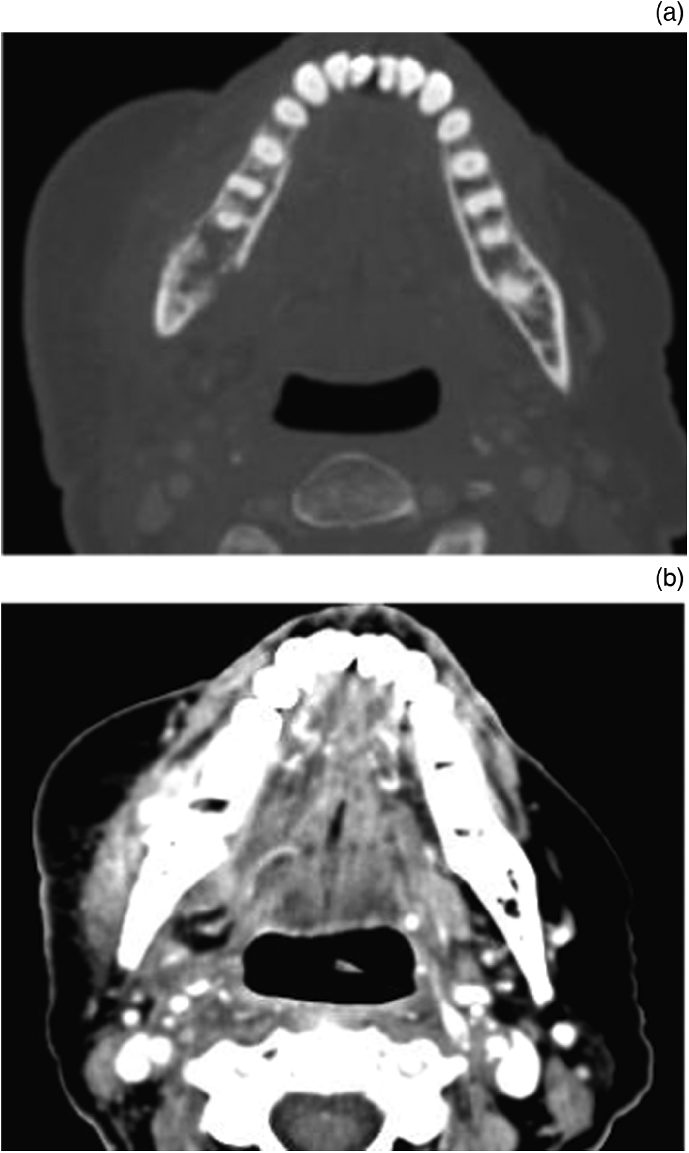
Six years after the completion of chemoradiation therapy, the patient presented with pain and swelling of the right side of her mandible. This coincided with an episode of streptococcal pharyngitis, but her symptoms did not resolve with a course of amoxicillin followed by amoxicillin and clavulanate potassium. After a draining cutaneous sinus developed on her right cheek, she was sent for a computed tomography scan (Figure 1). The scan revealed an ill-defined lytic lesion of the right mandible, associated with some soft tissue swelling in the masticator and submandibular spaces. A positron emission tomography scan showed hypermetabolic activity limited to the right mandible. Based on these findings, the patient was diagnosed with osteoradionecrosis and was then referred to our tertiary centre for treatment.
Axial computed tomography images of: (a) bone windows showing an ill-defined lytic lesion of the right mandible, and (b) soft tissue windows showing soft tissue oedema and inflammatory changes of the masticator muscle and submandibular space.

The patient underwent hyperbaric oxygen therapy using a standard protocol. This involved ‘dives’ (compressions) at 2.4 atmospheres absolute (ATA) for 90 minutes during each of her 30 sessions. During this period of treatment, she developed severe trismus and lost 9 kg, which necessitated supplemental feedings through a gastrostomy tube. The draining cutaneous sinus persisted and was unresponsive to oral antibiotics. A peripherally inserted central catheter was placed and the patient received parenteral antibiotics.
Following completion of the hyperbaric therapy, the patient underwent a composite resection, including right hemimandibulectomy and excision of the cutaneous fistula. This was followed by reconstruction of the defect with a fibula free flap. There were no surgical complications, and the patient recovered well post-operatively.
Histological analysis revealed that the tumour comprised a spindle cell component which stained negatively for squamous markers (p63 and cytokeratin 5/6), with focal high-grade osteosarcoma characterised by malignant osteoid and adjacent chondroblastic features (Figure 2). Radiation changes in the salivary gland tissue, a subcutaneous abscess, and osteoradionecrosis of the mandibular head and coronoid process were also evident. The final margins of resection were free of tumour involvement. Based on these findings, the patient was diagnosed with high-grade osteosarcoma.
Histology slides showing: (a) a representative area of multifocal deposition of osteoid by cytologically malignant cells, and (b) cartilaginous differentiation with cytological atypia and adjacent malignant spindle cells. (H&E; ×100)
Despite these negative resections, the patient's tumour recurred at seven months; at the time of writing, she was being evaluated for palliative options.
Discussion
This paper represents the first published case of radiation-induced sarcoma that initially presented in the setting of osteoradionecrosis. This dramatic change in diagnosis could have conceivably resulted in alterations in this patient's treatment plan, but appropriate treatment for osteoradionecrosis fortuitously assured that the initial treatment for the radiation-induced sarcoma was appropriate. While these two disease processes are usually easily differentiated, overlapping clinical, radiographical and pathological findings can, as in this case, result in an initial erroneous diagnosis. Thus, the differential diagnosis of such a lesion must include local tumour recurrence, bone metastasis, radiation-induced sarcoma, osteoradionecrosis and infection. If possible, treatment should be individually tailored to address possible changes in final diagnosis. Given this experience, the authors provide their insights into management of these respective disease processes.
Osteoradionecrosis is a condition of non-vital bone in a site of radiation injury. The absence of reserve reparative capacity is a result of the prior radiation injury. Even apparently innocuous forms of trauma, such as denture-related injury, ulcers or tooth extraction, can overwhelm the reparative capacity of the radiation-injured bone.
In 1983, Marx demonstrated successful resolution of mandibular osteoradionecrosis in 58 patients using a staged protocol with hyperbaric oxygen and surgery.Reference Marx4Hyperbaric oxygen transiently elevates tissue oxygen tension, and stimulates fibroblastic proliferation and oxygen-dependent collagen synthesis, allowing for angiogenesis in the radiated bed. Patients with refractory disease are often required to undergo sequestrectomy or saucerisation. Ultimately, patients who do not respond to these conservative measures require wide resection of all affected bone, with reconstruction using vascularised flaps.
Radiation therapy can induce a variety of tumours, including carcinomas and sarcomas. Because of the rarity of radiation-induced malignancy, with an estimated incidence of 0.03–1 per cent, prospective studies are not feasible. A review of 65 cases of radiation-induced malignancy showed that squamous cell carcinoma was the most common form, with malignancy most frequently involving the hypopharynx.Reference Gutierrez, Llorente Pendas, Coca Pelaz, Cabanillas Farpon and Suzarez Nieto3 Sarcomas were another common malignancy seen in post-irradiated areas, with radiation-induced sarcoma representing 12 per cent of all radiation-induced malignancy cases. The overall incidence of radiation-induced malignancy in patients exposed to ionising radiation has been reported as 0.03–0.3 per cent.Reference Gutierrez, Llorente Pendas, Coca Pelaz, Cabanillas Farpon and Suzarez Nieto3
Carcinomas occur after low-dose irradiation, whereas sarcomas occur after high doses of radiation with most cases being reported after exposure to 5500 cGy.Reference Maghami, St-John, Bhuta and Abemayor5 In a report of 59 patients with radiation-induced sarcoma, by Cai et al., the median radiation dose was 5500 cGy.Reference Cai, Wu, Li, Zhang, Xie and Wu6 However, cases of radiation-induced sarcoma have been reported to occur after radiation doses as low as 1340 cGy.Reference Gutierrez, Llorente Pendas, Coca Pelaz, Cabanillas Farpon and Suzarez Nieto3 New advances in radiation therapy, such as intensity-modulated radiation therapy and proton therapy, may have an effect on the development of secondary malignancies. However, these treatments have not been studied extensively and no conclusions can be made at this time.Reference Thariat, Italiano, Collin, Iannessi, Marcy and Lacout7 In one study, the latency time for development of radiation-induced sarcoma ranged from 3 months to 53 years, with a mean time of 17 years.Reference Maghami, St-John, Bhuta and Abemayor5 Other studies have reported mean latency times of 12.9, 13 and 13.3 years.Reference Gutierrez, Llorente Pendas, Coca Pelaz, Cabanillas Farpon and Suzarez Nieto3, Reference Wei-wei, Qiu-liang, Guo-hao, Zhi-hua and Zong-yuan8, Reference Chabchoub, Gharbi, Remadi, Limem, Trabelsi and Hochlef9
The diagnosis of radiation-induced sarcoma can be challenging.Reference Debnam, Guha-Thakurta, Mahfouz, Garden, Benjamin and Sturgis10 The tumour must meet specific criteria in order for radiation to be implicated as the inciting factor in sarcoma. These criteria were first described in 1948 by Chan et al., and revised in 1971 by Arlen et al. These criteria are as follows: (1) the patient must have received radiation, (2) there is a latency period of at least three years, (3) the histology of the tumour is different than in the first cancer, and (4) the tumour area is within the radiation field or adjacent to it.Reference Thariat, Italiano, Collin, Iannessi, Marcy and Lacout7, Reference Seethala, Sturgis, Raymond and Deavers11 Despite these criteria, the diagnosis can still be incorrect in up to 25 per cent of cases.Reference Thariat, Italiano, Collin, Iannessi, Marcy and Lacout7
Patients with Li–Fraumeni syndrome, Von Recklinghausen's disease, and those that harbour mutations in tumour suppressor genes such as p53 and retinoblastoma are more likely to develop radiation-induced sarcoma.Reference Chabchoub, Gharbi, Remadi, Limem, Trabelsi and Hochlef9 Mertens et al. reported that the most frequent mutations were alterations in chromosome 3p.Reference Mertens, Larramendy, Gustavsson, Gisselsson, Rydholm and Brosjo12 The age at which radiation occurred is an important factor, with the risk being greater in children than in adults (3.6 per cent vs 0.7 per cent).Reference Thariat, Italiano, Collin, Iannessi, Marcy and Lacout7 It appears that when chemotherapy is administered concurrently with radiation therapy, a synergistic effect occurs. The ratio of bone sarcoma incidence in irradiated tissue compared to the normal population after concurrent chemotherapy and radiotherapy was 113:1, versus 28:1 after chemotherapy alone and 19:1 after radiotherapy alone.Reference Menu-Branthomme, Rubino, Shamsaldin, Hawkins, Grumaud and Dondon13
As radiation-induced sarcoma is a rare malignancy, there are no disease-specific treatment guidelines. Radiation-induced sarcoma is currently treated using the accepted treatment protocols for primary osteosarcoma, with adjustments as needed based on individual cases.Reference Maghami, St-John, Bhuta and Abemayor5, Reference Thariat, Italiano, Collin, Iannessi, Marcy and Lacout7 In a retrospective case series of 80 radiation-induced sarcoma cases, the average survival rate for patients who underwent surgery was 48 months, compared with an average of 6 months for those who received chemotherapy alone.Reference Lagrange, Ramaioli, Chateau, Marchal, Resbeut and Richaud14 Another study showed 63 per cent survival at five years for patients who had undergone complete excision; in contrast, the survival rate was only 10 per cent for patients in whom at least one surgical margin was not cleared of tumour.Reference Bjerkehagen, Smeland, Walberg, Skeldal, Hall and Nesland15
Adjuvant therapy is limited in the treatment of radiation-induced sarcoma. As these tumours are radiation induced, they are generally considered to be radiation resistant, and adjuvant radiation therapy is limited by previous radiation.Reference Gutierrez, Llorente Pendas, Coca Pelaz, Cabanillas Farpon and Suzarez Nieto3 The role of chemotherapy is controversial. In 1997, a meta-analysis showed that chemotherapy adds a benefit in terms of relapse-free survival, but not in overall survival.Reference Thariat, Italiano, Collin, Iannessi, Marcy and Lacout7 A more recent meta-analysis did show a significant benefit in overall survival, with an absolute risk reduction of death of 6 per cent.Reference Thariat, Italiano, Collin, Iannessi, Marcy and Lacout7 Most recently, adjuvant chemotherapy has been advocated for use in those patients with high-grade features, such as high-grade histology, tumour size greater than 5 cm and positive margins.Reference Thariat, Italiano, Collin, Iannessi, Marcy and Lacout7
• The differential diagnosis in osteoradionecrosis cases should include local tumour recurrence, bone metastasis, radiation-induced sarcoma, osteoradionecrosis and infection
• Radiation-induced sarcoma can present as osteoradionecrosis
• Primary treatment of both osteoradionecrosis and radiation-induced sarcoma is wide surgical extirpation, with negative margins
• Chemotherapy and radiation therapy have a limited role in the treatment of radiation-induced malignancy
• Overall survival for radiation-induced sarcoma cases is poor
In conclusion, while osteoradionecrosis and radiation-induced sarcoma are usually easily differentiated, overlapping clinical, radiographical and pathological findings can result in an initial erroneous diagnosis. Thus, the differential diagnosis of such a lesion must include local tumour recurrence, bone metastasis, radiation-induced sarcoma, osteoradionecrosis and infection. If possible, treatment should be individually tailored to address possible changes in the final diagnosis. In many cases of osteoradionecrosis, this includes complete resection of all affected tissues, back to viable, healthy bone and soft tissues. Any decisions about adjuvant treatment should be made after the review of final pathology.