Introduction
During fertilization, intracellular Ca2+ increase in the oocyte occurs immediately after insemination, which is a universal phenomenon observed in the animal kingdom (Miyazaki, Reference Miyazaki2006; Whitaker, Reference Whitaker2006; Kashir et al., Reference Kashir, Deguchi, Jones, Coward and Stricker2013; Costache et al., Reference Costache, McDougall and Dumollard2014). Artificial induction of Ca2+ increase in the oocyte, in the absence of insemination, induced parthenogenetic activation (Steinhardt & Epel, Reference Steinhardt and Epel1974; Zucker et al., Reference Zucker, Steinhardt and Winkler1978). Therefore, intracellular Ca2+ increase in the oocyte is thought to be indispensable for oocyte activation and further development (Santella et al., Reference Santella, Lim and Moccia2004; Whitaker, Reference Whitaker2006).
There are several Ca2+ releasing mechanisms in a cell. Activation of voltage-dependent ion channels on the plasma membrane induces the influx of external Ca2+ into the cell (Shen & Buck, Reference Shen and Buck1993; McCulloh et al., Reference McCulloh, Ivonnet, Landowne and Chambers2000). Conversely, an intracellular Ca2+-mobilizing mechanism utilizes inositol 1,4,5-trisphosphate (IP3) to release Ca2+ from the endoplasmic reticulum (ER) through its specific receptors (Miyazaki et al., Reference Miyazaki, Shirakawa, Nakada and Honda1993; Berridge, Reference Berridge2009). In addition, ADP-ribose, a product of nicotinamide adenine dinucleotide (NAD), as well as cyclic ADP-ribose (cADPr), gates an ion channel in the plasma membrane of ascidian oocytes (Wilding et al., Reference Wilding, Russo, Marino, Grumetto, De Simone, Tosti and Dale1998a, Reference Wilding, Russo, Galione, Marino and Daleb). Furthermore, another Ca2+-mobilizing second messenger nicotinic acid adenine dinucleotide phosphate (NAADP) is produced from nicotinamide adenine dinucleotide (NADP) by the same enzyme that produces cADPr: ADP-ribosyl cyclase (ADPRC) (Lee et al., Reference Lee, Walseth, Bratt, Hayes and Clapper1989; Aarhus et al., Reference Aarhus, Graeff, Dickey, Walseth and Lee1995).
cADPr recognizes its receptor on the ER and releases Ca2+ through a different pathway independent of IP3 (Clapper et al., Reference Clapper, Walseth, Dargie and Lee1987). In sea urchin and starfish eggs, NAADP releases Ca2+ from acidic granules (Lee & Aarhus, Reference Lee and Aarhus1995) or induces Ca2+ influx into the egg through a plasma membrane Ca2+ channel (Santella et al., Reference Santella, Kyozuka, Genazzani, De Riso and Carafoli2000; Moccia et al., Reference Moccia, Lim, Nusco, Ercolano and Santella2003). Actin microfilaments in the egg cortex are also thought to be implicated in the intracellular Ca2+ increase (Lange, Reference Lange1999; Lim et al., Reference Lim, Lange and Santella2002; Chun et al., Reference Chun, Limatola, Vasilev and Santella2014). Thus, oocytes might recruit more than one pathway to release intracellular Ca2+ during the physiological events such as meiotic maturation and fertilization.
So-called ‘sperm factors’ identified as phospholipase C zeta (PLC zeta) in mammalian sperm and citrate synthase in Cynopus sperm trigger Ca2+ release through IICR in the oocyte (Saunders et al., Reference Saunders, Larman, Parrington, Cox, Royse, Blayney, Swann and Lai2002; Harada et al., Reference Harada, Kawazoe, Eto, Ueno and Iwao2011). The IICR mechanisms have been identified in the oocytes of amphibian (El-Jouni et al., Reference El-Jouni, Jang, Haun and Machaca2005), ascidian (Yoshida et al., Reference Yoshida, Sensui, Inoue, Morisawa and Mikoshiba1998), Annelida (Stricker, Reference Stricker1996), mollusc (Deguchi et al., Reference Deguchi, Osanai and Morisawa1996), and other animal groups (Jaffe et al., 2001). Intracellular Ca2+ release by mechanisms other than IICR have also been reported in oocytes. In sea urchins and starfishes, increased Ca2+ at fertilization is induced by cGMP, ryanodine, cADPr, and NAADP (Whalley et al., Reference Whalley, McDougall, Crossley, Swann and Whitaker1992: Galione et al., Reference Galione, McDougall, Busa, Willmott, Gillot and Whitaker1993: Lee & Aarhus, Reference Lee and Aarhus1995: Santella et al., Reference Santella, Kyozuka, Genazzani, De Riso and Carafoli2000: Nusco et al., Reference Nusco, Lim, Sabala and Santella2002: Matsumoto et al., Reference Matsumoto, Solzin, Helbig, Hagen, Ueno, Kawase, Maruyama, Ogiso, Godde, Minakata, Kaupp, Hoshi and Weyand2003). In Annelida, during fertilization in Polychaeta, two different Ca2+ sources contribute to the increase of cytosolic Ca2+ in the oocytes (Nakano et al., Reference Nakano, Deguchi and Kyozuka2014). Mammalian oocytes possess a cADPr-dependent Ca2+ increase system (Ayabe et al., Reference Ayabe, Kopf and Schultz1995). Therefore, intricately intertwined Ca2+ release mechanisms may be triggered by the fertilizing spermatozoon. However, the concerted mechanism by which these Ca2+ release systems are triggered by fertilizing sperm are not yet clear in most species. Echinoderms have been proved to be one of the most suitable models for analysis of fertilization, as it is easy to obtain isolated gametes and fertilize them in seawater (Santella et al., Reference Santella, Vasilev and Chun2012). Fully grown oocytes isolated from ovaries of starfish are immature and lack the ability to initiate development upon stimulation by sperm (Fujimori & Hirai, Reference Fujimori and Hirai1979). The IICR mechanism is not fully functional before GVBD (Chiba et al. Reference Chiba, Kado and Jaffe1990; Lim et al., Reference Lim, Ercolano, Kyozuka, Nusco, Moccia, Lange and Santella2003). For normal fertilization to occur, progression of oocyte maturation along with GVBD is necessary, which can be induced by the application of 1-methyladenine (1-MA), a maturation-inducing hormone of starfish (Kanatani et al., Reference Kanatani, Shirai, Nakanishi and Kurokawa1969). These matured eggs now manifest a normal Ca2+ response at fertilization. The optimal capability of releasing Ca2+ develops during oocyte maturation (Chiba et al., Reference Chiba, Kado and Jaffe1990). When immature oocytes are treated with subthreshold concentrations of 1-MA, they do not undergo complete oocyte maturation without reaching GVBD, i.e. only ‘cortically matured’ oocytes are obtained. Nonetheless, upon insemination, these eggs exhibit propagation of the sperm-induced Ca2+ wave and the elevation of the fertilization envelope like fully matured eggs (Hirohashi et al., Reference Hirohashi, Harada and Chiba2008), which are the signs of normal exocytosis of cortical granules (Vacquier, Reference Vacquier1975). The fact that oocyte maturation may proceed in two phases encouraged us to explore the possibility of separating the multiple Ca2+ release mechanisms that optimize during meiotic maturation to be fully functional at fertilization.
In this study, we report that NAADP and its metabolic precursor NADP can effectively induce Ca2+ increase before the IICR mechanism is optimized in the maturing oocytes. As the Ca2+ ionophore ionomycin and the media condition mimicking the intracellular pH changes in the oocytes exposed to 1-MA can potentiate the NADP-dependent Ca2+ release mechanism much before the time schedule of IP3 sensitization, it may well play a role in shaping the Ca2+ wave at the time of fertilization.
Materials and methods
Animals
Starfish (Asterina pectinifera) were collected in early September at the beginning of the breeding season near the Research Center for Marine Biology, Asamushi, facing the Mutsu Bay in Aomori, Japan. Some additional experiments were also performed before summer with the same species collected in Tokyo Bay, where the breeding season of the animals starts earlier. Females and males were kept in separate tanks. The temperature of seawater in the tank was maintained at around 15–18°C.
Handling of gametes
To obtain isolated oocytes, fully grown ovaries were excised from the female arms using forceps. These ovaries were dissected into pieces and filtered through a nylon mesh to remove tissue fragments from the dissected ovaries. Isolated oocytes were washed several times with filtered seawater and were maintained at 15°C as immature oocytes. For meiotic maturation, isolated oocytes were transferred to fresh seawater containing 1 μM 1-MA at 20°C. For experiments assessing oocyte maturation, aliquots of maturing oocytes were taken at certain time points after 1-MA application. For fertilization, sperm were obtained by cutting isolated testis from the males in seawater immediately before use.
Chemicals and reagents
The fluorescent Ca2+ indicator Calcium Green-1, conjugated with 10 kDa dextran (5 mg/ml in pipet, Molecular Probes, USA), was injected into immature oocytes using a high pressure gas injection system (NARISHIGE PM-300) and the fluorescence microscope (Olympus IX45, Japan) as described by Kishimoto (Reference Kishimoto1986). Oocyte maturation was then induced by the application of 1-MA. NADP, NAADP, and cADPr were dissolved in distilled water to prepare stock solutions and were injected after a dilution of 100-fold or more with the injection buffer (100 mM l-aspartic acid, 10 mM HEPES, pH 7.0). Caged inositol (1,4,5)-trisphosphate (caged IP3) was co-injected with calcium green dextran into the immature oocytes. The final concentration of caged IP3 in the microinjection pipet was 1 μM. All chemicals were purchased from Sigma-Aldrich, or from Wako, Japan, that provides equivalent chemicals with the same grade.
Measurement of intracellular calcium changes:
Oocytes were placed in the measurement chamber of a 3-cm plastic dish with a 1-cm hole at the bottom. The hole was sealed by double-layered coverslips, which had a 130-μm space between both sides to hold the oocytes between the small upper coverslip and the bottom coverslip. The chamber was then filled with 500 μl filtered seawater. The chamber with cells was placed on an inverted fluorescence microscope attached to the computer controlled wheel filters (Eclips Ti, Nikon, Japan) and a cooled charge coupled device (CCD) camera (Cool SNAP MYO, Photometrics, USA). For image acquisition and analysis, NIS-elements were used (Nikon, Japan). Calcium changes were shown by relative fluorescence values in reference to the basal level before experiments started.
Statistical differences were assessed by t-tests. Results showing the difference of P < 0.01 were considered statistically significant.
Results
Oocytes prior to GVBD already manifest high Ca2+ response at fertilization
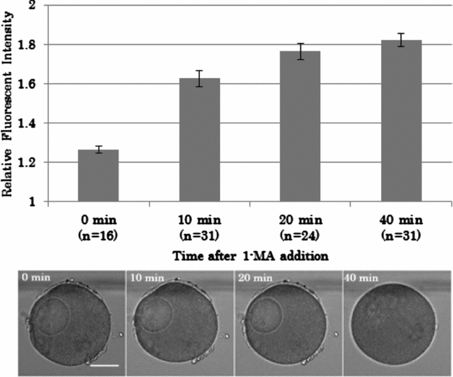
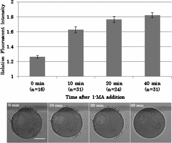
Oocyte maturation began after adding 1-MA, but GVBD did not occur until 20 min after the 1-MA application at 20°C (Fig. 1). After 20 min, the nuclear membrane became unclear, and then GVBD proceeded visibly. Nucleoplasm was mixed with the cytoplasm indistinguishably within 40 min. Sperm were added at 0, 10, 20, and 40 min after 1-MA application, and the Ca2+ response of the oocytes was examined. As previously shown (Chiba et al., Reference Chiba, Kado and Jaffe1990), the Ca2+ response in immature oocytes (not exposed to 1-MA) at fertilization was lower than that measured after GVBD (Fig. 1). The peak amplitude of the intracellular Ca2+ increase was 1.26 ± 0.02 RFU in the immature oocytes, while the value in the fully mature oocytes 40 min after the 1-MA treatment reached 1.82 ± 0.03 RFU (Fig. 1). However, Ca2+ response to sperm was comparably high in the ‘maturing’ oocytes at 10 min and 20 min after 1-MA application (1.63 ± 0.04 and 1.77 ± 0.04 RFU, respectively) when the germinal vesicle was still intact (Fig. 1).
Sperm-induced Ca2+ increase in the oocytes fertilized at different stages of maturation. Oocytes were inseminated at different intervals after application of 1-MA (0, 10, 20, and 40 min) and the peak levels of Ca2+ in the oocytes were examined as relative fluorescence intensities (top). The oocyte response without 1-MA addition is indicated by 0 min. The numbers below the time on the horizontal bar represent the number of oocytes examined. The error bars represent standard error. The images of the oocytes corresponding to the indicated time on the horizontal bar represent the meiotic stage after 1-MA application before insemination (bottom). Scale bar indicates 50 μm.
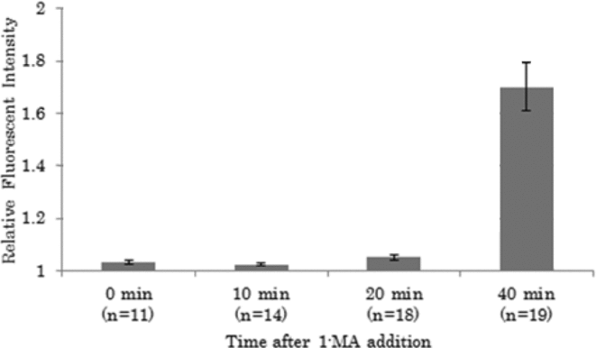
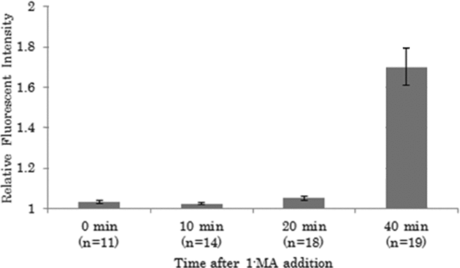
As the Ca2+ increase in matured oocytes during fertilization is suggested to be driven by IICR, the IP3 responses of oocytes at the three different meiotic stages were examined (Fig. 2). The Ca2+ release following IP3 uncaging in the oocytes cortex exposed to 1-MA addition for 40 min reached an amplitude of 1.70 ± 0.09 RFU. This is a significantly elevated level in comparison with the Ca2+ response to the same amount of IP3 uncaged in the immature oocytes (1.03 ± 0.01 RFU). Interestingly, IP3 uncaging in the maturing oocytes at the 10 or 20 min time points displayed no significantly higher Ca2+ response (1.02 ± 0.01 and 1.05 ± 0.01 RFP, respectively) in comparison with the average value scored by the immature oocytes P > 0.1), which is at striking variance with the aforementioned results obtained with fertilizing sperm.
Induction of Ca2+ response by IP3 in the oocytes during maturation. Oocytes preloaded with caged IP3 (1 μM in the needle) were induced to undergo maturation by 1-MA application. This was followed by uncaging of IP3 by ultraviolet (UV) light irradiation for 30 s at the indicated time after 1-MA treatment. The oocyte response without 1-MA addition is indicated by 0 min. Error bars indicate standard error.
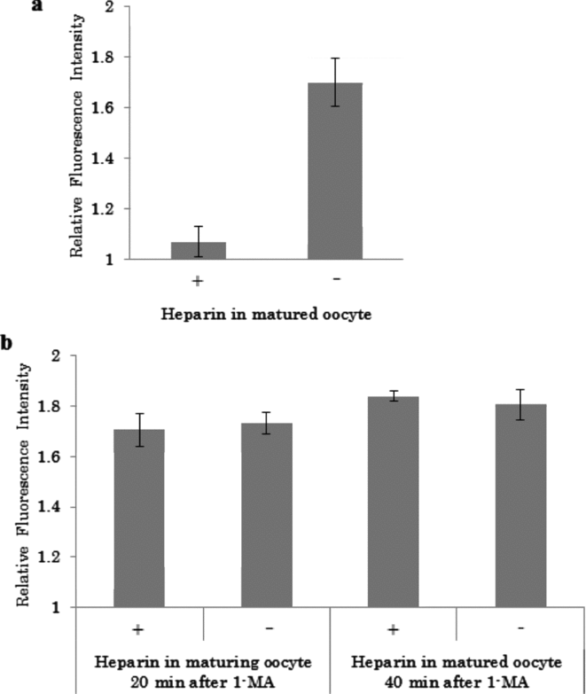
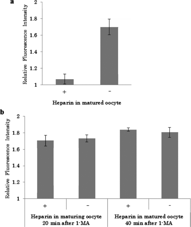
While heparin (10 mg/ml in pipette) pre-injected into matured oocytes 40 min after 1-MA application blocked the Ca2+ response by uncaged IP3 (Fig. 3 a), the same dose of heparin did not significantly affect the Ca2+ increase induced by fertilizing sperm in the maturing (20 min) and the matured (40 min after 1-MA application) oocytes (Fig. 3 b). Taken together, these results indicated that maturing oocytes might have developed a distinct Ca2+-releasing mechanism that is independent of IP3 before the IICR mechanism established itself.
The Ca2+ responses of maturing and matured oocytes to uncaged IP3 (a) and sperm (b) in the presence or absence of preloaded heparin. (a) Caged IP3 and calcium dyes were microinjected into oocytes either with (+) or without (−) heparin (10 mg/ml, concentration in the micropipette) Forty min after the exposure to 1-MA, the maturing oocytes with (n = 12) or without heparin (n = 13) were illuminated with UV (30 s) to photoactivate the caged IP3 (n = 12), and the peak value of Ca2+ increase was examined. (b) Oocytes were microinjected with calcium dyes with (+) or without (−) heparin and exposed to 1-MA to induce meiotic maturation. Either 20 min (maturing oocytes) or 40 min after the incubation with 1-MA (matured oocytes), oocytes microinjected with (n = 10) or without heparin (n = 27) were fertilized and the Ca2+ response of the eggs were examined. Error bars represent standard error.
Ca2+ responses to cADPr and NAADP in maturing oocytes
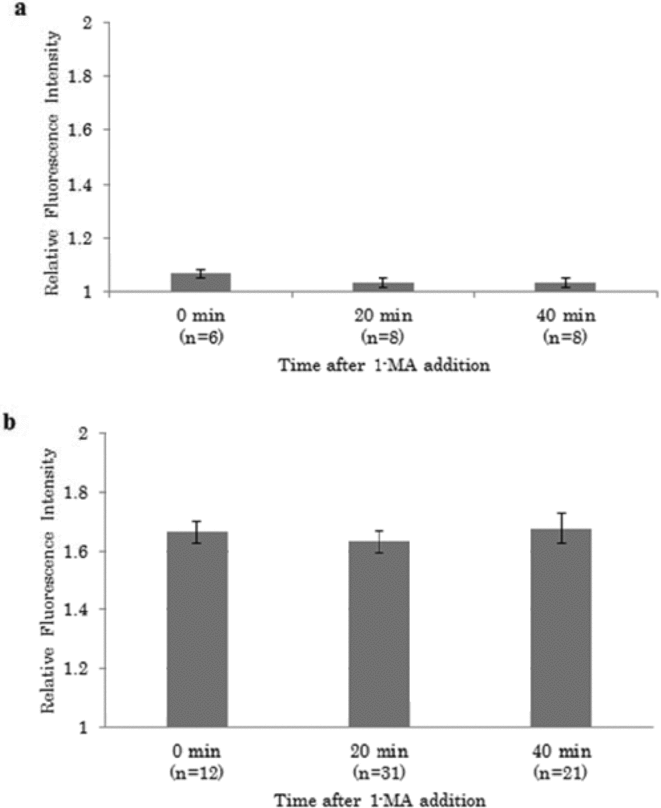
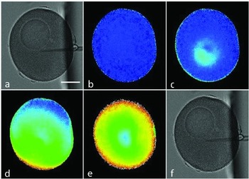
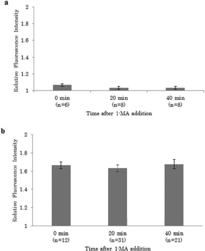
As aforementioned, the sperm-induced Ca2+ increase in maturing oocytes 20 min after 1-MA treatment was similar to that in matured oocytes (Fig. 1). As IICR was not yet fully operational in maturing oocytes, the Ca2+ increase in these oocytes could be attributed to other Ca2+-releasing second messengers. When cADPr (1 mM in pipette) was injected into immature oocytes (before 1-MA application), maturing oocytes (20 min after 1-MA application), or into matured oocytes (40 min after 1-MA application), all of these showed a faint Ca2+ increase without the occurrence of a Ca2+ burst (Fig. 4 a), suggesting that cADPr is unlikely to makes substantial contributions in shaping the Ca2+ wave at fertilization. In contrast, injection of another second messenger NAADP (100 μM in pipette) into immature, maturing and matured oocytes produced large amounts of Ca2+ release in all cases virtually to the same extent (Fig. 4 b). No significant differences were observed in the peak amplitudes of the Ca2+ increases after the microinjection of NAADP in immature (1.66 ± 0.04), maturing (1.63 ± 0.04), and matured oocytes (1.68 ± 0.05). The spatiotemporal pattern of the Ca2+ increase after NAADP injection in maturing oocytes was examined further (Fig. 5). During injection, NAADP was released from the tip of the micropipette near the centre of the oocyte (Fig. 5 a–c), and its diffusion was marked by a momentary Ca2+ increase near the micropipette tip. After a latent period of 20 s, however, a more robust Ca2+ increase started from the cortex (Fig. 5 d) and propagated to the centre of the oocyte (Fig. 5 e). The propagation pattern of Ca2+ after NAADP injection was similar to that in matured oocytes (Lim et al., Reference Lim, Kyozuka, Gragnaniello, Carafoli and Santella2001). We also confirmed that the propagation pattern of Ca2+ after NAADP injection in immature oocytes was similar to that in maturing or matured oocytes (data not shown). Thus, NAADP triggered a Ca2+ increase in the oocyte independently of the meiotic stage of the oocytes. Interestingly, the amplitude of the NAADP-induced Ca2+ increase in the immature oocytes was even higher than that induced by the fertilizing sperm.
Ca2+ responses of starfish oocytes to cADPr and NAADP at different stages of meiotic maturation. (a) cADPr (1 mM in pipet) was injected into the oocyte at 0, 20, and 40 min after 1-MA application, and the Ca2+ changes were measured. (b) NAADP (100 μM in pipet) was injected into the oocyte at 0, 20, and 40 min after 1-MA application and Ca2+ changes were measured. The oocyte response without 1-MA addition is indicated by 0 min. Error bars represent standard error.
Spatiotemporal changes of the intracellular Ca2+ increase after the microinjection of NAADP in maturing oocytes. NAADP (100 μM) was microinjected into maturing oocytes 20 min after 1-MA treatment. Bright field image (a) and fluorescence image (b) before microinjection. At 5 s after microinjection, a local Ca2+ increase occurred near the tip of the inserted needle (c). After a latent period, increased Ca2+ in the oocyte was observed from the cortex that propagated into the entire oocyte 20 s after injection (d) and 60 s after injection (e). Bright field image after injection (f). Scale bar indicates 50 μm.

Ca2+ responses to NADP, the precursor of NAADP, in the maturing oocytes
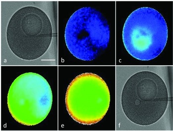
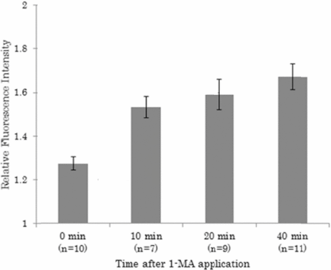
We noted that microinjection of NADP, the precursor of NAADP (Aarhus et al., Reference Aarhus, Graeff, Dickey, Walseth and Lee1995), also promoted a Ca2+ increase in the maturing oocytes. As shown in Fig. 6, the peak amplitudes of the Ca2+ response following the microinjection of NADP (5 mM in pipette) was progressively increased as the oocytes underwent meiotic maturation: immature (1.31 ± 0.04 RFU), maturing (at 10 min and 20 min after 1-MA application, 1.46 ± 0.06 and 1.55 ± 0.07 RFU, respectively), and matured oocytes (1.66 ± 0.06 RFU), ANOVA test (P = 0.00003). This result indicated that the Ca2+ store on which NADP acted was less sensitive in the immature oocytes not treated with 1-MA. Hence, the same amount of NADP releasing lesser intracellular Ca2+ in the immature oocytes than in the maturing or matured oocytes is at variance with what was observed with NAADP, which was equally effective regardless of the maturation status of the oocytes. In immature oocytes, NADP-induced a Ca2+ increase which was lower than that promoted by NAADP and higher of the sperm-induced Ca2+ release (see Fig. 1). The temporal and spatial analysis of the Ca2+ increase in maturing oocytes after NADP injection showed that it initially occurred at the centre of oocyte, but the major increase took place in the cortex after a certain latent period and then propagated centripetally (Fig. 7), a spatiotemporal pattern highly similar to that of NAADP-induced Ca2+ increases (Fig. 5).
Ca2+ responses of starfish oocytes to NADP at different stages of meiotic maturation. NADP (5 mM in pipet) was microinjected into the oocyte at different time points after 1-MA treatment, and the peak levels of Ca2+ increase after injection were examined as relative fluorescence intensities. The oocyte response without 1-MA addition is indicated by 0 min. Error bars represent standard error.
Spatiotemporal changes in intracellular Ca2+ after NADP injection in maturing oocytes. NADP (5 mM) was microinjected into maturing oocytes 20 min after 1-MA treatment. Bright field image (a) and fluorescence image (b) before the microinjection. After 5 s, a local Ca2+ increase occurred near the tip of the inserted needle (c). After a variable latent period, 20 s after injection (d) and 60 s after injection (e), the major Ca2+ increase started to be detected in an oocyte cortex and propagated into the entire oocyte. Bright field image after injection (f). Scale bar indicates 50 μm.

Development of the NADP-dependent Ca2+ release may be attributed to the maturation-associated increases in cytosolic Ca2+ and pH
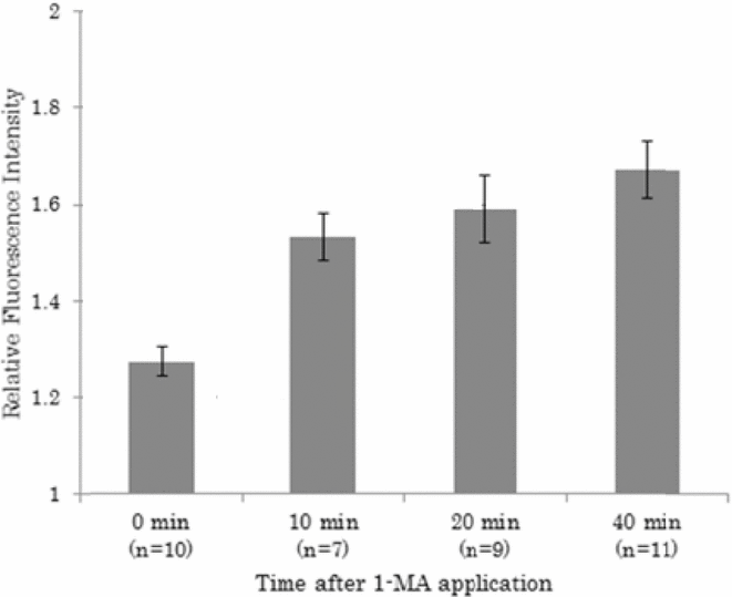
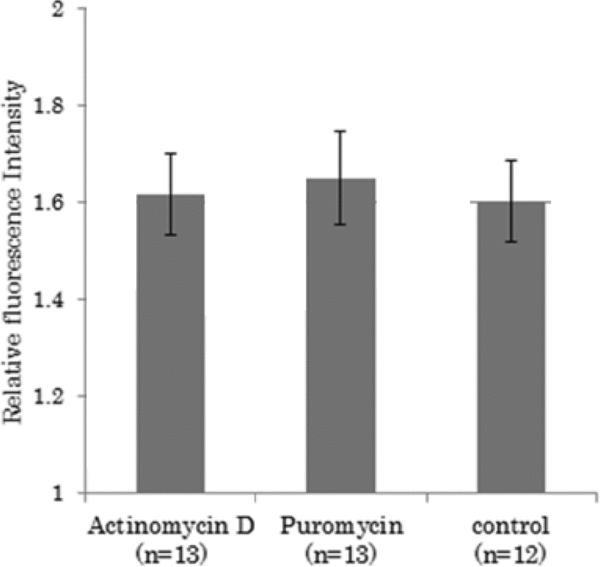
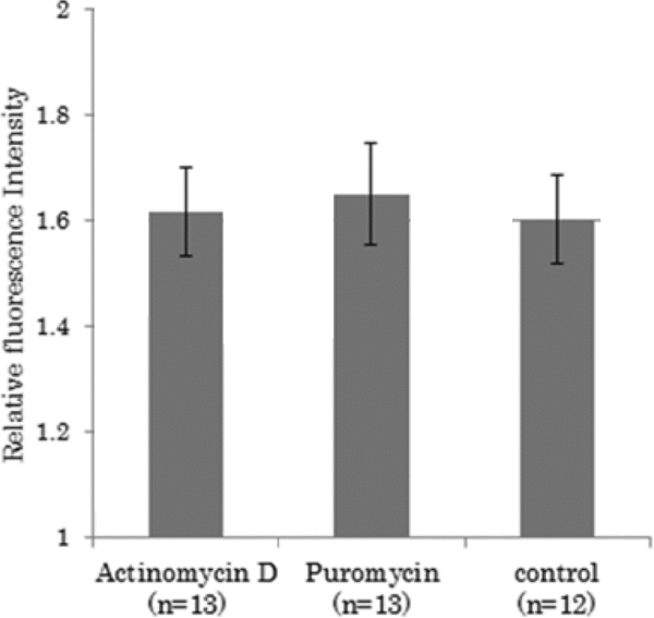
As NADP displayed only a low level of Ca2+ release in immature oocytes, a NADP-dependent Ca2+ release mechanism must develop during the oocyte maturation. We examined the necessity of protein synthesis for the activation of NADP-dependent Ca2+ release ability during oocyte maturation. When NADP was injected in oocytes matured for 40 min with 1-MA in the presence of actinomycin D (100 μg/ml) or puromycin (800 μg/ml) for 40 min, the increase in intracellular Ca2+ (1.64 ± 0.01 RFU and 1.60 ± 0.05 RFU, respectively) was as high as that of the control oocytes matured without the drugs (1.62 ± 0.07 RFU) (Fig. 8). These results indicate that protein synthesis was not required for the development of the NADP-dependent Ca2+ release mechanism.
Inhibition of protein synthesis had no effect on the sensitization of the maturing oocytes to NADP. NADP was injected in matured oocytes, 40 min after incubation in 1-MA seawater containing actinomycin D (100 μg/ml final), puromycin (800 μg/ml final), and 1-MA seawater.
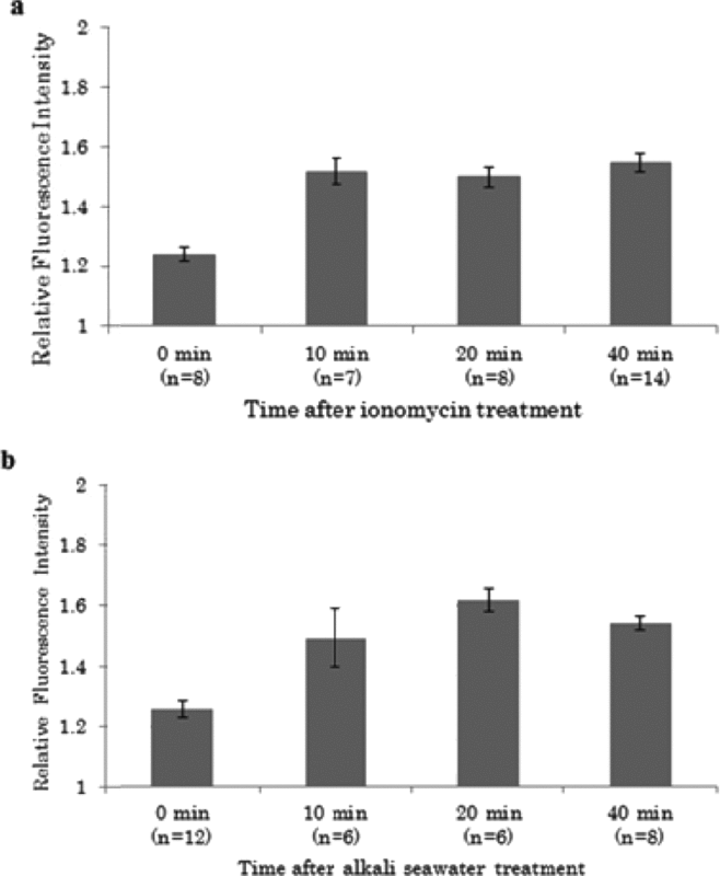
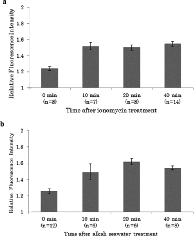
After the meiotic reinitiation with 1-MA, starfish oocytes manifest not only a rise of a Ca2+ transient but also a considerable increase in intracellular pH (Santella & Kyozuka, Reference Santella and Kyozuka1994; Harada et al., Reference Harada, Oita and Chiba2003). Thus, we then examined the effects of intracellular Ca2+ and pH increases on the activation of NADP-dependent Ca2+ release in the absence of 1-MA. To this end, immature oocytes were treated with ionomycin (IM; 1 μM final concentration) for 3 min, washed in seawater, and then injected with NADP at 0 min, 10 min, 20 min, and 40 min after the IM treatment (Fig. 9 a). Each measurement was started 1 min before injection and no significant increase of intracellular Ca2+ was observed during that period. The peak level of Ca2+ increase in the oocyte was already high 10 min after the treatment (1.52 ± 0.04 RFU versus 1.24 ± 0.02 RFU in the control without IM treatment, P < 0.01). The Ca2+ response upon NADP injection maintained high values such as 1.50 ± 0.04 at 20 min, and 1.55 ± 0.03 at 40 min after without 1-MA application. Similarly, we artificially increased the cytosolic pH by continuously incubating the oocytes in seawater containing 1 mM NH4Cl, pH 7.5. When NADP was microinjected in these immature oocytes, the intracellular Ca2+ increase in response to NADP was significantly enhanced (1.50 ± 0.10 RFU) after 10 min in comparison with the control oocytes (1.26 ± 0.03 RFU). The elevated responsive to NADP was attained after more than 40 min as the Ca2+ response to NADP remained high at 20 min (1.62 ± 0.04 RFU) and 40 min (1.54 ± 0.02 RFU) (Fig. 9 b). These results indicate that transient Ca2+ increase and gradual increase in intracellular pH after reinitiation of meiosis may be causal to maturation of the NADP-dependent Ca2+ releasing pathway.
Effect of increased Ca2+ (a) and pH (b) on the activation of NADP-dependent Ca2+ increase in immature oocytes. (a) Immature oocytes were pre-treated with 1 μM ionomycin for 3 min and then washed in seawater. After 10, 20, and 40 min, 5 mM NADP was injected into oocytes of each meiotic stage. The horizontal bar represents time after treatment and washing. The oocyte response without 1-MA addition, is indicated by 0 min. The vertical bar represents relative fluorescence intensity. Numbers below each time point on the horizontal bar indicate the number of oocytes examined. (b) Immature oocytes were treated with seawater containing 1 mM NH4Cl (pH 7.5). After 10, 20, and 40 min, 5 mM NADP were injected into oocytes of each meiotic stage. The horizontal bar represents the incubation time in NH4Cl-containing seawater. The oocyte response without the incubation in NH4Cl-containing seawater is indicated by 0 min. The vertical bar represents relative fluorescence intensity. The numbers below each time point on the horizontal bar indicate the number of oocytes examined.
Discussion
In the fertilized eggs of starfish, the major Ca2+ increase initiates from the region of sperm contact and propagates to the entire oocyte as a single Ca2+ wave (Stricker et al., Reference Stricker, Centonze and Melendez1994). Injection or uncaging of IP3 induces a Ca2+ increase in oocytes (Stricker, Reference Stricker1995; Lim et al., Reference Lim, Kyozuka, Gragnaniello, Carafoli and Santella2001) and the Src-PLC gamma pathway is involved in IP3 production (Carroll et al., Reference Carroll, Ramarao, Mehlmann, Roche, Terasaki and Jaffe1997; Giusti et al., Reference Giusti, Xu, Hinkle, Terasaki and Jaffe2000; Jaffe et al., Reference Jaffe, Giusti, Carroll and Foltz2001). IP3 receptors are detected in immature and matured oocytes (Iwasaki et al., Reference Iwasaki, Chiba, Uchiyama, Yoshikawa, Suzuki, Ikeda, Furuichi and Mikoshiba2002). Thus, the IICR mechanism might contribute to the intracellular Ca2+ wave in the fertilized eggs of starfish. The IICR mechanism is known to develop during the oocyte's maturation to the GVBD stage (Chiba et al. Reference Chiba, Kado and Jaffe1990; Lim et al., Reference Lim, Ercolano, Kyozuka, Nusco, Moccia, Lange and Santella2003), but here we have shown that the oocytes exposed to 1-MA merely for 10 min, thus not having reached the GVBD stage, already display a high level of Ca2+ increase when fertilized (see Fig. 1). In support of the idea that the fertilization Ca2+ wave in these partially matured oocytes may not be based on the IP3 pathway, application of heparin did not block sperm-induced Ca2+ increase in these oocytes (Fig. 3). Similar results questioning the exclusive involvement of IP3 in the Ca2+ waves have been reported in the fertilized eggs of sea urchin and starfish (Crossley et al., Reference Crossley, Whalley and Whitaker1991; Santella et al., Reference Santella, De Riso, Gragnaniello and Kyozuka1999; Puppo et al. Reference Puppo, Chun, Gragnaniello, Garante and Santella2008). We confirmed that preloading of heparin in maturing oocytes did not suppress the Ca2+ increase after insemination, suggesting that a Ca2+-mobilizing system independent of IP3 is already functional before the completion of the maturation process and may contribute to the generation of the Ca2+ wave at fertilization.
In this context, we directed our attention to other Ca2+-mobilizing second messengers, namely NAADP and cADPr. In our study, cADPr did not induce a Ca2+ increase in matured eggs of A. pectinifera (Kyozuka et al. Reference Kyozuka, Chun, Puppo, Gragnaniello, Garante and Santella2008). The concentration of injected cADPr (1 mM in the pipette) was 10 times higher than that required to induce a Ca2+ increase along with elevation of the vitelline envelope in sea urchin, L. pictus (100 μM, Lee et al., Reference Lee, Aarhus and Walseth1993) and Strongylocentrotus nudes (100 μM, data not shown). Hence, it is unlikely that the cADRr-dependent pathway contributes to the generation of the Ca2+ wave in the pre-GVBD oocytes at fertilization. At variance with A. pectinifera, in Astropecten aranciacus, cADPr is able to release Ca2+ in immature oocytes, but the peak level of Ca2+ increase is slightly low and the spatial pattern of Ca2+ increase is different from that in matured oocytes. Interestingly, the Ca2+ response following uncaging of cADPr in the matured oocytes of this starfish species is linked to the presence of Ca2+ in seawater (Nusco et al., Reference Nusco, Lim, Sabala and Santella2002). Conversely, there is a possibility that NAADP might be involved in this presence. In A. pectinifera, we found that the NAADP-dependent Ca2+ release mechanism was already functional in immature oocytes. As the Ca2+ increase in the NAADP-dependent pathway is not influenced by heparin, our observation of the heparin-insensitive Ca2+ wave in the maturing oocytes at fertilization raises the possibility that NAADP might be instrumental in this process. Curiously, we found that the NAADP-induced Ca2+ mobilizing mechanism does not become sensitized along with meiotic maturation of the oocytes, which was at variance with the IP3-dependent Ca2+-releasing mechanism. Nonetheless, it has been suggested that NAADP may play a role in shaping the Ca2+ wave in the fertilized eggs, especially at the initial stage. In sea urchins, it has been shown that sperm contains micromolar levels of NAADP, and it has been suggested that sperm could activate sea urchin eggs by injecting NAADP upon fusion (Billington et al., Reference Billington, Ho and Genazzani2002; Churchill et al., Reference Churchill, O'Neill, Masgrau, Patel, Thomas, Genazzani and Galione2003).
Unlike in the case of NAADP, the ability of the oocytes to respond to NADP with a Ca2+ was enhanced within 10 min after 1-MA application. Whatever it may mean, the changes underlying this shift did not require synthesis of new proteins. Actinomycin D (100 μg/ml) or puromycin (800 μg/ml) block protein synthesis in oocytes of starfish Marthasterias glacialis, but GVBD still proceeded in the presence of these inhibitors (Guerrier & Doree, Reference Guerrier and Doree1975). Similarly, our results using the same inhibitors concentration did not affect the development of NADP-dependent Ca2+ release. Thus, the oocytes’ sensitization to NADP seems to represent one of the changes related to meiotic resumption that do not require protein synthesis. Although it is not explainable how oocytes get sensitized to NADP during meiotic maturation, an analogy may be drawn from the case of IICR. In starfish oocytes IP3 receptors are already present inside immature oocytes, and their activities become enhanced along with oocyte maturation presumably by optimally re-positioning themselves in the subcellular domains (Iwasaki et al., Reference Iwasaki, Chiba, Uchiyama, Yoshikawa, Suzuki, Ikeda, Furuichi and Mikoshiba2002) Indeed, the NADP-induced Ca2+ releasing activity was potentiated in the cortical region of the oocyte within 10 min after 1-MA application. Immediately after application of 1-MA, surface topography of immature oocyte changes with reorganization of actin cytoskeleton in the cortex (Schroeder & Stricker, Reference Schroeder and Stricker1983; Chun & Santella, Reference Chun and Santella2009). In A. aranciacus, the increased sensitivity to IP3 is linked to the structural organization of the actin cytoskeleton occurring during oocyte maturation (Lim et al., Reference Lim, Ercolano, Kyozuka, Nusco, Moccia, Lange and Santella2003). In a future study, it would be interesting to know if sensitization of the Ca2+-releasing activity in response to NADP is also modulated by the actin cytoskeleton-related changes in the oocyte cortex. Alternatively, NADP might be differentially metabolized in the maturing oocytes to give a rise to a more active metabolite that evokes potent Ca2+ release, e.g. NAADP.
At the reinitiation of meiosis by 1-MA, a transient Ca2+ increase is observed both in cytoplasmic and nuclear domains. The Ca2+ wave starts about 2 min after 1-MA addition at the vegetal hemisphere of the oocyte cortex and propagates in 20 s to the animal hemisphere (Santella & Kyozuka, Reference Santella and Kyozuka1994; Santella et al., Reference Santella, De Riso, Gragnaniello and Kyozuka1998). In addition to this change, a novel Ca2+ spike was recently discovered in the maturing oocytes of starfish at the time of GVBD (Limatola et al., Reference Limatola, Chun, Kyozuka and Santella2015). Interestingly, both of these early and late Ca2+ responses were shown to be heavily affected by the structural organization of the actin cytoskeleton of the cortex (Kyozuka et al. Reference Kyozuka, Chun, Puppo, Gragnaniello, Garante and Santella2008, Santella et al., Reference Santella, Limatola and Chun2015). In parallel with the Ca2+ increase, the maturing oocytes of starfish display a gradual increase in cytosolic pH, which is shifted from 7.1 to 7.4 before GVBD and stays elevated for over 20 min (Harada et al., Reference Harada, Oita and Chiba2003). As activities of many intracellular proteins are modulated by Ca2+ and cytosolic pH, the changes of these two parameters during the 1-MA-induced meiotic maturation may play important roles as demonstrated by a brief episode of artificial Ca2+ surge in immature oocytes (Vasilev et al., Reference Vasilev, Chun, Gragnaniello, Garante and Santella2012).
To test if the increases of Ca2+ and pH that take place during meiotic maturation is responsible for the sensitization of the NADP-induced Ca2+ increase, we have artificially changed the cytosolic Ca2+ level and pH by use of an ionophore and alkaline conditioned seawater. Injection of NADP in immature oocytes within 10 min after ionomycin treatment induced a Ca2+ increase similar to that elicited by NADP in oocytes incubated in 1-MA. Similar to seawater conditioned with a weak alkali NH4Cl that was designed to increase the intracellular pH in oocytes (Deguchi & Osanai, Reference Deguchi and Osanai1994), microinjection of NADP induced a Ca2+ increase that mimicked the response in the oocytes treated with 1-MA. In both cases that artificially increased cytosolic Ca2+ and pH, the effect of sensitizing the oocytes to NADP was observed only during the first 10 min of incubation, while prolonged incubation had no additive effect. As this first 10 min overlaps the hormone-dependent period for meiotic maturation (Guerrier & Doree, Reference Guerrier and Doree1975; Nemoto, Reference Nemoto1982), it can be speculated that the Ca2+ and pH increase might act on the factors that are momentarily active in this period. As to the question of how fertilizing sperm initiates the Ca2+ wave in the egg, there are two main hypotheses: the ‘receptor hypothesis’ and the ‘sperm factor hypothesis’ (Miyazaki, Reference Miyazaki2006; Santella et al., Reference Santella, Lim and Moccia2004). In the former case, sperm bind to the specific receptor on the oocyte and transmit the activating signal to the oocyte interior and induce the Ca2+ increase. The other hypothesis is that sperm directly introduce molecules such as PLC directly into the oocyte after the fusion of the gamete membranes. Mammalian sperm introduce sperm-specific PLC, PLC zeta, into the oocyte and induce IP3 production (Saunders et al., Reference Saunders, Larman, Parrington, Cox, Royse, Blayney, Swann and Lai2002), but the isotype was not found in echinoderm, implying a different mechanism at work
Sea urchin sperm possess high levels of NAADP, raising the possibility that this potent Ca2+-mobilizing agent could serve as a sperm factor that directly activates the eggs when infused (Churchill et al., Reference Churchill, O'Neill, Masgrau, Patel, Thomas, Genazzani and Galione2003; Billington et al., Reference Billington, Ho and Genazzani2002). In this study, we have demonstrated that starfish oocytes are indeed highly receptive to NAADP even at the GV stage. While an increase in cytoplasmic Ca2+ by intracellular injection of sperm would provide strong evidence for introduction of molecules related to Ca2+ release directly into the oocyte, no such data have been reported to date on the Ca2+ burst following the sperm injection in starfish eggs. Nonetheless, it is noteworthy that NADP is synthesized in oocytes (Epel, Reference Epel1990), and that sperm sensitizes the nicotinamide nucleotides metabolic pathway in ascidian eggs at fertilization (Wilding et al., Reference Wilding, Russo, Marino, Grumetto, De Simone, Tosti and Dale1998a, Reference Wilding, Russo, Galione, Marino and Daleb). Furthermore, the enzymatic activity of NAD kinase, which is involved in the production of NADP, is controlled by Ca2+ (Epel et al., Reference Epel, Patton, Wallace and Cheung1981). In support of the idea that NADP may also play a role in generating the Ca2+ wave, it was reported that NAD kinase is activated within 30 s to 1 min after sperm incorporation in sea urchin eggs. When sperm bind to the egg surface, a cortical flash occurs, which is then followed by a Ca2+ wave propagating from the sperm–egg interaction to the opposite pole (Miyazaki, Reference Miyazaki2006; Moccia et al., Reference Moccia, Nusco, Lim, Kyozuka and Santella2006). Therefore, in theory, a cortical flash by the fertilizing spermatozoon might supply the priming Ca2+ to activate NAD kinase for NADP production.
An important aspect of this work is that the Ca2+ response induced by sperm is not initiated and propagated by IP3 and cADPr, respectively, as in immature oocytes the IP3-dependent Ca2+ stores are insensitive to this Ca2+-linked second messenger and cADPr does not release Ca2+ when injected into this species of starfish. Furthermore, Ca2+ stores sensitive to NAADP and NADP are functional in the cortex of immature oocytes. In the light of these findings, immature oocytes of the starfish A. pectinifera constitute an excellent experimental animal model for the study of the biochemical mechanisms of sperm-induced signal transduction and for the understanding of the nature of the Ca2+ stores from which Ca2+ is liberated at fertilization.
Acknowledgements
We thank Dr Ritsu Kuraishi (Keio University) for supply of animals from Tokyo and Drs Luigia Santella and Jong Tai Chun (Stazione Zoologica Anton Dohrn) for useful discussions and comments on the manuscript.