Case report
A 20-year-old female patient was referred to our cardiac centre from a peripheral clinic for cardiac evaluation. She had complaints of worsening dyspnoea on exertion and easy fatigability. Her physical examination revealed a thin built girl with displaced apex beat, grade III/VI pansyatolic murmur at upper left sternal edge, and grade III/VI pansystolic murmur at apex radiating to axilla and loud pulmonary component of second heart sound. There was no cyanosis or clubbing.
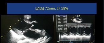
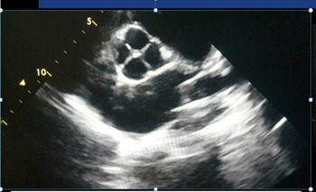
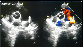
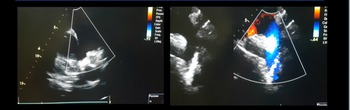
Her 12-lead electrocardiogram showed sinus rhythm and her chest X-ray revealed cardiomegaly. Definitive diagnosis was made on transthoracic echocardiogram that showed dilated left atrium and left ventricle with good biventricular function and no resting regional wall motion abnormality (Fig 1). She had a large patent ductus arteriosus with left to right shunt mainly in systolic phase, X shaped quadricuspid aortic valve with trivial aortic regurgitation (Figs 2 and 3) with pressure half-time was more than 500 ms. Quadricuspid aortic valve was classified as type A as four cusps were of equal sizes (Fig 2). Her left heart was volume loaded, and her M mode derived left ventricular diastolic dimension in parasternal long axis view was 72 mm (z score > 6) (Fig 1) along with a grade II/IV mitral valve regurgitation. She also had mild tricuspid regurgitation with peak instantaneous pressure gradient of 60 mmHg. After discussion with the patient and family, cardiac catheterisation and angiography and haemodynamic study were planned with view to offer her patent ductus arteriosus device closure if found suitable anatomically and haemodynamically.
Transthoracic echocardiography PLAX diastolic view: left ventricle M – Mode.

Transthoracic echocardiography parasternal short diastolic view: X shaped QAV.

Parasternal short-axis view showing large PDA with left to right shunt.

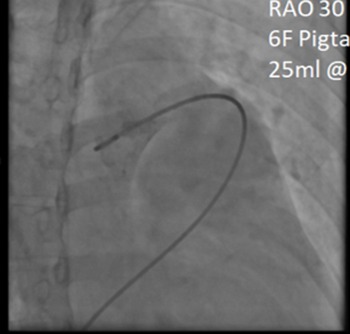
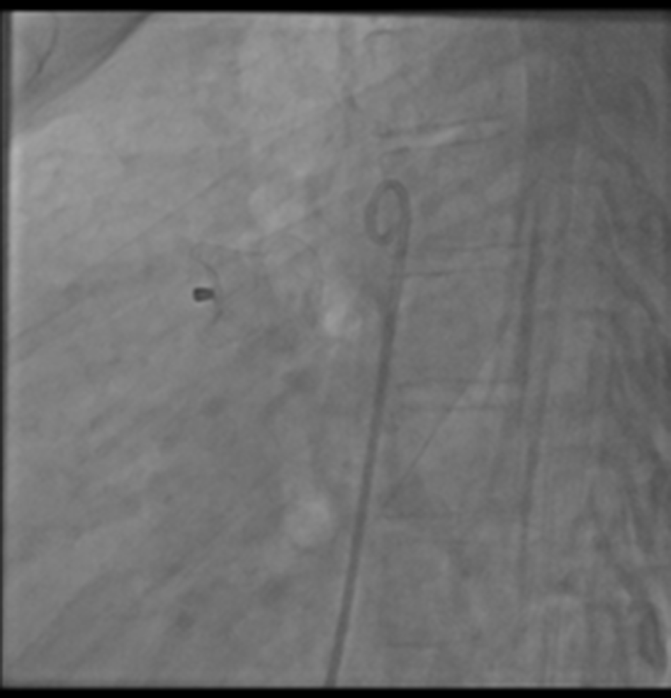
After written consent, she was taken to catheterisation lab. Right side of the groin was used to access her femoral artery and vein. Her haemodynamic study revealed aortic pressure of 135/62/85 mmHg, pulmonary artery – 96/60/72 mmHg, RV – 100/10 mmHg, RA – 10 mmHg, and left ventricle – 145/10 mmHg. Her ratio of pulmonary to systemic flow (Qp:Qs) was 2:1 in air. An aortogram in true lateral (LAO 90) and slight RAO view showed a large patent ductus arteriosus (Fig 4), though not optimally visualised in spite of few aortogram in various views, so echocardiographic measurements were repeated for size of duct and choice of occluder. As her pulmonary artery pressure was about two-third of the systemic pressures, therefore, it was decided to proceed with the device closure of patent ductus arteriosus. As her patent ductus arteriosus measured about 14–15 mm, so a large duct occluder was chosen. A 10F delivery sheath advanced from right femoral vein, crossing patent ductus arteriosus ante-grade and was parked in the descending aorta. A 20/18 mm patent ductus arteriosus device (Shashma, SHSMA Corporation, Shanghai, China) was loaded and advanced through the delivery sheath and was deployed across the patent ductus arteriosus. A repeat haemodynamic assessment was done while device remained attached to the delivery cable. Her pulmonary artery pressure dropped to almost half of systemic pressure, and after angiogram, transthoracic echocardiogram showed complete occlusion of the duct, the device was released (Fig 5). She was kept admitted in the hospital for the next 24 hours. Her echocardiogram at discharge and after 2 weeks showed complete occlusion of the patent ductus arteriosus with no residual leak and laminar flow across LPA and descending aorta (Fig 6). Her LV dimensions also improved and she reported improved health and exercise capacity.
Aortogram in RAO 30 view with 6 F pigtail catheter showing faintly visualised large PDA.

Post PDA device closure (LAO 90) showing well- placed device.

Post PDA device closure, transthoracic echo sowed well-placed PDA device with no residual leak and laminar flow in LPA and Aorta.

Discussion
Normally, aortic valve is tricuspid, separating left ventricle from the ascending aorta. However, in some people, aortic valve can be unicuspid, bicuspid, or rarely even quadricuspid. Reference Oladiran, Nwosu, Dhital and Ezioma1 Quadricuspid aortic valve is a remarkably rare congenital cardiac abnormality with definitive embryological explanation. Reference Sharif, Azzam and Sharif-Rasslan2–Reference Seol, Kim, Cho, Kim, Kim and Kim7 The estimated incidence of quadricuspid aortic valve is variable ranging from 0.008% to 1.46%, depending upon the studied population and imaging modalities used. Reference Oladiran, Nwosu, Dhital and Ezioma1,Reference Veronese, Brandão, Steffen, Pomerantzeff and Jatene8,Reference Hayakawa, Asai, Kinoshita and Suzuki9 Hayakawa et al from Japan reported an incidence of 0.48% among 627 patients undergoing aortic valve surgery; all cases had significant aortic regurgitation and required surgical aortic valve replacement. Reference Hayakawa, Asai, Kinoshita and Suzuki9
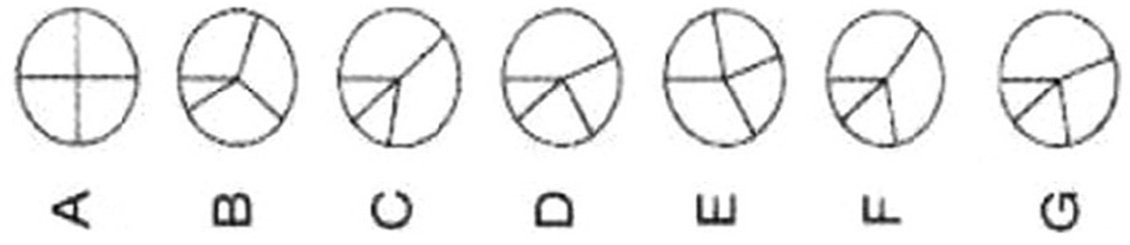
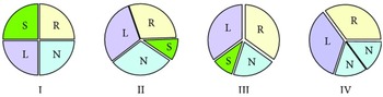
Quadricuspid aortic valve is described by the presence of four aortic valve leaflets, which can be either equal in size or variable in regard to each other. Reference Oladiran, Nwosu, Dhital and Ezioma1 Though, there is no definite gender preference, it is more commonly reported in female patients. Reference Sharif, Azzam and Sharif-Rasslan2 The clinical presentation depends upon functional status of the quadricuspid aortic valve, left ventricular systolic and diastolic function, and associated cardiac or coronary anomalies. Reference Yuan10 Prior to the widespread use of echocardiogram, most cases were diagnosed during cardiac surgery or at autopsy. Reference Hayakawa, Asai, Kinoshita and Suzuki9 Presently, with the use of two-dimensional/three-dimensional echocardiogram, cardiac CT, and cardiac MRI, quadricuspid aortic valve can be reliably diagnosed along with its sequel on heart. Reference Garg, Garg, Agrawal and Sharma11,Reference Suraci, Horvath, Urina, Rosen and Santana12 Quadricuspid aortic valve is straightforwardly visualised on transthoracic echocardiogram with a characteristic X pattern of aortic valve in diastole. Reference Garg, Garg, Agrawal and Sharma11 These patients generally remain asymptomatic without any significant auscultatory finding during childhood; hence, a diagnosis is rarely made in early years. Reference Brock, Tugertimur and Schwartz13 With the passage of time, these patients develop progressive aortic valve regurgitation as a result of fibrosis and incomplete coaptation and about half of them eventually require either aortic valve replacement or repair. Reference Lin, Yin and Wang5,Reference Seol, Kim, Cho, Kim, Kim and Kim7,Reference Hayakawa, Asai, Kinoshita and Suzuki9,Reference Garg, Garg, Agrawal and Sharma11,Reference Brock, Tugertimur and Schwartz13–Reference Pasanisi, Mazzanti and Sassone15 Aortic valve stenosis and aortic root dilatation are less common in comparison to patients with bicuspid aortic valve. Reference Sharif, Azzam and Sharif-Rasslan2,Reference Savino, Quintavalle and Ambrosio6 Quadricuspid aortic valves were classified into seven types by Hurwitz and Roberts (Fig 7). Reference Brock, Tugertimur and Schwartz13,Reference Hurwitz and Roberts16 Of these, types A, B, and C account for more than 85% of the cases. Reference L'Angiocola, Vetrano and Lardieri4 Nakamura et al designed a more simplified classification by focusing on the position of the supernumerary cusp (Fig 8). Reference Oladiran, Nwosu, Dhital and Ezioma1,Reference Nakamura, Taniguchi, Saiki, Morimoto and Yamaga17
Seven anatomic variations of QAV, as presented by Brock M et al. Reference Brock, Tugertimur and Schwartz13

Nakamura et al classification of QAV as presented by Oladiran O et al. Reference Oladiran, Nwosu, Dhital and Ezioma1

The association of quadricuspid aortic valve with patent ductus arteriosus was first reported in 1923 by Simonds as a postmortem finding. Reference Simonds18 Other associated anomalies are rare and includes coronary artery anomalies, Reference Hayakawa, Asai, Kinoshita and Suzuki9 non-obstructive cardiomyopathy, fibromuscular sub-aortic stenosis, Reference Seol, Kim, Cho, Kim, Kim and Kim7 rupture sinus of Valsalva, Reference Nakano, Waseda, Ando, Tanabe, Niwa and Amano19–Reference Akerem Khan, Tamin, Burkhart, Araoz and Young21 atrial septal defect, Reference Garg, Garg, Agrawal and Sharma11,Reference Saito, Yoshimura, Miyatsu and Ikeda22 partial anomalous pulmonary venous connection, Reference Doğan, Arısoy, Memiç and Dağlı23 ventricular septal defect, Reference Tatari, Dehaki, Omrani, Ghaheri, Al-Dairy and Mortezaeian24 transposition of great arteries, Reference Erdmenger, Vázquez-Antona and Becerra25 tetralogy of Fallot, Reference Stanescu and Branidou26,Reference Maheshwari, Tanwar and Mittal27 pulmonary valve stenosis, and congenital complete heart block. Infective endocarditis is also a potential complication Reference Seol, Kim, Cho, Kim, Kim and Kim7,Reference Garg, Garg, Agrawal and Sharma11,Reference Jackson, Sarwar, Hwang, Joglekar and Alsafwah28 with reported incidence of 1.4%. Reference Savino, Quintavalle and Ambrosio6
We are reporting a case of young girl with a quadricuspid aortic valve with mild aortic valve regurgitation, large patent ductus arteriosus, and volume-loaded left heart who had a successful device closure of her patent ductus arteriosus. To the best of our knowledge, such a case is being reported from Pakistan for the first time.
We successfully managed a young girl and quadricuspid aortic valve with mild aortic regurgitation and a large patent ductus arteriosus with volume-loaded LA and LV. It is a rare association as incidence of quadricuspid aortic valve itself varies from 0.008% to 0.043% depending upon the study population and diagnostic modality. Reference Savino, Quintavalle and Ambrosio6 More frequently, quadricuspid aortic valve is an isolated finding but may be associated with other heart lesions. Reference Savino, Quintavalle and Ambrosio6 Tsang et al reported their experience of 50 patients with mean age of 43.5 years at the time of diagnosis along with 32% associated findings including abnormalities of other cardiac valves, septal defects, persistent left superior caval vein, and patent ductus arteriosus. Reference Tsang, Abudiab and Ammash29 Patients with quadricuspid aortic valve should also be carefully evaluated for functional status of aortic valve, cardiac function, and other congenital abnormalities. Reference Seol, Kim, Cho, Kim, Kim and Kim7
Our case was of a young girl, who presented to us for the first time with complaints of worsening dyspnoea on exertion and fatigability. It is unfortunate to see grown-up patients with easily amenable congenital cardiac defects in developing countries due to lack of health education and infrastructure. The quadricuspid aortic valve was an incidental finding, her major issue was a large unrestricted patent ductus arteriosus with moderate pulmonary hypertension. Her patent ductus arteriosus was measured about 14–15 mm at its narrowest diameter on transthoracic echocardiogram (Fig 5). She underwent successful device closure of her patent ductus arteriosus as her haemodynamics was found to be favourable on cardiac catheterisation study (details are in the case report section). Her case again confirmed our observation and belief that large patent ductus arteriosus behaves unpredictably in different patients especially in regard to the development of severe or irreversible pulmonary hypertension. Our patient had type A quadricuspid aortic valve and had only mild aortic valve regurgitation. As aortic regurgitation is progressive in these patients, thereupon a close follow-up was recommended.
In contrast to bicuspid aortic valve, quadricuspid aortic valve progressively becomes regurgitant. The cases of aortic valve stenosis or ascending aorta dilatation are less common in this cohort. Reference Savino, Quintavalle and Ambrosio6 In a study by Cho et al, 26 patients with quadricuspid aortic valve were identified from 1994 to 2012. Aortic regurgitation was present in 20 of these patients (77%), and the most frequent valve morphology type was three equally sized cusps and one smaller cusp (n = 12; 46%), followed by two larger equally sized cusps and two smaller equally sized cusps (n = 6; 23%). Reference Cho, Son and Park30 The severity of aortic regurgitation appeared to be correlated with patient’s age and valve morphology.
In patients with a satisfactory echo window, transthoracic echocardiography can offer comprehensive diagnosis of quadricuspid aortic valve and associated anomalies. If transthoracic echo window is suboptimal, use of other diagnostic modalities including three-dimensional echocardiography, trans-esophageal echocardiography, CT, and MRI may be warranted. Reference Garg, Garg, Agrawal and Sharma11 We used only transthoracic echocardiogram to diagnose the index case. Echocardiography was also helpful in selecting the size of patent ductus arteriosus occluder as repeated aortogram failed to ideally visualise the patent ductus arteriosus in spite of high volumes of contrast (Fig 6).
In patients with quadricuspid aortic valve with significant aortic valve regurgitation, surgical aortic valve replacement, or repair may be indicated depending upon symptoms, echocardiography findings and relevant centre’s experience and preferences. Reference L'Angiocola, Vetrano and Lardieri4,Reference Lin, Yin and Wang5,Reference Pasanisi, Mazzanti and Sassone15,Reference Hasegawa, Tanaka and Okita31,Reference Kawashima, Komiya, Tsuneyoshi, Shimamoto, Sakai and Hiraoka32 More recently, there have been case reports of safe use of trans-catheter aortic valve implantation in selected patient. Reference Aoyama, Futami, Tanaka, Takeda, Nishimura and Tobaru33,Reference Benkemoun, Bramlage and Beaufigeau34 As patients with quadricuspid aortic valve have tendency to progressively develop aortic regurgitation, careful and regular follow-up in these patients is recommended.
Conclusion
Quadricuspid aortic valve is a rare congenital cardiac anomaly, and patent ductus arteriosus association is exceedingly rare. The importance of diagnosing quadricuspid aortic valve lies in the fact that many will require valve repair or replacement because of worsening aortic valve regurgitation or stenosis. Patients should undergo regular and close follow-up to avoid morbidity, mortality, and complications associated with LV decompensation.
Authors’ contributions
All authors contributed significantly in manuscript preparation.
Financial support
This research received no specific grant from any funding agency, commercial, or not-for-profit sectors.
Conflicts of interest
None.