Implications
Using an appropriately validated gelatin zymography, role played by gelatinases in normal discharge of the placenta at bovine parturition was implied based on our findings of higher gelatinase expressions in the area of the placenta closer to the umbilical insertion point and in the section of the umbilical cord closer to the rupture site. These findings observed on the non-retaining placenta of cows could serve as a control model when contrasting with the retaining placenta, and might help in strategic prevention of the retention of fetal membrane in dairy cows management.
Introduction
Bovine placenta is classified as ‘cotyledonary synepitheliochorial’ type (Wooding and Wathes, Reference Wooding and Wathes1980), which the trophoblast cells of bovine embryo fuse together to form multi-nucleated syncytium, with each nucleus remaining diploid (Klish et al., Reference Klish, Hecht, Pfarrer, Schuler, Hoffmann and Leiser1999). In contrast with the species of invasive placenta, bovine endometrium essentially remains intact as the syncytium chorioallantois grows and expands within the lumen of the uterus (Cross et al., Reference Cross, Nakano, Natale, Simmons and Watson2006). The syncytium chorioallantois lies in simple flat apposition with the uterine epithelium, except in the zonary fetomaternal unit named placentome, in which the epithelio-chorial becomes more intimate by interdigitating the cotyledonary villous projections (fetal compartment) with specialized recessive sites on the surface of bovine endometrium called caruncles (maternal compartment; Peter, Reference Peter2013). Ample blood circulates through cotyledons from branches of the umbilical arteries and veins. The amnion attaches over much of its surface to the syncytium chorioallantois and together referred to as the fetal membranes that are expelled after delivery.
Delayed release of fetal membrane at term parturition, or the retention of fetal membranes (RFM), is one of the major reproductive disorders in cattle. RFM is more precisely defined as those fetal membranes that are not expelled from the uterus within 12–48 h postpartum. Although hormonal change and prostaglandins might play an important role, the underlying mechanisms involved in RFM are barely understood. The proteolytic enzymes specifically degrading the extracellular matrix (ECM) components of the placenta are known to participate in this process (Dilly et al., Reference Dilly, Hambruch, Shenavai, Schuler, Froehlich, Haeger, Ozalp and Pfarrer2011).
Matrix metalloproteinases (MMP), especially MMP-2 (gelatinase A, 72-kDa type IV collagenase, EC 3.4.24.24) and MMP-9 (gelatinase B, 92-kDa type IV collagenase, EC 3.4.24.35), degrade major components of the basement membrane. They have been suggested to participate in the remodeling and ECM breakdown of the uterus during estrus cycle, placentation, implantation, gestation and periparturient in different species (Walter and Boos, Reference Walter and Boos2001; Uekita et al., Reference Uekita, Yamanouchi, Sato, Tojo, Seiki and Tachi2004; Demir-Weusten et al., Reference Demir-Weusten, Seval, Kaufmann, Demir, Yucel and Huppertz2007; Kizaki et al., Reference Kizaki, Ushizawa, Takahashi, Yamada, Todoroki, Sato, Ito and Hashizume2008; McNaughton and Murray, Reference McNaughton and Murray2009). Multiple forms of MMP-2 and -9 are present physiologically varying in molecular size and functionality, such as the low molecular weight MMP-2 species of 68, 64 and 60 kDa, respectively (Maj and Kankofer, Reference Maj and Kankofer1997; Kizaki et al., Reference Kizaki, Ushizawa, Takahashi, Yamada, Todoroki, Sato, Ito and Hashizume2008) and the high molecular weight MMP-9 species of 125 kDa (neutrophil gelatinase-associated lipocalin-MMP-9, NGAL-MMP-9), 140 kDa (MMP-9-tissue inhibitor of metalloproteinase, MMP-9-TIMP-1) and 220 kDa (MMP-9 dimmer), respectively (Fernandez et al., Reference Fernandez, Yan, Louis, Yang, Kutok and Moses2005; Roy et al., Reference Roy, Louis, Loughlin, Wiederschain, Kilroy, Lamb, Zurakowski and Moses2008; Provatopoulou et al., Reference Provatopoulou, Antonia, Eleni, Flora, Ioannis, Evgenios, Afroditi, Ioannis and George2009). NGAL or lipocalin 2 is a prominent member of the lipocalin family (Kjeldsen et al., Reference Kjeldsen, Johnsen, Sengelov and Borregaard1993) capable of binding hydrophobic molecules. NGAL was originally identified in complex form with MMP-9, presumably to prevent MMP-9 autodegradation, thereby increasing MMP-9 potency in vivo (Yan et al., Reference Yan, Borregaard, Kjeldsen and Moses2001).
Gelatin zymography, a substrate SDS-PAGE, allows fingerprinting gelatinase subtypes based on molecular size. Although gelatin zymography is not as suitable as immunohistochemical analysis in identifying tissue distribution (Beceriklisoy et al., Reference Beceriklisoy, Walter, Schafer-Somi, Miller, Kanca, Izgur and Aslan2007), it could specify the in situ forms of gelatinases, which would be otherwise not feasible using antibody-based assays. Gelatin zymography provides the activity/protein level of information for gelatinases in supplementary to the gene level of information. The purposes of the current study were to examine the topographic expression of gelatinase subtypes on non-retaining cow placenta and to access the implications to placenta remodeling at parturition. Placentomes and interplacentomal placenta located in different proximity to the umbilical insertion point, each from the fetal and maternal sides of the placenta were sampled for immediate gelatin zymographic assay. Our results provided the fingerprints of gelatinase multi-forms in situ and their levels of expression and the physiological implications were discussed.
Material and methods
Animals
Placental tissues were collected from Holstein cows (Bos Taurus; n=11, 24 to 56 months of age) at the University Farm of National Chung Hsing University (Taichung, Taiwan). The experimental procedures complied with Guide for Care and Use of Agriculture Animals of the university. Pregnant cows were retained in the same parturition house since their last trimester and fed twice per day the same dry cow concentrate (3000 kcal/kg metabolic energy, 16% crude protein, Lee Han Co. Ltd, Kao Hsiung, Taiwan) until labor, with free accessible water and pangola hay. All pregnancies were monitored by regular clinical examinations, and blood samples were taken and investigated for confirmation of stable plasma P4 levels throughout the last 3 weeks of gestation. In all experimental cows, parturition occurred physiologically without any problems and the placentas were delivered within 12 h after calving. The whole placentas, umbilical cord included, were successfully recovered from five primiparous and six multiparous cows within an interval of 3 months. None of the six multiparous cows suffered from RFM in earlier calvings. The placenta was thoroughly washed with sterile saline on cow side and was transported on ice to laboratory within 30 min.
Postpartum body fluids collection and separation
Different types of body fluids were sampled immediately following calving and placenta discharge, including cow blood samples from the tail vein, neonatal calf blood samples from the jugular vein, umbilical blood samples and fetal fluids from the ruptured fetal membranes. In each parturition, 20-ml blood samples were drained into 25-ml heparinized centrifuge tubes (200 IU heparine/ml 0.9% NaCl) for immediate plasma separation by 1700×g centrifuge for 10 min at 4°C. Meanwhile, about 20-ml fetal fluid were sampled and immediately spun under 800×g for 10 min at 4°C to recover the supernatant. In addition, about 50 ml of the very first colostrum was collected directly from teats and centrifuged under 1600×g for 30 min at 4°C. Plasma as well as the supernatants of fetal fluids and colosturm prepared from above was later stored in 1-ml aliquots under −80°C until future uses.
Sampling and preparation of placenta compartment
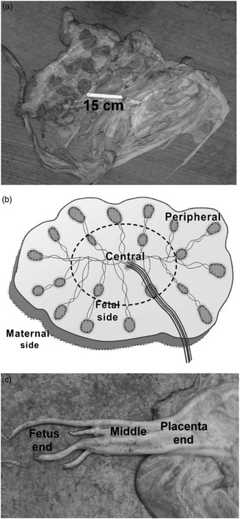
The whole, intact placenta was fully unfolded (Figure 1a) and stripped of blood with PBS. The dissection was performed in perpendicular to the luminal surface and done systematically as follows: three evenly spaced replicates of placentome and interplacentomal placneta, each of roughly 200-mg weight, were dissected from two topographical regions: the peripheral region closer to placental edge and the central region closer to the umbilical insertion point, each was further split into the fetal and maternal sides (Figure 1b). Umbilical cord was cross-sectioned into three topographical sections: the placenta section, the middle section and the fetus section (Figure 1c). The placentomes were later analyzed as a single feto (cotyledon)-maternal (caruncle) unit because no further separation was applicable. Preliminary zymographic analyses failed to reveal significant (P>0.05) replication variation in gelatinase expression for both placentomes and interplacentomal placenta. Hence, the triplicate tissues were pooled before processing as described below. Later gelatin zymography also indicated no difference between fresh and once-thawed samples.
Morphology of a non-retaining placenta from term Holstein cow with maternal side up (a), and the schematic assignation of topographic locations on placenta (b) and accessory umbilical cord (c).

All chemicals used for homogenization and extraction were from Sigma Chemical Co. (St Louis, MO, USA) unless specified otherwise. About 500-mg pooled tissues were minced to ∼0.5-cm edge length and then homogenized on ice in 45-ml 0.1 M Tris-HCl buffer (pH 7.5) containing 0.1% Triton X-100 (Maj and Kankofer, Reference Maj and Kankofer1997) with Ultra-Turrax IKA T10 (Module S10 N-10 G, IKA Corporation, Guangzhou, China) at 30 000 r.p.m. for 20 s. The homogenates were centrifuged by 200×g for 30 min at 4°C (Kubota 5800, Kubota Corporation, Tokyo, Japan) to collect the clear supernatants and stored in aliquots under −20°C until performing gelatin zymography within 1 week.
Protein concentration determination
Protein concentration of the supernatants of body fluids and tissue homogenates from above was determined by a Coomassie Brilliant Blue G-250 binding-based assay (Bradford, Reference Bradford1976; Bio-Rad Laboratories, Hercules, CA, USA), performing in microplate format (Multiskan Ascent, Thermo Labsystems, Helsinki, Finland) with a bovine serum albumin calibration curve.
Gelatin zymography
The gelatinolytic MMP subtyping was modified from that of Pugin et al. (Reference Pugin, Widmer, Kossodo, Liang, Preas and Suffredini1999) and Yu et al. (Reference Yu, Chen, Ho, Peh, Liu, Tiantonga, Nagahata and Chang2012) using SDS-PAGE (Minigel apparatus; Bio-Rad Laboratories, Inc. Hercules, CA, USA) with 7.5% acrylamide separating gel and 3% acrylamide stacking gel containing 0.1% bovine gelatin in Laemmli non-reducing system (Laemmli, Reference Laemmli1970). Fluid supernatants (7 µg protein/lane) or tissue extracts (10 µg protein per lane) were simply mixed with the sample buffer containing 2% SDS, but no reducing agent and directly subjected to electrophoresis. Afterwards, the gels were incubated in renaturing solution (2.5% (v/v) TritonX-100) for 30 min, followed by thorough distilled H2O dripping, and then developed in pH 7.4, 50 mM Tris-base buffer containing 200 mM NaCl, 0.02% Brij-35 and 5 mM CaCl2 overnight at 37°C to allow gelatinolysis. The next day the gels were stained with 0.5% Coomassie Brilliant Blue R-250 in 15% acetic acid, 25% methanol in distilled water and de-stained in 15% acetic acid, 25% methanol in water. The proteolytic activity was indicated by clear bands, visualizing both the pro-enzyme and the smaller active form, on a blue background. Dried gels were scanned with Epson Stylus TX130 (Seiko Epson Corporation, Nagano, Japan) and band volumes were determined using software Image J, version 1.45 (National Institutes of Health, Bethesda, MD, USA). Human plasma prepared from clinically healthy volunteers was used as a positive control as reference of the latent forms of MMP-2 and MMP-9, and a 125-kDa NGAL-proMMP-9 complex (Kolkenbrock et al., Reference Kolkenbrock, Hecker-Kia, Orgel, Kinawi and Ulbrich1996; Makowski and Ramsby, Reference Makowski and Ramsby2003).
Statistical analyses
Statistics for this study consulted that of Thomas et al. (Reference Thomas, Thomson, Sephton, Cowan, Wood, Vince, Kingsland and Lewis-Jones2002) and Provatopoulou et al. (Reference Provatopoulou, Antonia, Eleni, Flora, Ioannis, Evgenios, Afroditi, Ioannis and George2009) and the Student t-test of SAS Institute (2008) was performed to compare the difference in gelatinase level between topographic regions. P-value<0.05 was regarded statistically significant.
Results
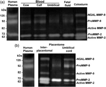
Representative gelatin zymographic images of five different body fluids (Figure 2a) and different compartments of non-retaining placenta (Figure 2b) showed similar molecular size between the body fluids and placenta tissues for latent or proenzyme forms of MMP-2 (72 kDa) and MMP-9 (92 kDa), the NGAL-MMP-9 complex (~125 kDa), as well as the active forms of MMP-2 (~62 kDa) and MMP-9 (~82 kDa).
Representative gelatin zymographic images for supernatants of body fluids (a) and extracts of term placenta (b) of non-retaining Holstein cows and neonatal calves.

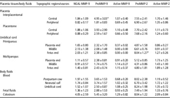
After densitometric scanning, each digested band based on the specified molecular size and dividing the band area by 1000, means and standard errors (n=11, five primiparous and six multiparous) of the levels of five major gelatinase subtypes in placenta compartments and body fluids are expressed in arbitrary unit in Table 1. No significant interaction was observed between parity or fetal/maternal side with different placental topographic regions. No comparison was attempted between different body fluids either. Overall, topographic effect on gelatinase level were observed for the significantly (P<0.05) higher proMMP-9 level in the central area than in the peripheral area of the interplacentomal placenta, whereas levels of the rest of the gelatinase subtypes were noticed with similar trend but no significance (P<0.1; Table 1). In addition, significantly (P<0.05) higher proMMP-2 level was observed in the fetus end than in the placenta end of the umbilical cord for primiparous cows, whereas active MMP-2 level was noticed with similar trend but no significance (P<0.1; Table 1).
Levels of gelatinase subtypes in the non-retaining placenta tissue and body fluids of Holstein cows and their calves (arbitrary unit after dividing the densitometric results by 1000)

NGAL=neutrophil gelatinase-associated lipocalin; MMP=matrix metalloproteinases.
* Significantly (P<0.05) different from the counterparts.
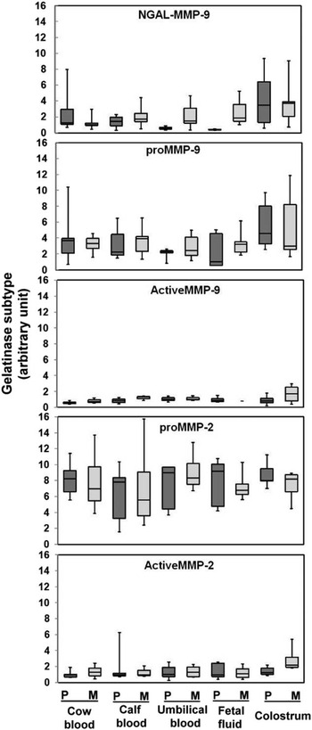
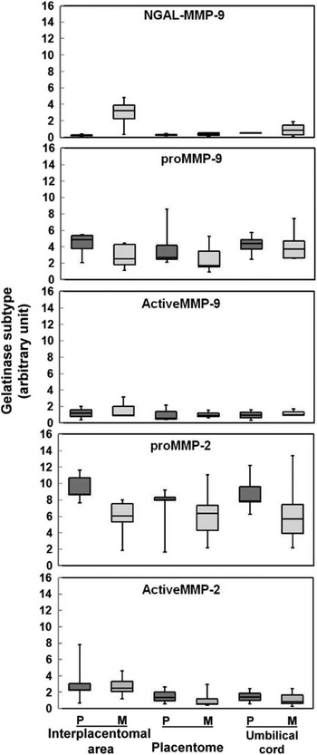
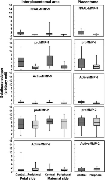
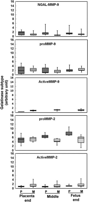
Levels of the five major gelatinase subtypes in the placenta compartments and body fluids were also depicted in box-plots to reveal the distribution and range for individual animals (Figures 3 to 6). Overall, parity apparently exerted no effect on the expression of the five major gelatinase subtypes on either body fluids (Figure 3) or placenta compartments, except for NGAL-MMP-9 complex on the interplacentomal area of the placenta (Figure 4). On the other hand, the topographic region seemed to exert some effect on the expression of proMMP-9, in both the placentomal and interplacentomal compartments of the placenta, as well as in the fetal/maternal side (Figure 5). Different umbilical sections also tended to affect the expression of proMMP-2 but not other gelatinase subtypes (Figure 6).
Levels of gelatinase subtypes in body fluids collected at term parturition from Holstein cows and their neonatal calves depicted as box-plots. (Boxes represent the interquartile range; lines inside boxes represent the median value; whiskers represent 5th and 95th percentiles.) Arbitrary unit was the densitometric results divided by 1000. P=primiparous cows; M=multiparous cows.

Levels of gelatinase subtypes in compartments of term placenta from Holstein cows depicted as box-plots. (Boxes represent the interquartile range; lines inside boxes represent the median value; whiskers represent 5th and 95th percentiles.) Arbitrary unit was the densitometric results divided by 1000. P=primiparous cows; M=multiparous cows.

Levels of gelatinase subtypes in different topographic locations of term placenta from Holstein cows depicted as box-plots. (Boxes represent the interquartile range; lines inside boxes represent the median value; whiskers represent 5th and 95th percentiles.) Arbitrary unit was the densitometric results divided by 1000.

Levels of gelatinase subtypes in different topographic sections of the accessory umbilical cord of term placenta from Holstein cows depicted as box-plots. (Boxes represent the interquartile range; lines inside boxes represent the median value; whiskers represent 5th and 95th percentiles). Arbitrary unit was the densitometric results divided by 1000. P=primiparous cows; M=multiparous cows.

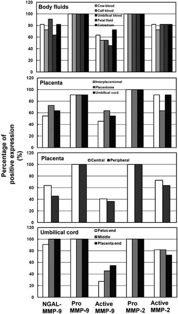
Positive expression of gelatinase subtypes on body fluids and different topographic regions of the placenta compartments was compiled in Figure 7. About 90% of participant cows were detected for the expressions of proMMP-9 and proMMP-2 in all body fluids and on every topographic region of the placenta compartments. In the other extreme, active MMP-9 has the lowest expression rate among all gelatinase subtypes in either body fluids or placenta compartments. Notably, an increasing rate of active MMP-9 expression was observed on the umbilical cord with increasing distance to the rupture site of umbilical cord (Figure 7).
Positive expression for gelatinase subtypes in body fluids and placenta compartments collected at term parturition from Holstein cows.

Discussion
The tight connection between the complementary interdigitation of fetal villous trees with maternal crypts during gestation must be terminated after expulsion of the fetus to ensure a healthy puerperium. The association of members of MMP family with the spatial regulation of ECM degradation in the bovine placenta for subsequent release of fetal membranes postpartum have been explored using different approaches, such as immunohistochemical analysis, gene expression analysis and zymography (Walter and Boos, Reference Walter and Boos2001; Takagi et al., Reference Takagi, Yamamoto, Ohtani and Miyamoto2007; Kizaki et al., Reference Kizaki, Ushizawa, Takahashi, Yamada, Todoroki, Sato, Ito and Hashizume2008; Dilly et al., Reference Dilly, Hambruch, Shenavai, Schuler, Froehlich, Haeger, Ozalp and Pfarrer2011). In this study, we used gelatin zymography to measure the expression level of gelatinase subtypes on different topographic locations of non-retaining cow placenta. Because the high expression level of MMP in tissues is generally accompanied with high MMP level in body fluids (Roy et al., Reference Roy, Louis, Loughlin, Wiederschain, Kilroy, Lamb, Zurakowski and Moses2008; Provatopoulou et al., Reference Provatopoulou, Antonia, Eleni, Flora, Ioannis, Evgenios, Afroditi, Ioannis and George2009), we concomitantly estimated the gelatinase subtypes in different body fluids from cows and neonatal calves. The methodology has been verified for optimal visualization of most of the gelatinase species on zymograms (Figure 2), where 7-µg protein equivalent of fluid volume or 10-µg protein equivalent of tissue extracts were subjected to analysis.
We have found that the proenzyme forms of MMP-2 and MMP-9 were the most abundantly and frequently detected gelatinase species of all in both fluids and placenta tissues. On the other hand, active MMP-2, despite being much less abundant than proMMP-2, has apparently higher expression level than active MMP-9 in most samples. As for NGAL-MMP-9 complex, it seemed more prevalently detected in both fluids and placenta tissues than active MMP-9. Our reports of differential gelatinase expressions on bovine placenta largely agreed with other reports on similar terms using different estimation tools (Walter and Boos, Reference Walter and Boos2001; Kizaki et al. Reference Kizaki, Ushizawa, Takahashi, Yamada, Todoroki, Sato, Ito and Hashizume2008; Dilly et al., Reference Dilly, Hambruch, Shenavai, Schuler, Froehlich, Haeger, Ozalp and Pfarrer2011). Nevertheless, the physiological implications were yet inconclusive. Maj and Kankofer (Reference Maj and Kankofer1997) suggested that this high proMMP-2/low active MMP-2 in bovine placenta might adversely affect the hydrolysis of collagen, and therefore might increase the rigidity of the intravillous ECM and, consequently, result in the etiology of placental retention. On the other hand, Kizaki et al. (Reference Kizaki, Ushizawa, Takahashi, Yamada, Todoroki, Sato, Ito and Hashizume2008) recommended proMMP-2 to be a more favorable gelatinase than MMP-9 in endometrial remodeling for prepartum cows. McNaughton and Murray (Reference McNaughton and Murray2009) included TIMP into consideration and suggested that high ratio of MMP-9/TIMP-2 expression in placentome might be associated with bovine RFM because the interaction of MMP-9 with TIMP-2 helps prevent epithelial separation within the placentome. They further implied that lower MMP-9 expression might be favorable for the timely release of term placenta. Dilly et al. (Reference Dilly, Hambruch, Shenavai, Schuler, Froehlich, Haeger, Ozalp and Pfarrer2011) reported that placentomes collected from cows with or without RFM expressed predominantly proMMP-2 but only small amounts of active MMP-2. The balancing between MMP and TIMP is apparently an important aspect that warrants extensive exploration.
Most MMP are secreted as inactive pro-enzymes, which become activated upon cleavage of an N-terminal propeptide. The latent forms of MMP-2 and MMP-9 are widely distributed in various tissues. Beceriklisoy et al. (Reference Beceriklisoy, Walter, Schafer-Somi, Miller, Kanca, Izgur and Aslan2007) summarized the distribution of MMP in the reproductive organ of bitches such as: MMP-2 in the endothelium and smooth muscles of blood vessels, the myometrium and the surface epithelium of the oviduct, whereas MMP-9 was present in the smooth muscle cells and epithelia of blood vessels, the maternal surface epithelial cells, uterine crypts and glands. Likely owing to their very low expression levels, the role of active MMP-9 and NGAL-MMP-9 complex in placenta remodeling during different reproduction stages is rarely discussed in the literature. In our study, both were the least frequently detected and, if detectable, the lowest expression level among all gelatinase species. Nonetheless, NGAL-MMP-9 complex seemed to express relatively higher in fluids (Figure 3) and the umbilical cord (Figure 6) with greater frequency (Figure 7) compared with the placenta tissue itself. Serum and urine levels of NGAL-MMP-9 complex have been evaluated for their diagnostic and prognostic values in a variety of diseases (Fernandez et al., Reference Fernandez, Yan, Louis, Yang, Kutok and Moses2005; Roy et al., Reference Roy, Louis, Loughlin, Wiederschain, Kilroy, Lamb, Zurakowski and Moses2008; Provatopoulou et al., Reference Provatopoulou, Antonia, Eleni, Flora, Ioannis, Evgenios, Afroditi, Ioannis and George2009). The frequency of detection of NGAL-MMP-9 complex, along with other high molecular weight MMP species, was significantly higher in urine from prostate and bladder cancer groups than controls (Roy et al., Reference Roy, Louis, Loughlin, Wiederschain, Kilroy, Lamb, Zurakowski and Moses2008). Women with invasive ductal carcinoma exhibited significantly increased blood levels of MMP-9, NGAL and NGAL-MMP-9 complex compared with healthy controls (Provatopoulou et al., Reference Provatopoulou, Antonia, Eleni, Flora, Ioannis, Evgenios, Afroditi, Ioannis and George2009). NGAL-MMP-9 complex was detected in over 80% of the urine samples from breast cancer patients but none in those from healthy age- and sex-matched controls (Fernandez et al., Reference Fernandez, Yan, Louis, Yang, Kutok and Moses2005). Our zymographic results provided the preliminary information about the expression of NGAL-MMP-9 complex in blood, fetal fluidsand colosturm of parturient cows and calves (Figure 3), and the physiological implications deserve further investigation.
The current study seems not to reveal characteristic gelatinase expression patterns among the placentome, interplacentomal area and umbilical cord (Figure 4). Nonetheless, the central area of the interplacentomal placenta, fetal and maternal sides combined or separated, had greater proMMP-9 expression than the peripheral counterpart (Figure 5). In addition, the rupture end (fetus end) of the umbilical cord tended to express greater proMMP-2 than the placenta end of the umbilical cord (Figure 6). As described in ‘Material and methods’ section, to avoid disturbing animals, we collected only the spontaneously released term placentas. No separation of the fetal or maternal side was applicable for our placentome-like structures. When sheep placenta was surgically obtained during the last third of gestation, caruncles, intercaruncles, cotyledons and intercotyledon were separately measured. Caruncle was found to have greater MMP-2 level than the other parts, and lower MMP-9 in the intercotyledon than in the other components (Vagnoni et al., Reference Vagnoni, Zheng and Magness1998). Takagi et al. (Reference Takagi, Yamamoto, Ohtani and Miyamoto2007) manually collected bovine placentomes from the uterus via the vagina postpartum. In the study by Dilly et al. (Reference Dilly, Hambruch, Shenavai, Schuler, Froehlich, Haeger, Ozalp and Pfarrer2011), cow placentomes were collected through cesarean section, and high expression of MMP-2 was found in the fetal compartment,concluding a role of MMP-2 in the timely release of fetal membranes in cattle. Their ways of placentome collection were different from ours. Because our placenta compartments were manually dissected, they were less homogenous and more subjective. However, as we have mentioned earlier, gelatin zymography could provide information of in situ forms (latent, active or complex) of each gelatinase, which would be impossible using antibody-based assays.
In the literature, the results regarding the gelatinase expression on different placenta compartments of ruminant placenta were conflicting. Takagi et al. (Reference Takagi, Yamamoto, Ohtani and Miyamoto2007) quantitatively measured the mRNA levels of MMP-2 and MMP-9 in the caruncle and cotyledon of bovine placenta and reported ∼10 times higher in the caruncle than in the cotyledon and marked contrasting changes between prepartum and postpartum periods. Uekita et al. (Reference Uekita, Yamanouchi, Sato, Tojo, Seiki and Tachi2004) estimated the expression of MMP-2 in addition to other MMP in the endometrium and placenta of sheep using quantitative RT-PCR analysis, in situ hybridization, immunoblotting, gelatin zymography and immunohistochemical analysis. They found that MMP-2 expression was detected both in the cotyledonal trophoblast cells and the subsyncytial stromal cells during all stages of gestation. We speculate that one of the reasons for the above inconsistent results was caused by lack of distinguishing among the multiple forms of gelatinases.
Taken human as an example, Demir-Weusten et al. (Reference Demir-Weusten, Seval, Kaufmann, Demir, Yucel and Huppertz2007) measured the expressions of MMP-2 and MMP-9 in terms of the placentas in the amnion, basal plate, chorionic plate, decidua, chorionlaeve, Nitabuch’s stria, umbilical cord and placental villi by immunohistochemical analysis and zymography. They reported that active MMP-2 expressed the highest in the decidua, followed by the chorionlaeve and umbilical cord, whereas the lowest in the amnion and chorionic villi. On the other hand, the most distinctive expression of proMMP-9 was observed for the Nitabuch’s stria, followed by the decidua, chorionicplate, chorionlaeve and basal plate at nearly the same integrated optical density levels. There was no detectable expression for the umbilical cord. They suggested that MMP-9 is more important than MMP-2 in separating the placenta from the uterine wall during labor. The histological structures of the human and bovine placentae are different: the human placenta belongs to the invasive hemochorialis type, bovine non-invasive syndesmochorialis type. Therefore, MMP may play a different role in the separation processes of the placenta in humans and bovines.
Our observations of differential topographic expressions for proMMP-9 and proMMP-2 imply that their expressions might be related to the proximity to blood supply, suggesting a regulatory role of blood-born factors. A study by Wischral et al. (Reference Wischral, Verreschi, Lima, Hayashi and Barnabe2001) has pointed out that placental retention in cows was related to both estrogen and PGF2α deficiency, but not progesterone. Measuring the placenta concentrations of related hormones might be able to provide evidences necessary for justifying our results.
When we segregated our results to primiparous and multiparous cows, we found that parity seemed to exert little effect on the expression of gelatinase subtypes, except that multiparous cows seemed express slightly greater NGAL-MMP-9 and lower proMMP-2 on interplacentomal placenta (Figure 4) and express slightly lower proMMP-2 on the fetus end of the umbilical cord (Figure 6) compared with the primiparous cows. The physiological implication of the parity effect is yet to be understood.
In conclusion, multiple forms of gelatinases, including complex form and both the latent and active forms were detected using gelatin zymography on non-retaining placentas of Holstein cows. High expressions of proenzyme forms and low expressions of active forms of MMP-2 and MMP-9 were frequently observed. The central placenta and fetus end of the umbilical cord expressed greater proenzyme forms of MMP-9 and MMP-2, respectively, than the counter parts, suggesting roles played by gelatinases in the detaching of fetal membrane at term. Gelatin zymography was proven feasible, sensitive and specific for fingerprinting the multiple forms of gelatinases in situ.
Acknowledgements
The authors acknowledge Professor P.C. Tang of National Chung Hsing University for providing constructive comments when revising the manuscript.