Introduction
The nematode fauna of Indonesian birds is poorly studied. So far only three spiruroid nematodes have been reported from Indonesian birds, i.e. Cheilospirura hamulosa (Dies, 1851) from Gallus gallus (Linnaeus) (family Phasianidae) of Makasar, Squamofilaria otusi Purwaningsih & Purnomo, Reference Purwaningsih and Purnomo1994 from Otus bakkamoena lempiji (Horsfield) (family Strigidae) of West Java and Synhimantus (Dispharynx) nasuta (Rudolphi, 1819) from Pycnonotus goiavier (Scopoli) (family Pycnonotidae) of East Kalimantan (Yamaguti, Reference Yamaguti1955; Purwaningsih & Purnomo, Reference Purwaningsih and Purnomo1994; Dewi et al., Reference Dewi, Irham, Prawiradilaga and Kawakami2006).
In this study we describe two new species of spiruroid nematodes that represent a new species of Diplotriaena Railliet and Henry, 1909 from Anthreptes malacensis malacensis (Scopoli) (family Nectariniidae) and a new species of Acuaria Bremser, 1811 from Dicrurus hottentottus jentincki (Vordeman) (family Dicruridae). The two new species were obtained from an ongoing bird biodiversity inventory project in Kangean Island, Indonesia.
Materials and methods
In this study, we examined 44 Kangean birds representing 18 species. Of these, 16 are resident species while the remaining two are migratory. All birds were trapped using mist-nets of 12 × 2.6 m in March 2006. The captured birds were euthanized using ether alcohol and then necropsied in the laboratory to discover nematodes. Nematodes were fixed and stored in 70% ethanol. Later, the worms were examined using a compound Olympus BH series microscope with a drawing tube and JEOL JSM5310LV scanning electron microscopy (SEM) at an accelerating voltage of 20 kV. For light microscopic studies, the specimens were cleared in glycerine. Measurements were made with an ocular micrometer. For SEM studies, the specimens were post-fixed in glutaraldehyde, dehydrated through an ethanol series and freeze dried. The dried specimens were then coated with gold at 5–8 mA for 5 min. Measurements were made by using software ImageJ, version 3.7 (http://rsb.info.nih.gov/ij/). Measurements (range, followed by mean in parentheses) are given in micrometres unless otherwise stated.
Diplotriaena anthreptis sp. nov.
Type host. Anthreptes malacensis malacensis (Scopoli) (Aves; Passeriformes; Nectariniidae).
Site. Abdominal cavity.
Type locality. Kangean Island, Indonesia (06°52′41.6″S and 115°25′12.7″E).
Collector. M. Irham and Alwin Marakarmah (MZB, Indonesia).
Type material. Four specimens (one male and three females). Type specimens deposited in Museum Zoologicum Bogoriense, Indonesia (MZB Na 418): holotype one male; allotype one female and paratype two females.
Etymology. The new species is named after the host.
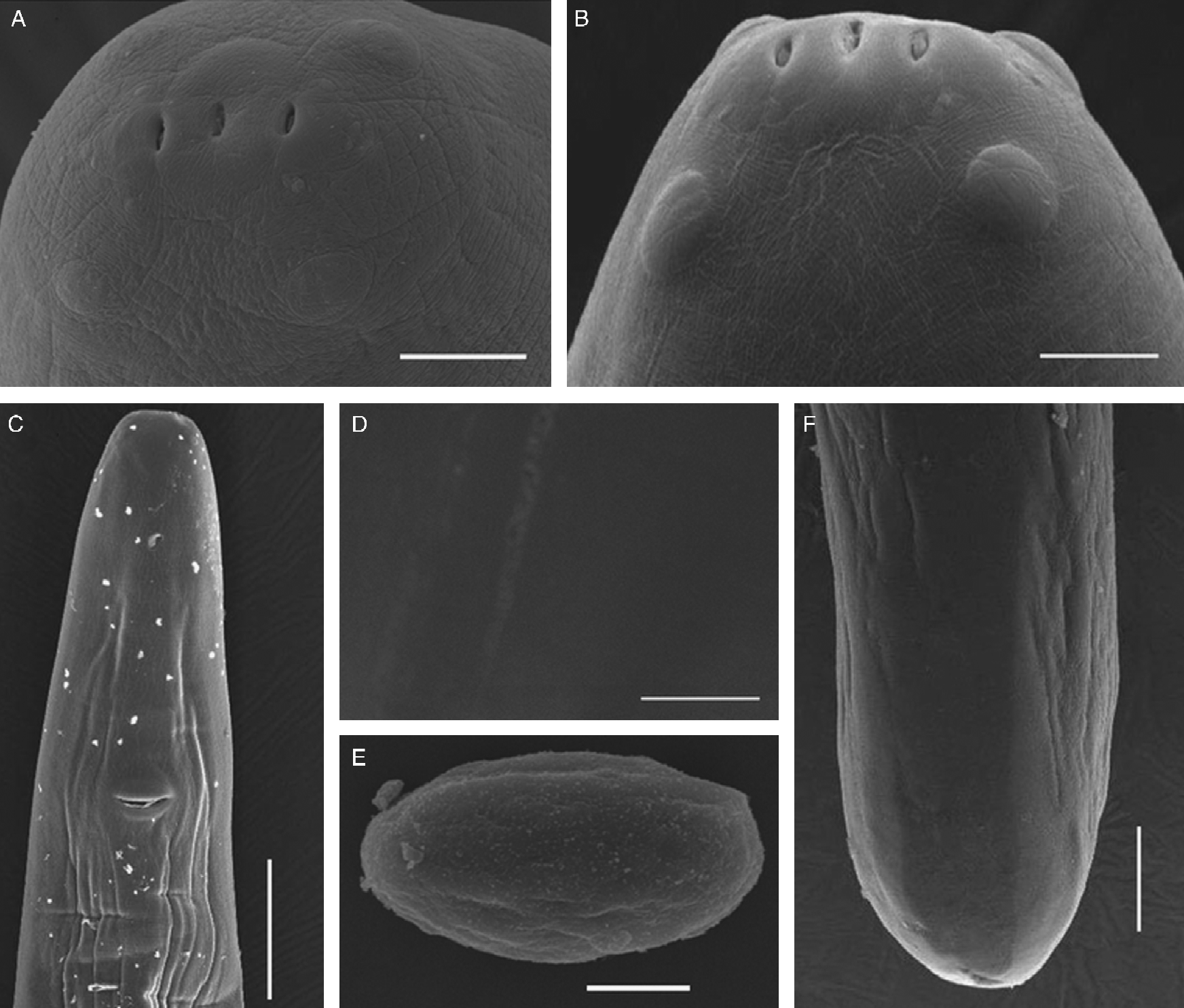
Description. Long and slender worms. Mouth small, opening rectangular, dorso-ventrally elongate, without lips. Eight submedian cephalic papillae arranged as four large, outer papillae and four small, inner-circle papillae (fig. 1A and B). A pair of lateral amphids on the inner circle. Two oval openings connected with manubria of tridents present on sides of oesophagus. Manubrium apex of trident concave (fig. 2B). Two lateral branches of tridents with bluntly rounded posterior end, middle one with flat posterior end. Oesophagus divided into short, slender anterior muscular part and very long, stouter posterior part (fig. 2A). Cuticle with smooth striations (fig. 1D).
Diplotriaena anthreptis sp. nov. A, Anterior end of female, apical view; B, anterior end of female, ventral view; C, anterior end of female with vulva, ventral view; D, body showing the cuticle with smooth striations; E, egg; F, posterior end of female. Scale bars: A = 30 μm; B = 25 μm; C = 200 μm; D, E = 10 μm; F = 100 μm.

Diplotriaena anthreptis sp. nov. A, Anterior end of female, lateral view; B, trident of female; C, posterior end of male, ventral view; D, anterior part of right spicule; E, posterior end of female, ventral view; F, egg. Scale bars: A = 200 μm; B = 50 μm; C, E = 100 μm; D, F = 25 μm.
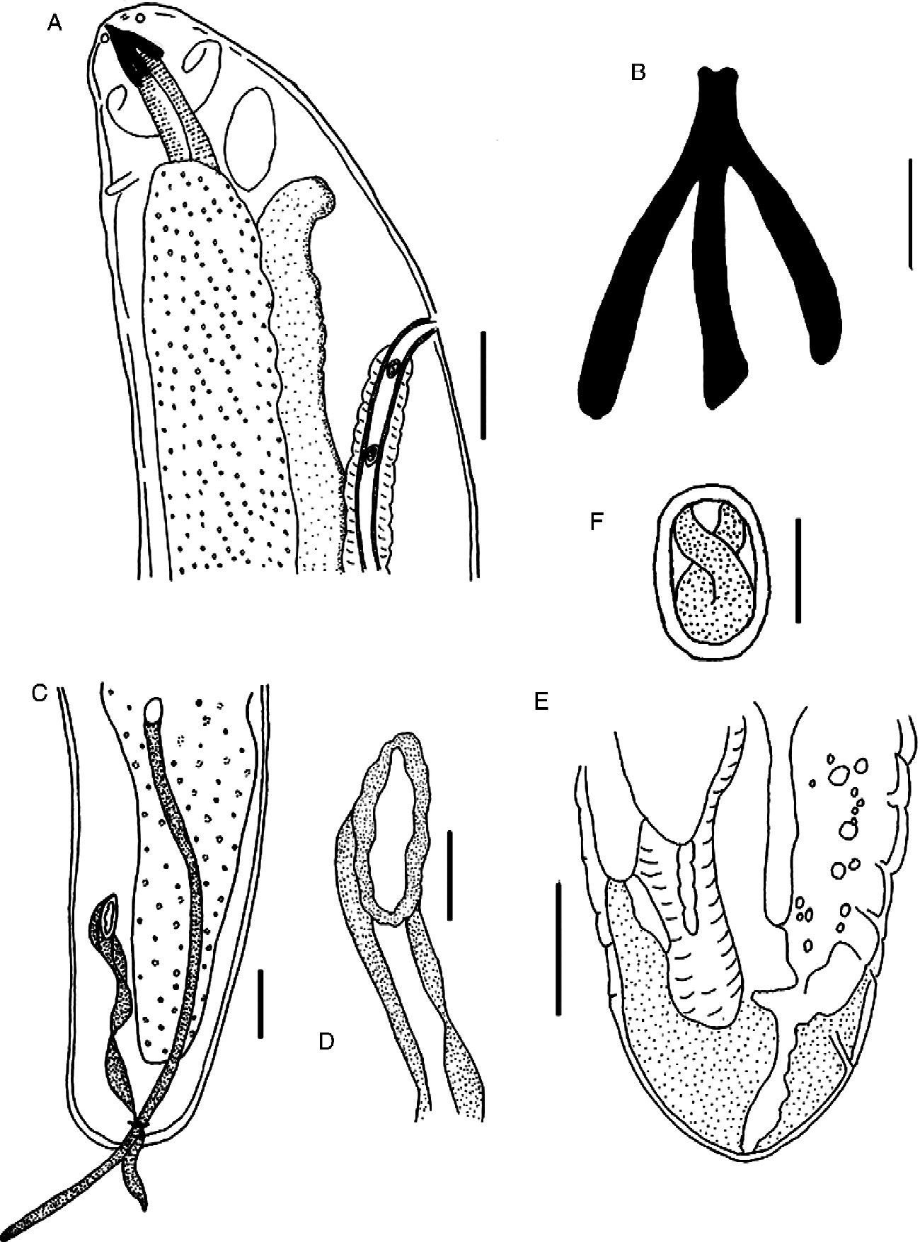
Male (holotype). Total body length 24.9 mm, maximum width 430. Head width 80. Trident 100 long. Nerve ring 140 from anterior end. Muscular oesophagus 190 long, 90 wide; glandular oesophagus 1.53 mm long, 140 wide. Spicules unequal and dissimilar. Left spicule long and simple, with pointed distal end, 910 long. Right spicule 560 long, twisted three times, with two twisted alae (fig. 2C and D). Ratio of right spicule:left spicule 1:1.6. Tail short, bluntly rounded, 60 long. Caudal papillae and caudal alae absent.
Female (one allotype, one paratype and one female used for SEM observation). Total length 83–102 (92) mm, maximum width 490–580 (510). Head width 90–130 (110). Trident 120–130 (123) long. Nerve ring 200–240 (220) from anterior end. Muscular oesophagus 300–350 (327) long, 90–100 (97) wide; glandular oesophagus 1.6–1.9 (1.77) mm long, 220–250 (233) wide. Vulva in anterior part of body, 560–750 (673) from anterior end (fig. 1C). Vagina runs posteriorly. Vagina vera long 1100–1200 (1160), vagina uterine not seen. Uterine branches initially parallel then divergent. Eggs oval, thick-shelled, embryonated in uteri, 36–42 × 29–31 (fig. 1E and F). Anus subterminal. Posterior end rounded (figs 1F and 2E).
Remarks. All species of the genus Diplotriaena are nematodes parasitizing the air sacs, lungs and body cavity of birds. Anderson (Reference Anderson1959) revised the genus and he recognized 54 as valid species, while Sonin (Reference Sonin and Skrjabin1968) recorded 61 valid species. Since then, more than 20 species have been added to this genus (Tadros, Reference Tadros1966; Soota & Chaturvedi, Reference Soota and Chaturvedi1968, Reference Soota and Chaturvedi1971; Pinto et al., Reference Pinto, Vicente and Noronha1970; Pinto & Noronha, 1970a, b, 1981; Soota, Reference Soota1970; Olsen & Braun, Reference Olsen and Braun1971; Webster & Speckmann, Reference Webster and Speckmann1976; Bain & Mawson, Reference Bain and Mawson1981; Wong et al., Reference Wong, Anderson and Frimeth1983; Lakshmi et al., Reference Lakshmi, Rao and Shyamasundari1985; Liu, Reference Liu1985, Reference Liu1987).
The new species is similar to Diplotriaena ozouxi Railliet and Henry, 1909; D. obtusa Henry and Ozoux, 1909, D. bargusinica Skrjabin, 1917, D. isabellina Koroliowa, 1926 and D. delta Johnston & Mawson, Reference Johnston and Mawson1940, in the size of the tridents and the length of the spicules. However, the new species differs from the similar species in the structure of the tridents (the apex of the manubria concave (versus not concave), the middle branch of the trident with a flat posterior (versus not flat)). Diplotriaena anthreptis sp. nov. can be distinguished from D. ozouxi by having two alae in the right spicule (versus one ala), and in the size of eggs (36–42 × 29–31 versus 50 × 40). The new species is also different from D. bargusinica in having a longer left spicule (910 versus 700), in the right spicule having two rather than one alae, in the spicule ratio (1:1.6 versus 1:1.1–1.3), and in the size of eggs (36–42 × 29–31 versus 70 × 50). The new species differs from D. delta in the structure of the trident (not bosses versus bosses). The new species can be separated from D. isabellina by having two rather one alae in the right spicule, and in the spicule ratio (1:1.6 versus 1:1.1–1.2). Finally, D. anthreptis differs from D. obtusa in having a shorter left spicule (910 versus 1064–1232) and a different spicule ratio (1:1.6 versus 1:1.8).
Acuaria irhami sp. nov.
Type host. Dicrurus hottentottus jentincki (Vordeman, 1893) (Aves; Passeriformes; Dicruridae).
Site. Under the lining of the gizzard.
Type locality. Kangean Island, Indonesia (06°52′41.6″S and 115°25′12.7″E).
Collector. M. Irham and Alwin Marakarmah (MZB, Indonesia).
Type material. Two males. Type specimens deposited in Museum Zoologicum Bogoriense, Indonesia (MZB Na 419): holotype one male; paratype one male (observed with SEM).
Etymology. The new species is named after M. Irham (MZB) for his kind help in providing the nematode specimens to us during his research project in Kangean Archipelago.
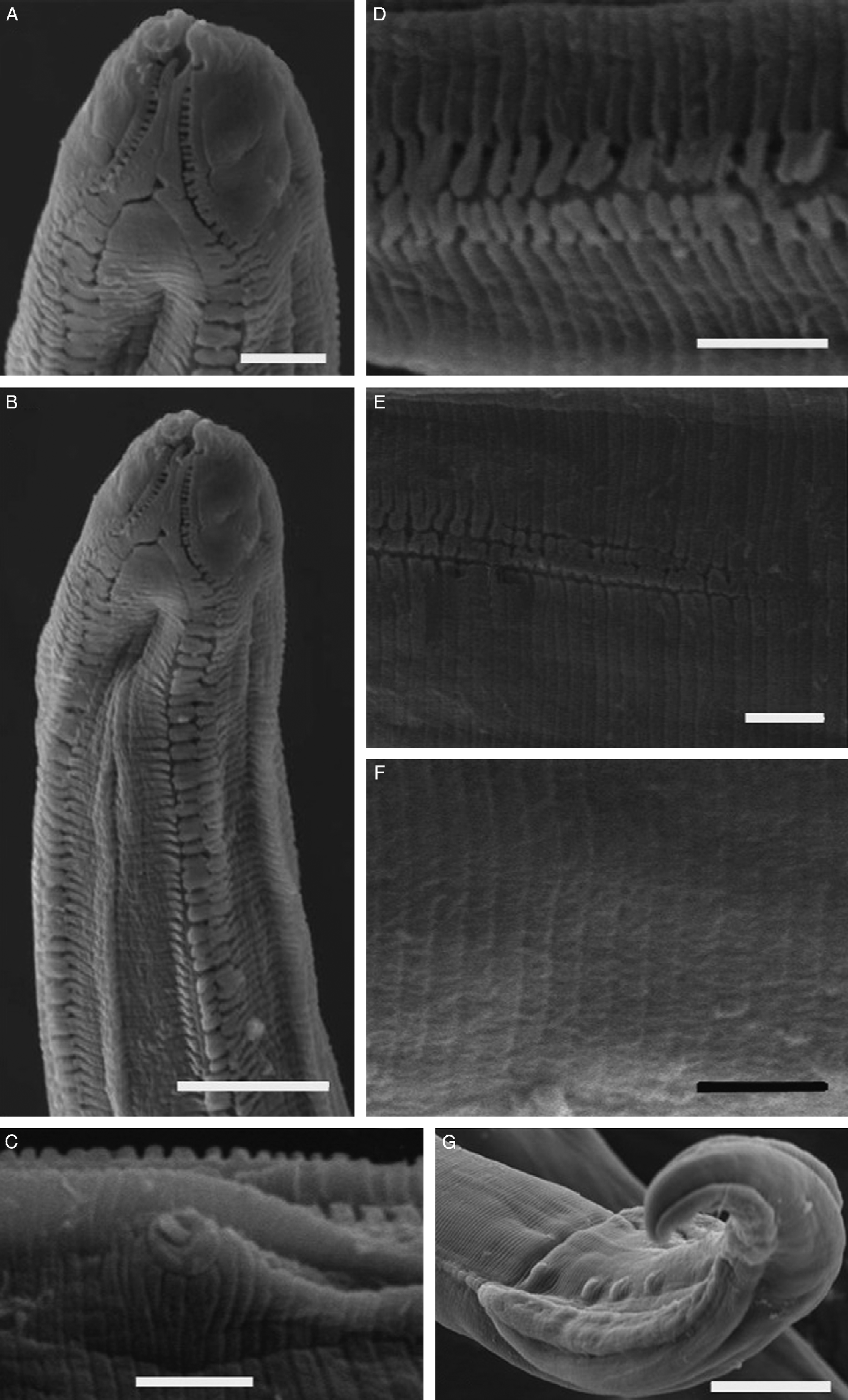
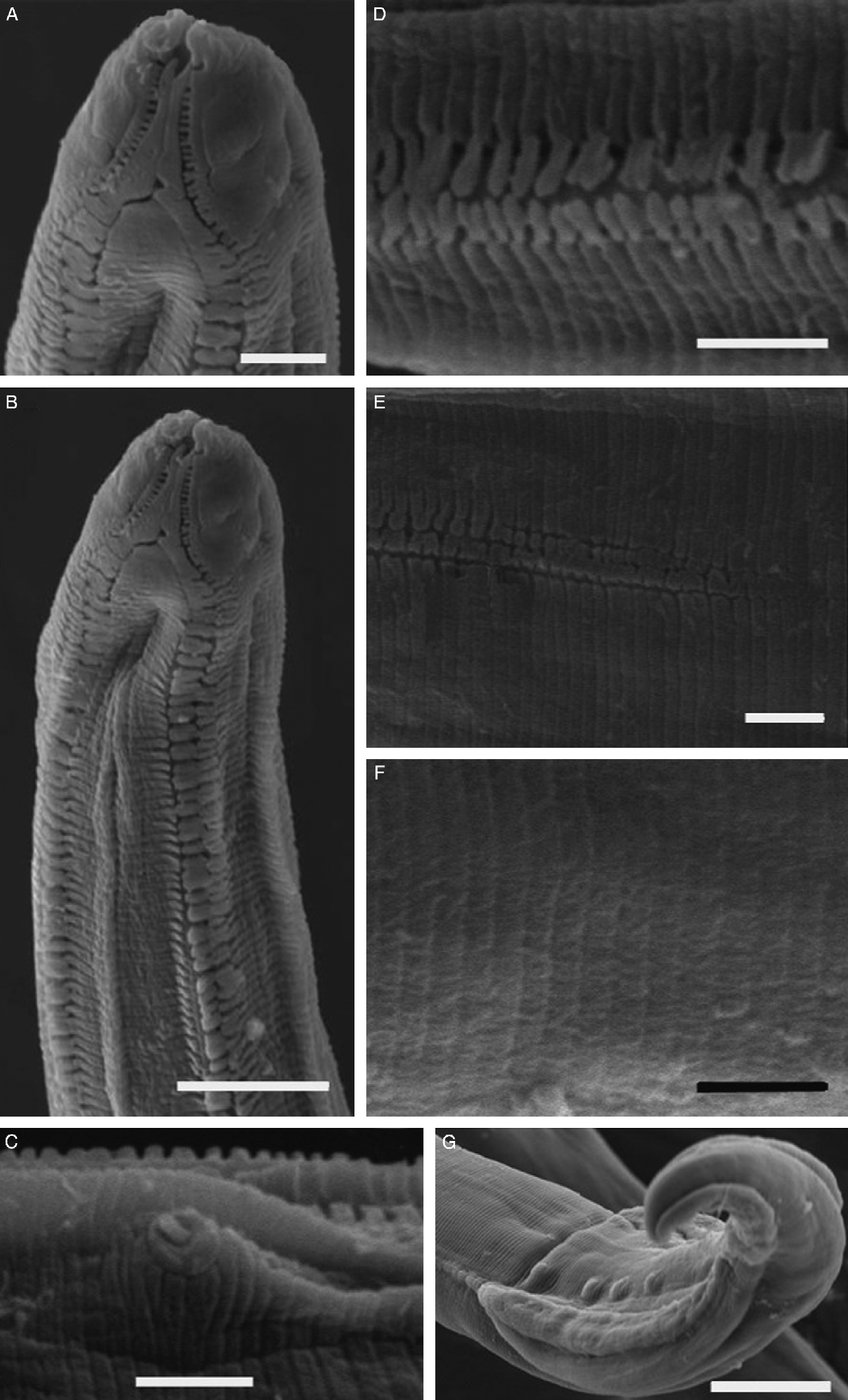
Description. Body small with transverse striations. Two pseudolabia at lateral of mouth, each bearing a pair of large cephalic papillae and one inconspicuous amphid. Cordons straight, non-anastomosing and non-recurrent (fig. 3B), originating at dorsal and ventral sides of oral opening (fig. 3A), continuing posteriorly to middle part of muscular oesophagus. Structure of cordon simple, consists of two longitudinal sections separated by a groove; the shape of both longitudinal sections is the same, blunt-shaped end (figs 3D and 4B), gradually narrowing to posterior (fig. 3E). Buccal capsule long and slender, transversely striated. Oesophagus clearly divided into a short muscular region and a long glandular region. Nerve ring located at level of anterior end of muscular oesophagus. Cervical papillae posterior to nerve ring. Excretory pore posterior to nerve ring (fig. 4A).
Acuaria irhami sp. nov. A, Anterior extremity, ventral view; B, anterior end of male, ventral view; C, cervical papilla; D, cordons; E, the end of cordons; F, dorsal striations; G, posterior end of male, ventral view. Scale bars: A, C–F = 10 μm; B = 25 μm; G = 50 μm.
Acuaria irhami sp. nov. A, Anterior end of male, lateral view; B, a part of cordons; C, posterior end of male, ventral view; D, spicule. Scale bars: A, C = 100 μm; B = 5 μm; D = 50 μm.

Male (n = 2) (the first measurement is of holotype, and the second is of paratype). Body small with transverse striations, 7.16 and 8.10 mm long, 100 and 110 wide. Buccal cavity 90 and 100 long. Cordons 235 and 250 long, 7.5 wide. Nerve ring 135 and 150 from anterior end; cervical papillae 173 and 185 from anterior end (fig. 3C); excretory pore 220 and 242 from anterior end. Oesophagus divided into two parts. Muscular region 300 and 340 long, 40 maximum width, glandular region 960 and 1080 long, 70 maximum width. Transverse striations 2.4–2.7 apart (fig. 3F). Spicules with rounded distal end, equal and similar, without longitudinal groove throughout the length, 180 and 210 long (fig. 4D). Tail 240 and 250 long. Caudal alae well developed, 380 and 410 long, maximum width 50 and 55 (fig. 3G). Ten pairs of pedunculate caudal papillae, four pairs preanal and six pairs postanal. Postanal papillae undivided into two distinct groups, arranging progressively closer together toward the tip of the tail (fig. 4C).
Remarks. The genus Acuaria has a wide geographical distribution. This genus is the richest genus of acuarid nematodes, with more than 70 nominal species (Zhang et al., Reference Zhang, Brooks and Causey2003). Adult Acuaria occur in the oesophagus, the crop and the gizzard of birds, where they are found free or burrowing, in or under the lining (Baylis, Reference Baylis and Sewell1939). Almost all known Acuaria species are from passerine birds of the order Oscires (see Mawson, Reference Mawson1972). The important taxonomic characters in this genus are the cordon lengths in both sexes, the shape, the size, the length and the ratio of the two spicules, and the number and the arrangement of the caudal papillae of the male (Mawson, Reference Mawson1972).
This new species can be easily distinguished from all other Acuaria species except A. microecae Mawson, Reference Mawson1972 by having equal and similar spicules (versus subequal and dissimilar spicules) (Cram, Reference Cram1927; Williams, Reference Williams1929; Baylis, Reference Baylis and Sewell1939; Rasheed, Reference Rasheed1960; Chabaud & Petter, Reference Chabaud and Petter1961; Hsu, Reference Hsu1963; Skrjabin et al., Reference Skrjabin, Sobolev and Ivashkin1965; Gupta & Jehan, Reference Gupta and Jehan1972; Mawson, Reference Mawson1972; Chabaud, Reference Chabaud, Anderson, Chabaud and Willmott1975; Wang, Reference Wang1976; Gupta & Kumar, Reference Gupta and Kumar1977; Zhang, Reference Zhang1990; Zhang & Wang, Reference Zhang and Wang1993; Zhang et al., Reference Zhang, Brooks and Causey2003). The new species differs from A. microecae in having six pairs of postanal papillae (versus seven), in the absence of a median preanal papilla (versus present), and in the cordon of the male reaching the middle of the muscular oesophagus (versus reaching the end of the muscular oesophagus).
Some species of Acuaria have a spicule ratio close to 1:1, i.e. A. alli Rasheed, Reference Rasheed1960; A. brevispicula Maplestone, 1932; A. galliardi Chabaud & Petter, Reference Chabaud and Petter1961; A. kungi Singh, 1948; A. longicaudata Hsu and Hoeppli, 1931; A. minor Williams, Reference Williams1929; A. minuta Williams, Reference Williams1929; A. ornata (Gendre, 1912); A. pattoni Williams, Reference Williams1929; A. mirafrae Mawson, Reference Mawson1972; and A. upupa Rasheed, Reference Rasheed1960. However, A. irhami sp. nov. can be distinguished from A. alli in the spicule length (180–210 in two spicules versus 110 in the left spicule and 100 in the right spicule). The new species differs from A. brevispicula in having three pairs instead of four pairs of preanal papillae and in the shape of spicules (spicules straight versus spicules with distal end bent ventrally). The present species is different from A. kungi in the shape of spicules (similar versus dissimilar shaped) and in the numbers of postanal papillae (six pairs versus seven pairs). Acuaria longicaudata, A. ornata and A. pattoni can be distinguished from the new species in the arrangement of postanal papillae (postanal papillae divided into two distinct groups in the former versus postanal papillae undivided into groups in the latter) and in the spicules with alae instead of spicule without alae. Acuaria irhami sp. nov. differs from A. minor and A. minuta in the arrangement of postanal papillae (two groups instead of one group), and in having spicules without expanded distal end instead of spicules with expanded distal end. Acuaria mirafrae can be differentiated from the new species by having postanal papillae in two distinct groups versus undivided groups, and in the structure of cordons (the right end of each section cordon is circular versus having a blunt tip) (Mawson, Reference Mawson1972). The new species differs from A. upupa in having six pairs instead of seven pairs of postanal papillae, and in the structure of spicules (spicules with rounded tip versus spicules with alate tip).
In having cordons reaching the middle of the muscular oesophagus, the new species resembles A. cordata (Mueller, 1789); A. excubitori Gupta & Kumar, Reference Gupta and Kumar1977; A. mayori Lent, Freitas & Proenca, 1945; A. muelleri Gupta & Kumar, Reference Gupta and Kumar1977; A. srivastavai Gupta & Kumar, Reference Gupta and Kumar1977; and A. subula (Durjadin, 1845). The new species differs from A. cordata by having six pairs instead of seven pairs of postanal papillae. Acuaria irhami sp. nov. is different from A. excubitori in having ten pairs instead of 13 pairs of caudal papillae. The new species can be easily distinguished from A. mayori, A. muelleri, A. srivastavai and A. subula in the spicule ratio (1:1 versus 1:1.5–1.7 in A. mayori, 1:1.5–1.9 in A. muelleri, 1:1.8–2.6 in A. srivastavai and 1:1.7 in A. subula) (Baron, Reference Baron1967; Zhang et al., Reference Zhang, Brooks and Causey2003). The new species is also different from A. gracilis (Gendre, 1912) paraziting the same bird family, Dicruridae, in spicule ratio (1:1 versus 1:1.1–1.4) and in the shorter cordon (reaching to middle structure of muscular oesophagus versus cordon longer than muscular oesophagus).
Discussion
Anthreptes malacensis malacensis, the host of Diplotriaena anthreptis sp. nov., is distributed throughout the Java Island and South-East Asia, while Dicrurus hottentottus jentincki, the host of Acuaria irhami sp.nov., is recorded in the area of Kangean, Bali, East Java, Nusa Penida and small islands on the Java Sea (Dickinson, Reference Dickinson2003). It is possible that the two new species have the same geographic distribution as their hosts.
In this study, we found five other nematodes besides the two new species, i.e. Viguiera dicrurusi Gupta, 1960 from D. h. jentincki, Contracaecum sp. from Tringa hypoleucos (Linnaeus) (family Scolopacidae), and three different spiruroid nematode species. These three spiruroid nematodes are members of the family Habronematidae, which parasitize Orthotomus ruficeps (Lesson) (family Sylviidae), Pycnonotus goiavier and D. h. jentincki. The specimens could not be identified due to poor preservation.
Acknowledgements
We would like to express our sincere thanks to M. Irham and A. Marakarmah (Zoology Division, RC Biology-LIPI), for their kind help in collecting and identifying the bird host specimens. Thanks are also due to Professor I. Beveridge (Department of Veterinary Science, The University of Melbourne, Australia), Dr D. Wowor, Ir. E. Purwaningsih, Dr D. Peggie (Zoology Division, RC Biology-LIPI) for revisions and suggestions about the manuscript. This study was a part of an avifauna diversity study in the Kangean Archipelago sponsored by NAGAO.