Introduction
In 1868, Paul Langerhans identified a non-pigmented dendritic cell present in the epidermis. These cells were subsequently identified as part of the immune system, functioning as a peripheral Major histocompatibility complex II (MHC-II) expressing antigen presenting cell.Reference Langerhans1
Several syndromes were subsequently described, and their common origin from Langerhans' cells was identified in 1953.Reference Lichtenstein and Histiocytosis2 In 1987, the Histiocyte Society combined the previously used terms ‘eosinophilic granuloma’ (single organ involvement), ‘Hand–Schuller–Christian's disease’ (classically a triad of exophthalmos, diabetes insipidus and bone lesions), ‘Letterer–Siwe's disease’ (an aggressive, multisystemic form involving the liver and spleen in young children), and ‘histiocytosis X’ under the term ‘Langerhans’ cell histiocytosis'.Reference Chu, D'Angio, Favara, Ladisch, Nesbit and Pritchard3
There is a wide spectrum of Langerhans' cell histiocytosis, varying from isolated lesions to multisystemic disease with significant mortality.Reference Arico, Girschikofsky, Genereau, Klersy, McClain and Grois4 Spontaneous disease regression has been described.Reference Broadbent, Davies, Heaf, Pincott, Pritchard and Levinsky5 The exact aetiology of Langerhans' cell histiocytosis is not known, although smoking is recognised as the main risk factor for pulmonary Langerhans' cell histiocytosis.Reference Vassallo, Ryu, Colby, Hartman and Limper6 The cells within lesions from various forms of Langerhans' cell histiocytosis have been shown to be of monoclonal origin.Reference Willman, Busque, Griffith, Favara, McClain and Duncan7
The majority of Langerhans' cell histiocytosis cases occur in children, but presentation in adults is becoming increasingly recognised.Reference Arico, Girschikofsky, Genereau, Klersy, McClain and Grois4 Langerhans' cell histiocytosis can affect the lungs, bones, skin and mucous membranes, lymphoreticular system and central nervous system (especially the hypothalamo-pituitary axis), with rarer reports of disease in the genitourinary and gastrointestinal tracts and thyroid.Reference Howarth, Gilchrist, Mullan, Wiseman, Edmonson and Schomberg8 The definitive diagnosis is established by biopsy of the tissue and positive staining for CD1 antigen, or observation of Birbeck granules on electron microscopy. A presumptive diagnosis may be made if the histological evidence points towards a clonal proliferation of Langerhans' cells in the biopsied tissue.Reference Chu, D'Angio, Favara, Ladisch, Nesbit and Pritchard3 Staging of the disease should include a bone scan, radiographs of any suspicious bony lesions, chest radiograph, routine laboratory tests and possibly abdominal ultrasound.Reference Stockschlaeder and Sucker9
Osseous involvement is common in Langerhans' cell histiocytosis. A 50-year series of 314 Langerhans' cell histiocytosis cases (adult and paediatric) from the Mayo Clinic showed the occurrence of purely osseous disease to be 36 per cent, with a further 24 per cent of the total having osseous disease as part of multisystemic disease; similar results were found in another case series.Reference Arico, Girschikofsky, Genereau, Klersy, McClain and Grois4, Reference Howarth, Gilchrist, Mullan, Wiseman, Edmonson and Schomberg8 In the Mayo Clinic series, 94 of the 314 patients had disease in the bones of the skull: 28 parietal, 28 frontal, 26 temporal and 12 occipital.Reference Howarth, Gilchrist, Mullan, Wiseman, Edmonson and Schomberg8
Case report
A 49-year-old woman presented with a four-month history of left aural fullness and a three-week history of vertigo and disequilibrium.
The patient's otoscopic examination was normal. She had right-beating nystagmus and mild cerebellar signs, with a mean left-sided hearing loss of 50 dB.
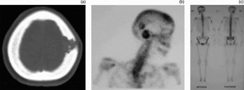
Magnetic resonance imaging (MRI) revealed a normal labyrinth but a lytic lesion in the left parietal bone. Computed tomography (CT) confirmed this lesion, and also identified diffuse abnormalities of the left petrous bone, including a focal defect in the squamous temporal bone, and thickened mastoid trabeculations (Figure 1a).
(a) Axial computed tomography scan showing lytic left parietal bone lesion. This had a bevelled edge and was felt to be a typical Langerhans' cell histiocytosis lesion. (b), (c) Skeletal scintigraphy demonstrating increased uptake of Tc99 m methylene-diphosphonate (MDP) in the left parietal bone, and also in the right temporal bone and left humerus.

Biopsy of the parietal bone lesion confirmed Langerhans' cell histiocytosis. The patient was subsequently found to have diabetes insipidus, confirmed with a water deprivation test.
Skeletal scintigraphy identified other lesions in the right temporal and parietal bones, as well as the left humerus (Figures 1b and 1c).
The patient was treated with prednisolone and vinblastine for three months, as well as desmopressin (DDAVP). Her hearing loss did not improve, and she was left with a residual mean left-sided hearing loss of 68 dB.
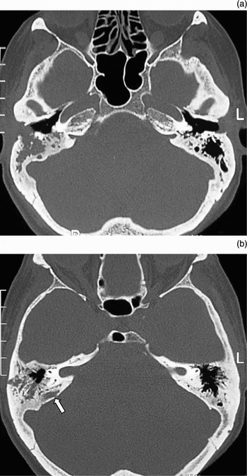
The patient re-presented two years later with a right sensorineural hearing loss (SNHL) of 43 dB. Repeat imaging revealed several areas of bone destruction in the right petrous bone, although there were no lesions in the otic capsule (Figure 2). There was involvement of the external auditory canal with a polyp visible otoscopically, and bone erosion on CT. The left temporal bone appeared normal.
(a) Axial, high resolution computed tomography scan of the temporal bones, demonstrating a lytic lesion centred on the posterior aspect of the right external auditory meatus and the mastoid air cells, with a soft tissue mass in the canal. (b) A second lesion is seen posterior to the right lateral semicircular canal (arrow). At this stage, there is no erosion into this structure.

The patient was treated with prednisolone, aciclovir, aspirin and carbogen, and her right-sided hearing returned to normal over two weeks. The aural polyp settled with topical steroid treatment over the same time period. The patient subsequently received disease-modifiying chemotherapy with azathioprine and methotrexate. This led to clinical remission, with a normal bone scintogram.
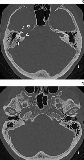
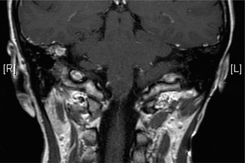
Three years later, the patient re-presented with a 10-day history of vertigo and hearing loss in the right ear. She had a mean right-sided hearing loss of 50 dB, and was commenced on prednisolone, betahistine and aciclovir on admission. Imaging revealed new destructive lesions in the posterior aspect of the otic capsule, adjacent to the crus commune, as well as the petrous apex adjacent to the internal auditory meatus (Figures 3a and 4). The previous bone changes identified on imaging three years previously had largely resolved (Figure 3b). Serial pure tone audiometry showed transient improvement in hearing over the next three days, but a further worsening two days later.
(a) Axial, high resolution temporal bone computed tomography scan, demonstrating a new lesion in a similar location to the lesion shown in Figure 2(b), but this time causing erosion of the right lateral semicircular canal (solid arrow). A further new lesion of the right petrous apex is also seen (hollow arrowheads). (b) The lesion centred on the posterior aspect of the right external auditory meatus is seen to have resolved.

Post-gadolinium, enhanced, coronal, T1 magnetic resonance image, showing enhancement of the right otic capsule lesion seen on computed tomography.

The patient was referred for urgent radiotherapy and received a course of 14 Gy in seven fractions to her right temporal bone. Her hearing transiently worsened following radiotherapy and then improved to a mean hearing level of 26 dB, although most of this hearing loss was at the high frequencies (70 dBHL at 4 kHz). Her vertigo settled at the same time.
Discussion
The otorhinolaryngological manifestations of Langerhans' cell histiocytosis include aural discharge, localised swelling, vertigo, deafness, cutaneous lesions and cervical lymphadenopathy.Reference Irving, Broadbent and Jones10 Sensorineural hearing loss is a rare manifestation, with the hearing loss usually being conductive secondary to obstruction of the external auditory meatus.Reference Cunningham, Curtin, Jaffe and Stool11, Reference McCaffrey and McDonald12 Involvement of the middle ear is most commonly due to secondary infection. This case demonstrated involvement of the otic capsule bone as well as, on the second presentation, the external auditory canal. The origin of temporal bone lesions is often around the mastoid and spreading anteriorly, and a site of origin in the endolymphatic sac has been proposed.Reference Mosnier, Rondini-Gilli, Crosara, Belmatoug, Gyna-Corse and Cazals-Hatem13 The otic capsule is often spared in Langerhans' cell histiocytosis, possibly because of its particularly dense bone structure, and disturbances in hearing and balance may be secondary to a perfusion defect due to pressure from surrounding granulation tissue.Reference Nanduri, Pritchard, Chong, Phelps, Sirimanna and Bailey14 There are five reported cases of sensorineural hearing loss in adults with little improvement in the sensorineural component of the hearing loss following treatment, including radiotherapy treatment in one case.Reference Mosnier, Rondini-Gilli, Crosara, Belmatoug, Gyna-Corse and Cazals-Hatem13, Reference Hore, Mitchell, Radcliffe, Quiney and Walker15, Reference Bauer, MacDonald, Domanowski and Fuleihan16 A paediatric case report detailed partial recovery of SNHL following a course of prednisolone.Reference Nanduri, Pritchard, Chong, Phelps, Sirimanna and Bailey14
The diagnosis depends on imaging, but confusion may arise as early imaging findings may appear similar to inflammatory disease. Computed tomography and MRI are useful in demonstrating anatomical detail in the involved bone and soft tissues. On CT, lytic destruction is common, with irregular margins, especially after treatment. Enhancement within the lesion may be homogeneous or may occur only in the periphery.Reference Cunningham, Curtin and Butkiewicz17 As seen in the present case, the CT appearance of destructive lesions of the temporal bone may resolve with treatment (Figures 2a and 3b). On MR imaging, similar enhancement may be seen, and additional information may be gained about marrow and soft tissue involvement.Reference Bonafe, Joomye, Jaeger, Fraysse and Manelfe18 The MRI appearance, however, is not specific. Bone scintigraphy at diagnosis and on follow up usually reveals the sites of active disease, especially when the involvement is polyostotic, but this imaging modality does not have sufficient resolution within the temporal bone to demonstrate the exact anatomical relations to the complex structures contained within the temporal bone.
Treatment options for Langerhans' cell histiocytosis include watchful waiting, surgery, radiotherapy, and local and systemic chemotherapy. Treatments for severe cases with end-organ damage have included liver, lung and allogeneic bone marrow transplantation.Reference Stockschlaeder and Sucker9
Radiotherapy is one treatment option, with a dosage of 5–20 Gy for osseous lesions advocated.Reference Howarth, Gilchrist, Mullan, Wiseman, Edmonson and Schomberg8 In a series of 63 patients treated with radiotherapy, local disease control was achieved in 82 per cent (median follow up was 44 months), as compared with 64 per cent disease control following chemotherapy (results from literature review).Reference Seegenschmiedt, Micke, Olschewski, Bruns, Heyd and Schaefer19 The dosage used was 3–50 Gy (median 12 Gy), and some of the patients also received surgery or chemotherapy.
Treatment of temporal bone lesions remains controversial. Surgery is required to make a histological diagnosis in cases of temporal bone Langerhans' cell histiocytosis, but does not clearly improve outcome. A recent report of three adults with labyrinthine involvement found that extensive surgery of temporal bone lesions improved conductive but not sensorineural hearing loss.Reference Mosnier, Rondini-Gilli, Crosara, Belmatoug, Gyna-Corse and Cazals-Hatem13 Extensive surgery such as mastoidectomy in cases of temporal bone Langerhans' cell histiocytosis has been shown to have a higher rate of complications, including conductive and sensorineural hearing loss, post-auricular fistula, and transient facial nerve palsy, compared with a conservative approach including a combination of watchful waiting, polypectomy, intralesional steroids, systemic chemotherapy and radiotherapy (10 Gy).Reference Irving, Broadbent and Jones10 Other authors have also reported good local disease control with low-dose radiotherapy.Reference McCaffrey and McDonald12, Reference Anonsen and Donaldson20 In the present case, in which SNHL did not respond to steroids, we found that prompt treatment with radiotherapy to the affected temporal bone salvaged hearing.
• Langerhans' cell histiocytosis is a rare, multisystemic disease that often affects the skull, including the temporal bone
• Hearing loss in Langerhans' cell histiocytosis is usually conductive but may be sensorineural
• Treatment options include intralesional steroids, systemic steroids, systemic chemotherapy, radiotherapy and surgical curettage
• Surgery is advocated mainly for biopsy of involved tissue to minimise complications
• In the presented case, low-dose radiotherapy appeared to result in significant improvement in sensorineural hearing loss (SNHL) secondary to temporal bone Langerhans' cell histiocytosis
• Osseous Langerhans' cell histiocytosis of the temporal bone may completely resolve radiologically, without improvement in SNHL
Acknowledgement
We thank Dr Susan Blaser, Neuro-radiologist, Hospital for Sick Children, Toronto, and Dr Pieter Pretorius, Neuro-radiologist, John Radcliffe Hospital, Oxford, for their advice.