





Introduction
Hypofractionation has changed adjuvant breast radiotherapy by shortening treatment while maintaining oncological outcomes for appropriately selected patients. The FAST-Forward trial established 26 Gy in 5 fractions as non-inferior to 40 Gy in 15 fractions for early breast cancer, and the regimen has subsequently been widely adopted as standard practice for many patients receiving adjuvant breast radiotherapy in the UK.Reference Brunt, Haviland and Wheatley1 However, national guidance also recognises that comorbidity and frailty may justify alternative schedules and local audit.2 In parallel, radiographer-led skin-care guidance emphasises the importance of consistent assessment, practical self-care advice and timely escalation for treatment-related reactions.3 These principles become particularly important when underlying dermatologic disease may amplify risk or complicate interpretation of acute change.
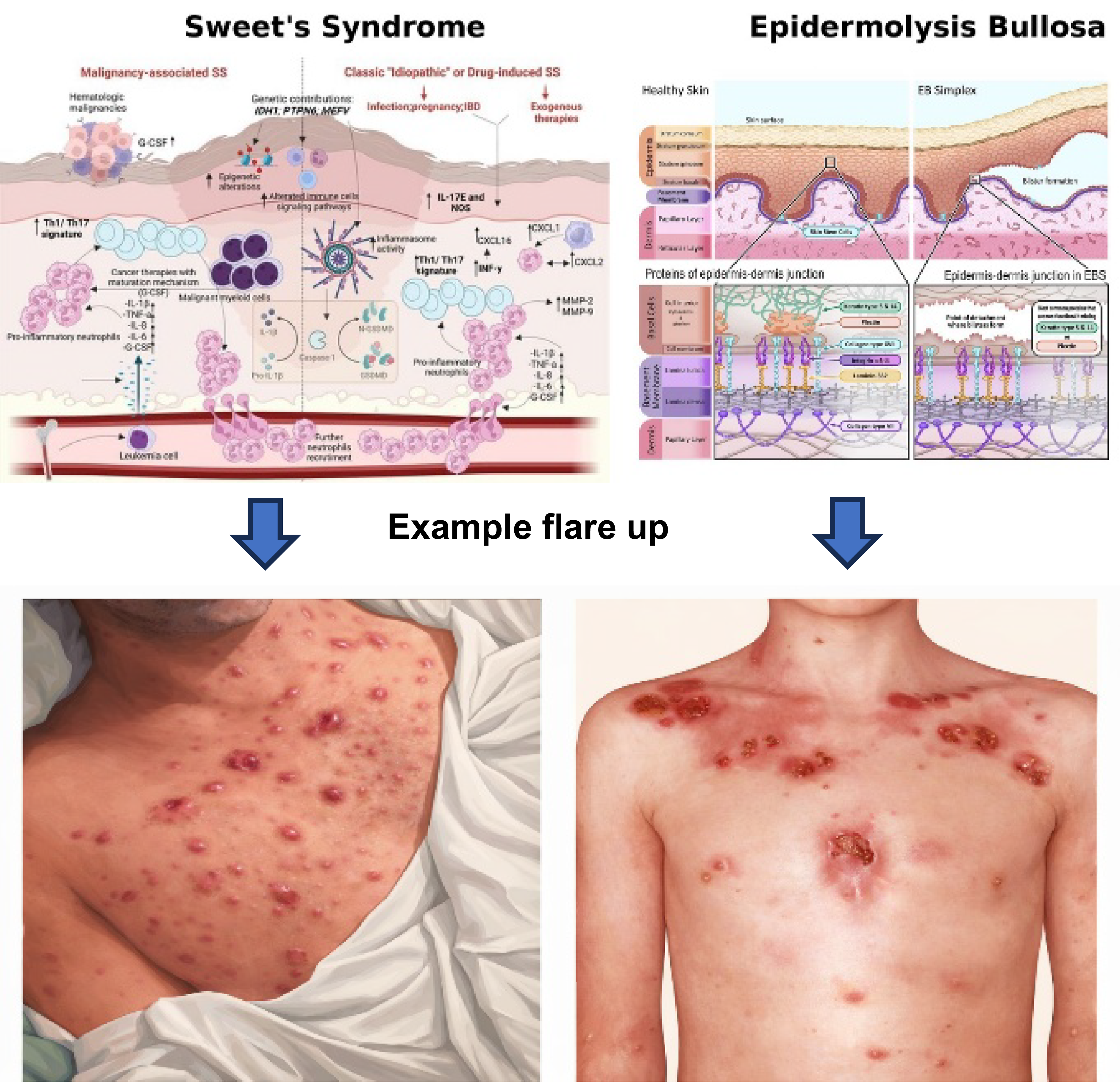
In patients with rare dermatologic conditions, the risk assessment is more complex because acute skin change may represent expected radiation dermatitis, inflammatory flare, infection or fragility-related injury. Sweet’s syndrome is a rare neutrophilic dermatosis that can be triggered by malignancy, drugs and radiotherapy.Reference Ollivier, Renaud and Gouders4 It is characterised by the acute onset of painful erythematous plaques, fever and neutrophilic infiltration of the dermis. Although uncommon, pathergy and exaggerated inflammatory responses following skin trauma have been reported, creating concern regarding radiotherapy tolerance and the potential for treatment-induced flare. Inherited epidermolysis bullosa (EB) is characterised by marked skin fragility, raising concern about breakdown and delayed healing during treatment (Figure 1).Reference Koulis, Herring, Smith and Verhaeghe5 Inherited EB comprises a heterogeneous group of genetic disorders characterised by skin fragility and blister formation following minimal trauma. Patients frequently experience chronic wounds, scarring and impaired healing, creating uncertainty regarding tolerance of radiotherapy-related skin reactions and treatment-associated friction. Published radiotherapy experience in both conditions is limited, and many departments will encounter these patients only rarely. We therefore report two prospectively managed cases to evaluate the feasibility and acute toxicity of adjuvant breast radiotherapy in patients with rare dermatological comorbidities. In doing so, we describe the multidisciplinary decision-making processes and practical management considerations that may assist departments when individualising treatment in situations where published evidence is limited.
Physiology and example flare up of Sweet’s syndrome and EB.

Governance approval for this two-patient case report was granted by the host NHS Trust radiotherapy research and development group.
Case Report
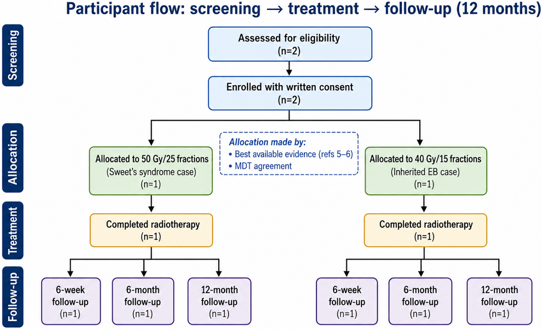
Two adult patients receiving adjuvant breast radiotherapy were managed under a prespecified multidisciplinary protocol with governance oversight. Written informed consent was obtained from both patients for the use of anonymised clinical information and publication of anonymised clinical photographs. Eligibility was pragmatic: confirmed Sweet’s syndrome or inherited EB, clinically indicated breast radiotherapy and agreement that treatment was deliverable with enhanced precautions. Both patients were treated on an Elekta Synergy linear accelerator using the department’s standard breast planning technique of forward planned intensity modulated radiotherapy (IMRT). Treatment was delivered with a tangential pair and mono isocentre, with daily cone beam computed tomography (CT) verification. The participant flow from screening and allocation to treatment completion and follow-up is shown in Figure 2.
Participant flow (screening → treatment → follow-up).

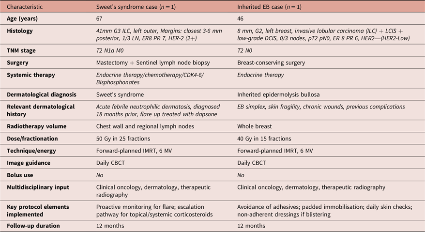
The first patient had Sweet’s syndrome and required chest wall and regional nodal radiotherapy. A conventional schedule of 50 Gy in 25 fractions was selected following multidisciplinary discussion between clinical oncology, dermatology and therapeutic radiography teams. Alternative hypofractionated schedules were considered, but concerns regarding inflammatory flare and the limited published evidence base led the multi-disciplinary team (MDT) to favour a conventional schedule. In addition to delivering a lower dose per fraction, the longer treatment course allowed more frequent clinical review and provided greater opportunity for early intervention should signs of flare occur.Reference Ollivier, Renaud and Gouders4 The second patient had inherited EB and received whole-breast radiotherapy to 40 Gy in 15 fractions. Ultra-hypofractionated treatment was considered; however, concerns regarding skin fragility and the limited available evidence led the MDT to favour moderate hypofractionation. This schedule was considered an appropriate balance between treatment efficacy, patient convenience and the ability to monitor and respond to emerging skin toxicity during treatment.Reference Koulis, Herring, Smith and Verhaeghe5 Fractionation decisions were agreed through multidisciplinary discussion involving clinical oncology, dermatology and therapeutic radiographers, with wound care input available if required. Baseline and treatment characteristics are summarised in Table 1.
Baseline and treatment characteristics

Table 1. Long description
A table comparing characteristics of two medical cases. The table has 14 rows and 3 columns. The columns are labeled ‘Characteristic’, ‘Sweet’s syndrome case (n = 1)’, and ‘Inherited EB case (n = 1)’. The rows include Age, Histology, TNM stage, Surgery, Systemic therapy, Dermatological diagnosis, Relevant dermatological history, Radiotherapy volume, Dose/fractionation, Technique/energy, Image guidance, Bolus use, Multidisciplinary input, Key protocol elements implemented, and Follow-up duration. Each row provides specific details for both cases, such as age, histology details, TNM stage, type of surgery, systemic therapy, dermatological diagnosis, relevant dermatological history, radiotherapy volume, dose/fractionation, technique/energy, image guidance, bolus use, multidisciplinary input, key protocol elements implemented, and follow-up duration.
The local protocol included careful handling, structured toxicity review and predefined escalation pathways. Weekly reviews were undertaken by therapeutic radiographers and clinical oncology staff throughout treatment, with dermatology input available when required. For suspected Sweet’s syndrome flare, prompt discussion with the treating oncologist and dermatology team regarding topical or systemic corticosteroids was incorporated into the pathway. For EB, emphasis was placed on minimising friction, avoiding traumatic dressings or adhesives where possible, maintaining infection surveillance and facilitating rapid review of any new blistering or skin breakdown. Escalation criteria included evidence of progressive blistering, suspected infection, signs of dermatological flare or development of grade 2 or higher toxicity. Clinical review findings were recorded prospectively using case report forms and serial medical photography. Acute toxicity was assessed using Radiation Therapy Oncology Group criteria. In the Sweet’s syndrome case, C-reactive protein was monitored during treatment to detect possible systemic inflammatory flare.
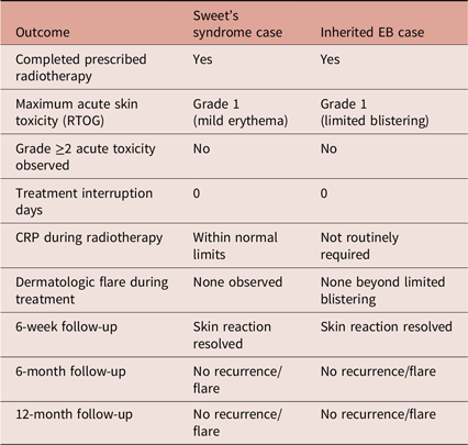
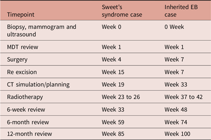
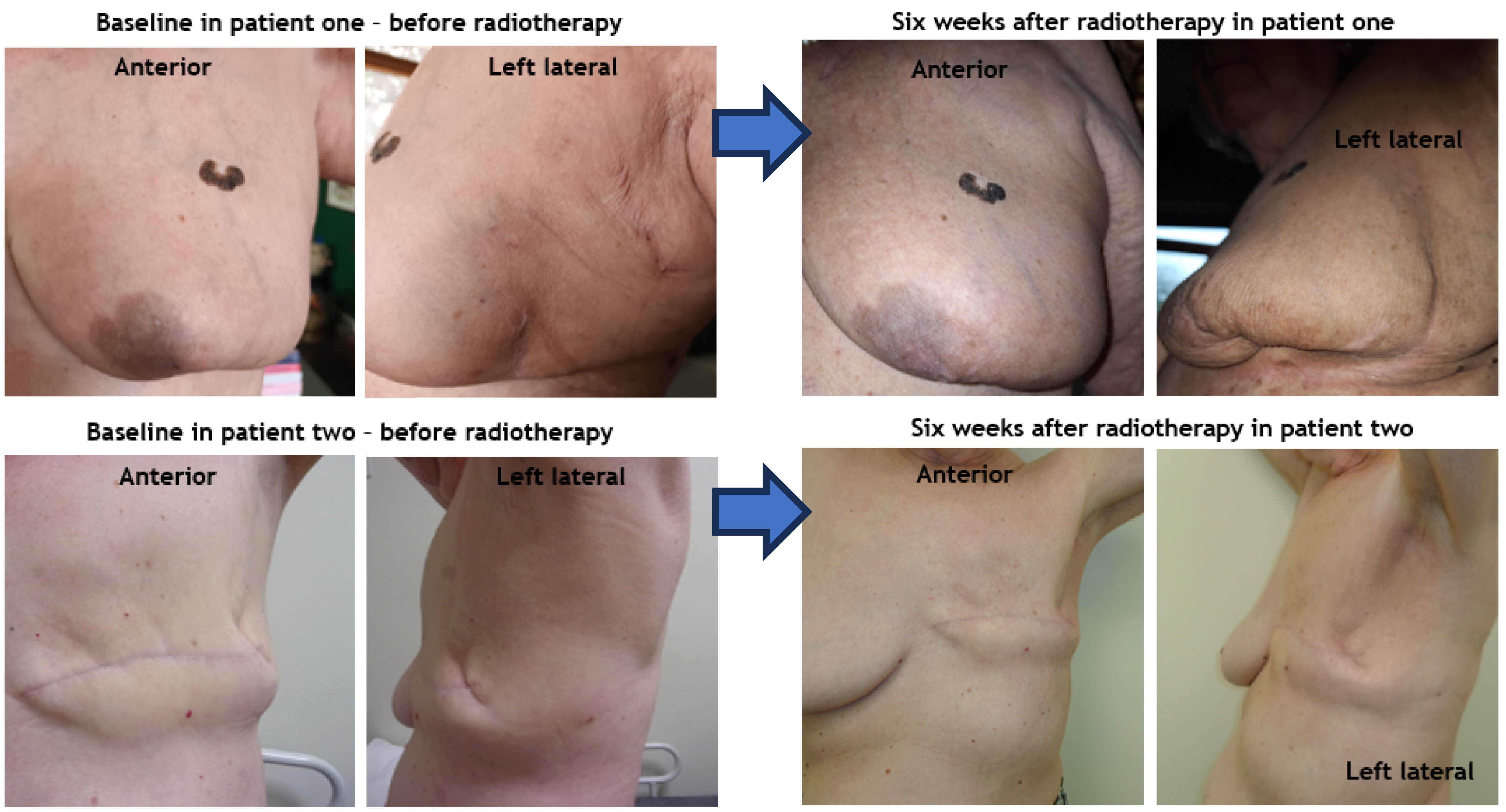
Both patients completed the prescribed course without interruption or permanent discontinuation. The Sweet’s syndrome case developed mild erythema only, graded as RTOG 1, with no biochemical or clinical evidence of systemic flare. The EB case developed limited blistering, also graded as RTOG 1, without extensive breakdown or infection. In both patients, the acute reaction had resolved by 6 weeks. No grade 2 or higher toxicity was observed. At 6 and 12 months, neither patient reported recurrence of radiotherapy-related skin reaction nor dermatologic flare. Feasibility and acute or early outcomes are summarised in Tables 2 and 3.
Side effects during and after radiotherapy

Table 2. Long description
A table comparing side effects during and after radiotherapy for two patients, one with Sweet’s syndrome and one with inherited EB. The table has 8 rows and 3 columns. The columns are labeled Outcome, Sweet’s syndrome case, and Inherited EB case. The rows list different outcomes and their respective details for each patient. Row 1: Completed prescribed radiotherapy, Yes, Yes. Row 2: Maximum acute skin toxicity (RTOG), Grade 1 (mild erythema), Grade 1 (limited blistering). Row 3: Grade >= 2 acute toxicity observed, No, No. Row 4: Treatment interruption days, 0, 0. Row 5: CRP during radiotherapy, Within normal limits, Not routinely required. Row 6: Dermatologic flare during treatment, None observed, None beyond limited blistering. Row 7: 6-week follow-up, Skin reaction resolved, Skin reaction resolved. Row 8: 6-month follow-up, No recurrence/flare, No recurrence/flare. Row 9: 12-month follow-up, No recurrence/flare, No recurrence/flare.
Clinical timeline

Table 3. Long description
A table comparing clinical timelines for Sweet’s syndrome case and Inherited EB case. The table has 8 rows and 3 columns. The columns are labeled Timepoint, Sweet’s syndrome case, and Inherited EB case. The rows are labeled as follows: Biopsy, mammogram and ultrasound: Week 0, 0 Week; MDT review: Week 1, Week 1; Surgery: Week 4, Week 7; Re excision: Week 15, Week 7; CT simulation/planning: Week 19, Week 33; Radiotherapy: Week 23 to 26, Week 37 to 42; 6-week review: Week 33, Week 48; 6-month review: Week 59, Week 74; 12-month review: Week 85, Week 100.
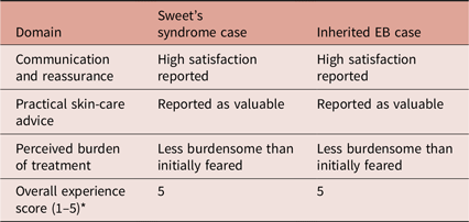
Patient feedback highlighted the value of clear communication, reassurance and practical skin-care advice during treatment, with a summary provided in Table 4.
Patient experience summary

Table 4. Long description
A table comparing patient experiences with Sweet’s syndrome and inherited EB cases across four domains. The table has four rows and three columns. The columns are labeled Domain, Sweet’s syndrome case, and Inherited EB case. The rows are labeled Communication and reassurance, Practical skin-care advice, Perceived burden of treatment, and Overall experience score (1-5). Row 1: Communication and reassurance, High satisfaction reported, High satisfaction reported. Row 2: Practical skin-care advice, Reported as valuable, Reported as valuable. Row 3: Perceived burden of treatment, Less burdensome than initially feared, Less burdensome than initially feared. Row 4: Overall experience score (1-5), 5, 5.
Baseline and six-week post-radiotherapy photography is shown in Figure 3.
Baseline and six-week post radiotherapy photography.

Discussion
These two cases suggest that rare dermatologic conditions do not automatically preclude curative-intent breast radiotherapy when treatment is individualised and supported by proactive multidisciplinary care. The principal finding was feasibility: both patients completed treatment, toxicity remained mild and early recovery was uncomplicated. This is reassuring because both Sweet’s syndrome and inherited EB raise understandable concern for exaggerated acute reactions, treatment interruption and impaired healing. In practical terms, the report supports a structured approach in which radiotherapy is neither withheld reflexively nor delivered using routine pathways without modification.
The Sweet’s syndrome case adds to limited published evidence showing that radiotherapy can be delivered safely when there is careful monitoring for flare and readiness to escalate treatment if required.Reference Ollivier, Renaud and Gouders4 The absence of systemic inflammatory activation in our case is clinically important because concern about flare can influence whether radiotherapy is offered at all. Likewise, the EB case supports earlier case experience showing that adjuvant breast radiotherapy can be feasible in selected patients with meticulous technique and skin precautions.Reference Koulis, Herring, Smith and Verhaeghe5 Our report extends that literature by describing a protocolised workflow rather than isolated treatment completion alone and by capturing short-term follow-up beyond end-of-treatment toxicity.
A key practice message is that standard hypofractionation policy should not be applied uncritically in unusual comorbidity contexts. In both cases, fractional dose formed only one component of the decision-making process. MDT discussions considered the potential benefits of longer treatment schedules, not only from a radiobiological perspective but also because they provided additional opportunities for clinical assessment and timely intervention should toxicity emerge. This was particularly relevant given the uncertainty surrounding the risk of inflammatory flare in Sweet’s syndrome and skin breakdown in EB. The final treatment approaches therefore reflected a balance between maintaining oncological effectiveness, minimising toxicity risk, respecting patient preferences and ensuring adequate opportunities for ongoing review. Hypofractionation remains the standard for most patients, but national guidance allows flexibility where comorbidity, frailty or tolerance concerns justify an alternative approach.2 In our report, the decision to use 50 Gy in 25 fractions for Sweet’s syndrome and 40 Gy in 15 fractions for EB was based on the specific risk profile of each patient rather than on a single institutional default. This is particularly relevant for departments under pressure to standardise, because rigid adherence to the shortest schedule may not always represent the most responsible practice in rare or poorly evidenced situations. Individualisation does not mean abandoning evidence-based practice; rather, it involves integrating the best available evidence with clinical expertise and patient preferences when managing uncommon clinical scenarios for which robust evidence may be lacking.
The cases also reinforce the importance of supportive radiographer-led skin care and early escalation. Breast radiotherapy skin reactions are common even in routine practice, and consistent assessment and advice are central to safe management.3 In rare inflammatory or fragility disorders, the threshold for review may need to be lower, not because severe toxicity is inevitable, but because the consequences of misclassification can be greater. A structured pathway helps staff distinguish expected treatment effects from condition-specific deterioration and supports timely intervention. The patient feedback in this report, although descriptive, also suggests that communication and anticipatory guidance are clinically meaningful aspects of care in situations where anxiety about skin toxicity may be high.
The main limitation is obvious: this is a two-patient report from a single centre, so findings cannot be generalised beyond carefully selected cases. The contribution is therefore practical rather than definitive. Even so, rare conditions are unlikely to generate large prospective datasets quickly, and concise case reporting remains important where it informs day-to-day decisions in radiotherapy departments. Our experience supports a cautious but not exclusionary approach: individualise the fractionation, minimise treatment-related trauma and ensure multidisciplinary review from planning through follow-up. Departments that encounter similar patients may benefit from treating these pathways as service protocols that can be refined prospectively as additional cases emerge.
Although based on only two patients, these cases demonstrate that breast radiotherapy can be delivered safely in selected patients with Sweet’s syndrome and inherited EB when supported by multidisciplinary planning, enhanced toxicity monitoring and individualised fractionation decisions. Further case reporting and prospective data collection are required to strengthen the evidence base for these rare clinical scenarios.
Recommendations for Practice
-
• Early multidisciplinary review should be arranged for any patient with rare inflammatory or fragility-related skin disease being considered for breast radiotherapy.
-
• Fractionation should be individualised rather than determined solely by routine default pathways.
-
• Departments should standardise practical handling precautions and escalation pathways for suspected flare, infection or skin breakdown.
-
• Prospective documentation of toxicity and patient experience should be encouraged to strengthen the evidence base for future cases.
Financial support
None.
Competing interests
The authors declare no conflicts of interest.

 Open access

