Introduction
Fibromyalgia (FM) is a chronic widespread pain syndrome associated with fatigue, sleep disturbance, psychological disorders, and cognitive dysfunction.Reference Clauw 1 Etiology and pathogenesis of FM remain unclear. Abnormal central pain processing is hypothesized to play a main role in pathophysiology of FM.Reference Petzke, Clauw, Ambrose, Khine and Gracely 2 Many neuroimaging studies have demonstrated alterations in the brain structure of people with FM. These abnormalities are associated with not only pain processing but also cognitive deficits and mood impairments in patients with FM.Reference Shi, Yuan, Dai, Ma and Sheng 3 Kuchinad et al demonstrated that patients with FM had significantly less total gray matter volume and 3.3 times greater age-associated reduction in gray matter volume than healthy controls did.Reference Kuchinad, Schweinhardt, Seminowicz, Wood, Chizh and Bushnell 4 Nevertheless, the mechanism underlying accelerated neurodegeneration in patients with FM remains relatively unknown.
Meta-analyses have demonstrated that cognitive function, including working memory, attention, and executive function, may be impaired in patients with FM.Reference Wu, Huang, Fang, Ko and Tsai 5 Furthermore, a cohort study demonstrated that patients with FM had a higher risk of all types of dementia than other patients did after adjustments for age, gender, income, and comorbidities.Reference Tzeng, Chung and Liu 6 Although stress, psychological disorders, and inflammation may be the mechanisms underlying neurocognitive impairmentReference Diaz-Piedra, Guzman, Buela-Casal and Catena 7 the mechanism underlying neurodegeneration in patients with FM has not yet been fully understood.
Sleep disturbance is common among patients with FM.Reference Bigatti, Hernandez, Cronan and Rand 8 Fang et al analyzed the associations among the pain severity, sleep quality, and cognitive dysfunction in patients with FM and suggested that sleep plays a key role in cognitive dysfunction.Reference Fang, Wu, Chen, Teng and Tsai 9 Notably, decrements in brain volume in areas such as the hippocampus and left orbitofrontal cortex have been observed in people with insomnia and the extent of brain volume loss was related to insomnia severity.Reference Altena, Vrenken, Van Der Werf, van den Heuvel and Van Someren 10 Therefore, sleep abnormalities in patients with FM may play a crucial role in neurocognitive impairment and neurodegeneration.
Tau and beta-amyloid (Aβ) proteins are located in the central nervous system (CNS) and are involved in the pathomechanism of neurological degeneration and may serve as biomarkers for neuron damage. Studies have observed increases in Aβ accumulation in the brains of patients with Alzheimer’s disease (AD).Reference Andreasen, Hesse and Davidsson 11 Moreover, tau and Aβ levels have been associated with sleep disruption in both animals and humans.Reference Shokri-Kojori, Wang and Wiers 12 The concentrations of these proteins in blood were increased in people with obstructive sleep apnea.Reference Motamedi, Kanefsky and Matsangas 13 Furthermore, an animal study demonstrated increased Aβ and tau deposits in the cortex of mice with chronic sleep deprivation.
Sleep disturbance can increase neurocognitive disorder risk through the accumulation of tau and Aβ proteins in the brain. However, the association between sleep disruption and neurodegeneration in patients with FM has not been investigated. We hypothesized that neurodegeneration in patients with FM may be related to sleep disturbance via tau and Aβ associated pathway. This study analyzed serum tau and Aβ levels and their relationships with clinical assessments of patients with FM. These data are valuable for guiding clinical evaluation and treatment of patients with FM.
Methods
Participants and blood sample
Patients who met the 2010 ACR criteria for FMReference Wolfe, Clauw and Fitzcharles 14 were recruited from clinics in Taipei Medical University Hospital. The diagnosis of FM was established by an experienced clinical specialist. Patients were excluded if theyReference Clauw 1 had malignancyReference Petzke, Clauw, Ambrose, Khine and Gracely 2; had active or chronic infections;Reference Shi, Yuan, Dai, Ma and Sheng 3 had dementia, CNS disease, and/or head traumaReference Kuchinad, Schweinhardt, Seminowicz, Wood, Chizh and Bushnell 4; had endocrine system diseasesReference Wu, Huang, Fang, Ko and Tsai 5; had major autoimmune disorders; orReference Tzeng, Chung and Liu 6 were pregnant. The control group was recruited from healthy people without diseases or drug use. The controls had no pain complaint more than 1 month prior to inclusion. To eliminate the effects of medications, patients with FM were asked to stop using medications except acetaminophen at least 2 weeks before conducting clinical evaluations and a blood test. All patients were fully briefed on the research procedure and provided their informed consent prior to inclusion. This study was approved by the Joint Institutional Review Board of Taipei Medical University. The first patient was recruited on 12 March 2018. All participants had 40 mL of blood from a forearm vein after waking up in the morning before eating. Protease was immediately added to the blood sample to minimize protein degradation. After collection, the whole blood was left undisturbed at room temperature for 30 minutes. Clots were removed by centrifuging at 1500 to 2000×g for 10 minutes in a refrigerated centrifuge. The serum was then transferred to fresh 1.5-mL tubes (1 mL of serum per tube) and stored at −80°C.
Widespread pain index and symptom severity scale
The widespread pain index (WPI) is a checklist consisting of 19 specific regions of the body with scores of 0 to 19. The score is calculated on the basis of whether the patient had pain in the specific pain locations in the past 1 week. The symptom severity scale (SSS) includes three main symptoms: fatigue, waking up unrefreshed, and cognitive symptoms. The severity of each of these symptoms is measured by score ranging from 0 (no problems) to 3 (severe). The maximum total score is 12; the higher this score, the severer is the associated symptom.
Fibromyalgia Impact Questionnaire
We used the Fibromyalgia Impact Questionnaire (FIQ)—a self-administered questionnaire that evaluates health status, progress, and outcomes of patients with FM and includes 10 items with 20 questions regarding physical functioning, work status, pain, fatigue, morning tiredness, stiffness, anxiety, and depression.Reference Burckhardt, Clark and Bennett 15 The highest possible score is 100; the higher the FIQ score, the greater is the impact or disability.
Pittsburgh Sleep Quality Index
The Pittsburgh Sleep Quality Index (PSQI), composed of 19 self-administrated questions, was used to evaluate sleep quality.Reference Buysse, Reynolds, Monk, Berman and Kupfer 16 It comprises seven components, namely subjective sleep quality, sleep latency, sleep duration, habitual sleep efficiency, sleep disturbances, sleeping medication use, and daytime dysfunction over the last month. Each component score ranges from 0 (no difficulties) to 3 (severe difficulty). The total score is calculated by summing the scores of each component (range of 0-21). A summed score of ≥5 is considered to be indicative of “poor” sleep quality.Reference Buysse, Reynolds, Monk, Berman and Kupfer 16
Beck Depression Inventory II
The Beck Depression Inventory-II (BDI-II), a self-reported questionnaire with 21 multiple-choice items,Reference Abdel-Rahman, Balogun and Kepple 17 assesses depression severity. The maximum score is 63, with each item being rated from 0 to 3 based on the severity. Depression severity is categorized into four levels based on the total score of the questionnaire: no (0-13), mild (14-28), moderate (29-35), and severe (≥36) depression.Reference Osman, Barrios, Gutierrez, Williams and Bailey 18
Beck Anxiety Inventory
The Beck Anxiety Inventory (BAI) is a brief assessment tool for measuring anxiety severity.Reference Beck, Epstein, Brown and Steer 19 Similar to BDI, this scale also has 21 multiple-choice items related to common anxiety symptoms. Each item is rated from 0 to 3 based on the effects of each symptom. This scale has a maximum score of 63: normal (0-9), mild (10-18), moderate (19-29), and severe (30-63) anxiety.Reference Julian 20
Visual analog scale
The visual analog scale (VAS) was used to evaluate pain level. This scale is a ruler on which the start point (0) corresponds to “no pain” and the endpoint (10) corresponds to “the worst imaginable pain.” Patients were asked to indicate the point corresponding to their mean pain level in the past 1 week.
Pressure pain threshold test
To evaluate the pain sensitivity in patients with FM, the patients’ minimum pressure pain threshold (PPT) was assessed using a handheld pressure gauge (Algometer, Paintest, Wagner Inc., Greenwich, CT, USA). The assessment sites were the nine paired tender points of the diagnostic criteria for FM defined by ACR in 1990 located at the occiput, low cervical spine, trapezius, supraspinatus, second rib, lateral epicondyle, gluteus, greater trochanter, and knee. During measurement, the patient was in a relaxed sitting position. The midpoint of the trapezius muscle, muscles under the occipital bone, muscles of the scapula, muscles near the spine, lateral muscles above the buttocks, posterior muscle of the greater trochanter and the chest lock, base of the mastoid muscle, second rib, the upper elbow, and inner edge of the knee were measured three times on each side and averaged.
Immunomagnetic reduction assay
We used immunomagnetic reduction (IMR) assay—a method to probe the associations of magnetic nanoparticles and target biomarkers with their magnetic reagents—to measure the levels of tau protein (tau) and Aβ1-42 in blood. IMR assay is an ultra-high sensitivity technology and referred to as a superconducting quantum interference device based IMR assays.Reference Chieh, Yang and Jian 21 The low limit of detection for Aβ and tau proteins of IMR was noted to be 1 to 10 pg/mL.Reference Yang, Yang and Chieh 22 Therefore, IMR can be used as a validate tool to measure the biomarkers for early diagnosis of Alzheimer’s disease.Reference Chiu, Yang and Horng 23 The magnetic reagents MF-AB2-0060 and MF-TAU-0060 (MagQu Co., Ltd. New Taipei City, Taiwan) were used to assay the biomarkers Aβ1-42 and t-tau. Xacpro-S (MagQu Co., Ltd.) was used as a detector to analyze the IMR signal of reagents in this study. The IMR signal is defined as the percentage reduction in the alternating current magnetic susceptibility of the mixture (including reagents and serum samples) caused by the association among Aβ1-42, t-tau, and magnetic reagents. After examination, the IMR signals were converted to serum tau and Aβ1-42 concentrations using characteristic curves. The mixtures of reagents and serum were used. For t-tau, t-tau reagents (MF-TAU-0060) and serum samples were mixed at a ratio of 2:1. For Aβ1-42 peptides, a 1:1 ratio of Aβ1-42 reagents (MF-AB2-0060) to serum samples was used. After mixing, each mixture was put into a SQUID-based alternating current magnetosusceptometer (XacPro-S, MagQu Co., Ltd.). Each sample was divided into three parts for which IMR signals were detected individually. Thus, each sample was measured three times, and three IMR signals were obtained. The mean, standard deviation (SD), and coefficient of variation (inter-run) of the IMR signals were analyzed. The products of serum t-tau and Aβ1-42 levels (t-tau × Aβ1-42) were computed by multiplying the levels of serum t-tau and Aβ1-42.
Statistical analysis
Data are presented as mean ± SD or number (%). A Mann–Whitney test was conducted to compare the results of questionnaires and protein levels between two groups. A chi-square test was used to analyze the difference among categorical variables between two groups. Pearson and partial correlations were used to evaluate the correlations of questionnaires with serum t-tau and Aβ1-42 concentrations in patients with FM. Statistical analyses were performed with SPSS (version 19.0 for Windows). The significance level was set at P < .05.
Results
Demographic characteristics and clinical profiles
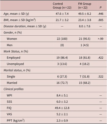
In total, 22 patients with FM and 22 age-matched healthy participants were enrolled. Table 1 presents the baseline characteristics and clinical profiles of the participants in the current study. No significant differences were observed in the baseline characteristics of age, gender, BMI, work status, or marital status between people with and without FM. The FM group had mean symptom duration of 7.96 years. Four patients with FM had known traditional cardiovascular risks, such as hypertension, hyperlipidemia, and diabetes mellitus, etc. In the FM group, the mean WPI score was 8.4 ± 5.1 and mean SSS score was 6.0 ± 3.2; 90% of patients had mild to moderate levels of pain, and the mean VAS score for pain was 5.2 ± 2.1. The mean PPT was 2.3 ± 0.9 kg/cm2. The mean FIQ score was 49.4 ± 12.8 in patients with FM, indicating moderate disability.
Demographic Characteristics and Clinical Profiles of Study Population.

Abbreviations: BMI, body mass index; FIQ, Fibromyalgia Impact Questionnaire; FM, fibromyalgia; n, number of participants; PPT, pressure pain threshold; SD, standard deviation; SSS, symptom severity scale; VAS, visual analog scale; WPI, widespread pain index; y, years.
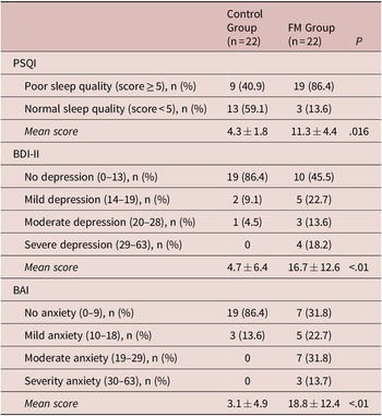
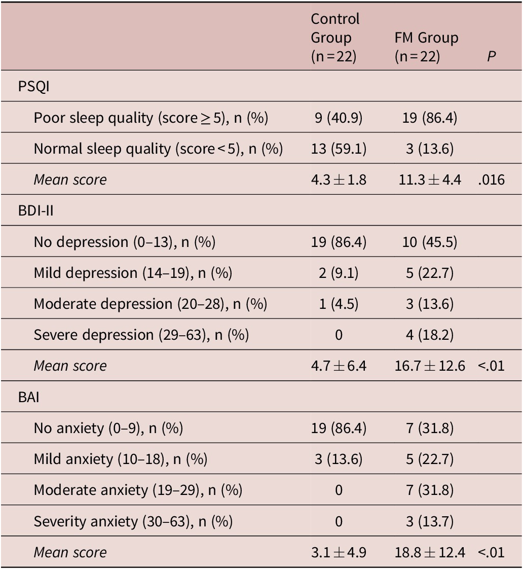
The PSQI, BDI-II, and BAI scores are presented in Table 2. Nineteen patients with FM (86%) had a PSQI score of ≥5, which indicated poor sleep quality. Poor sleep quality was noted in nine participants in the control group (40.9%).
Comparison of Sleep Quality, Depression, and Anxiety Between FM and Control Groups.
Abbreviations: BAI, Beck Anxiety Inventory; BDI, Beck Depression Inventory; FM, fibromyalgia; PSQI, Pittsburgh Sleep Quality Index.
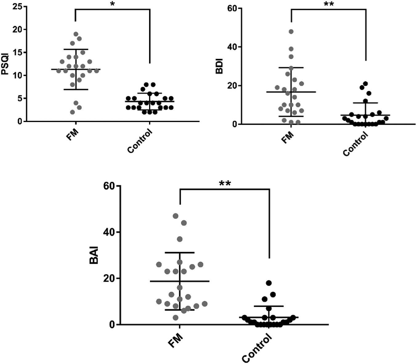
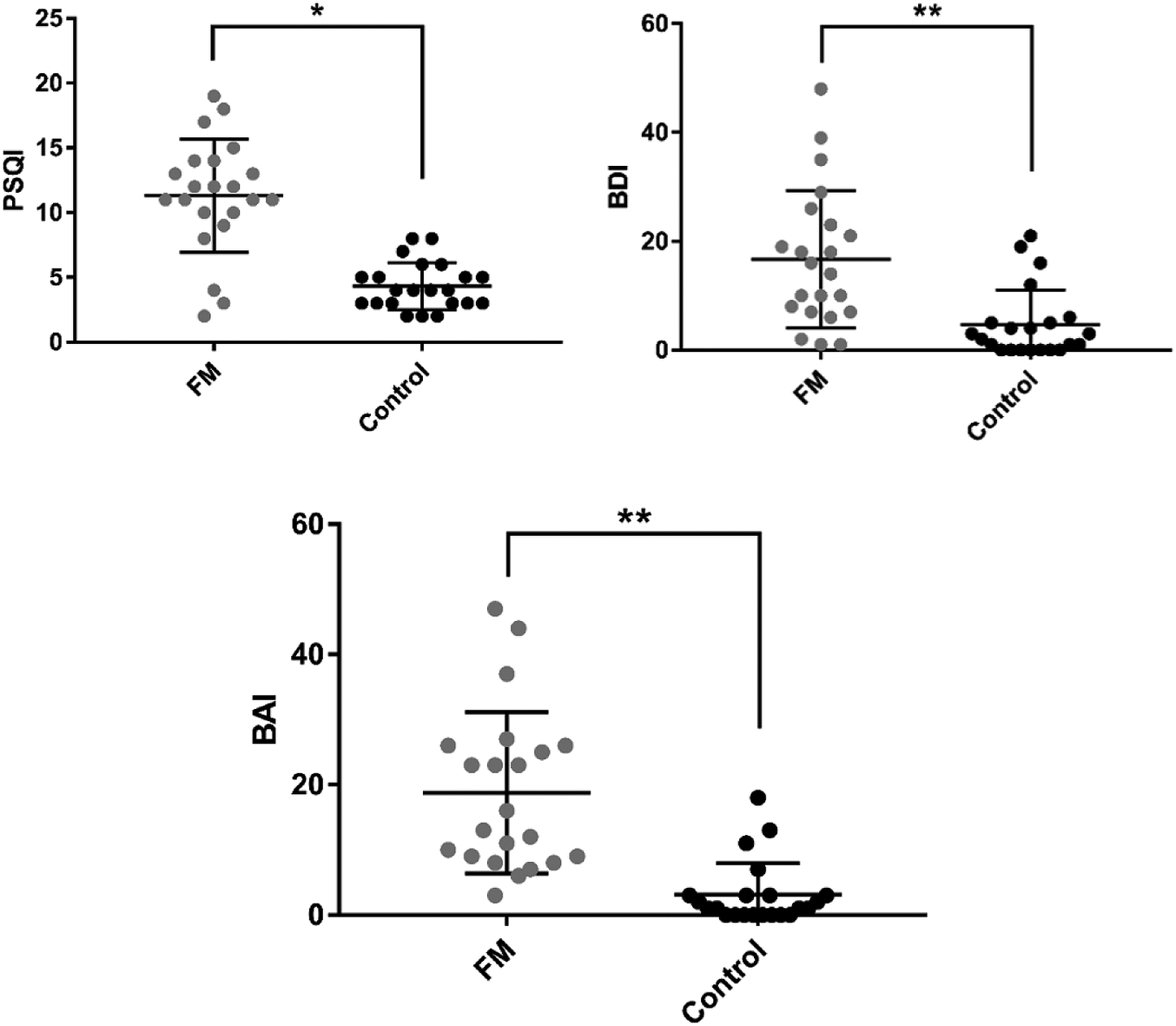
The mean total PSQI score of the FM group was significantly higher than that of the healthy group (P < .01; Figure 1A). The BDI-II and BAI scores indicated that 55% and 68% of patients with FM had depression and anxiety, respectively. In the control group, only 14% and 14% of participants presented with depression and anxiety, respectively. The mean BDI-II scores of patients with FM were higher than those of the healthy group (P < .01, Figure 1B). The mean BAI scores were also higher in the FM group than in the control group (P < .01, Figure 1C).
Comparison of clinical assessments regarding sleep, depression, and anxiety between FM and control groups. FM group had significantly higher levels of (A) sleep disturbance (PSQI score, FM: 11.3 vs CG: 4.3), (B) depression (BDI score, FM: 16.7 vs CG: 4.7), and (C) anxiety (BAI score, FM: 18.8 vs CG: 3.1) compared with controls. BAI, Beck Anxiety Inventory; BDI, Beck Depression Inventory; CG, Control group; FM, fibromyalgia; PSQI, Pittsburgh Sleep Quality Index. *P < .05; **P < .01.
Serum t-tau and Aβ1-42 levels in FM and healthy groups
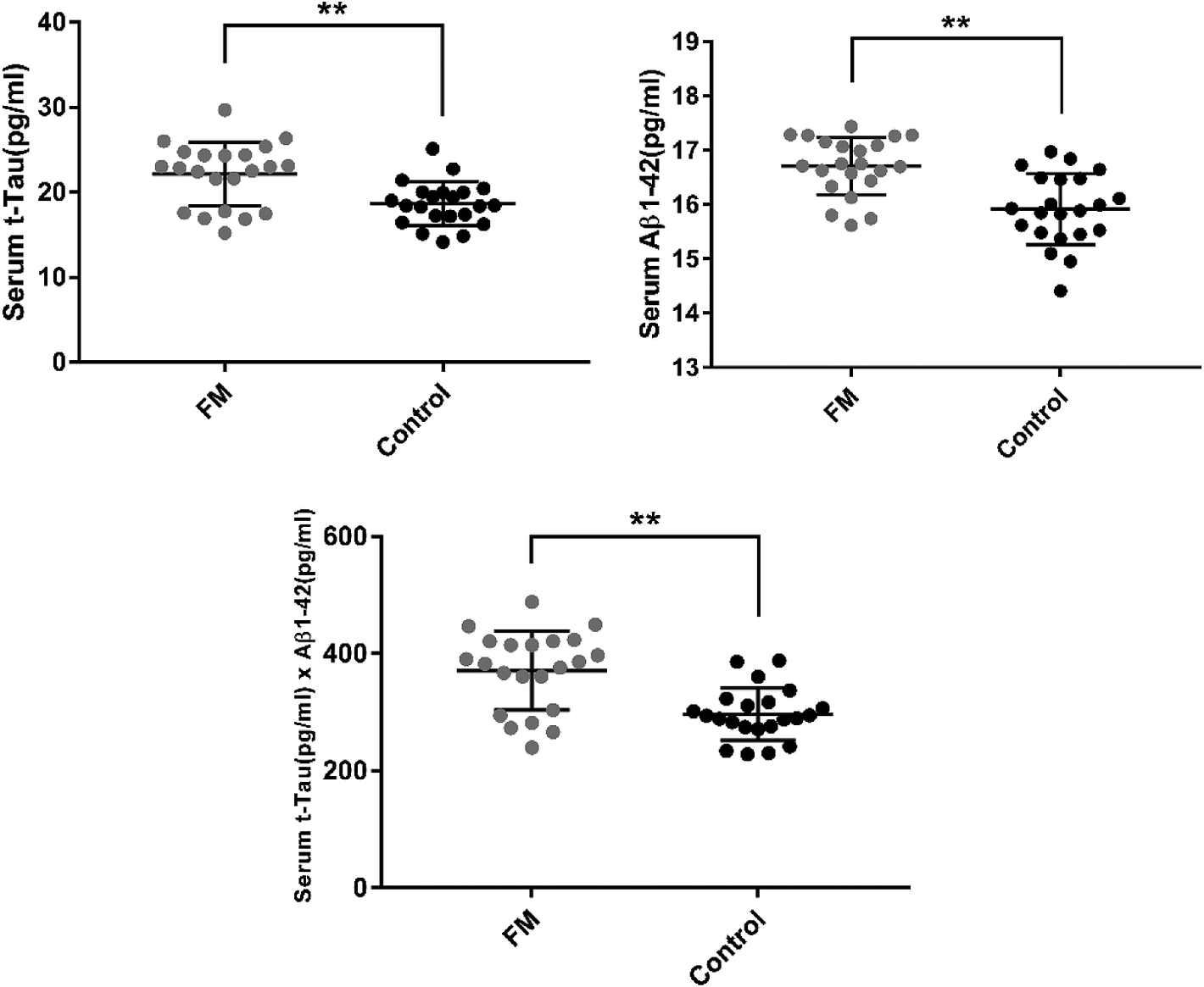
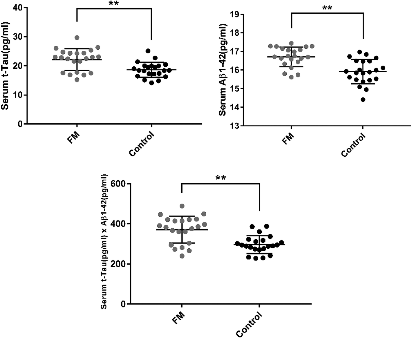
Significant differences were observed in the mean levels of serum tau and Aβ1-42 proteins between the two groups. Serum t-tau and Aβ1-42 levels were significantly higher in the FM group than in the healthy group (P = <.01 and .01, respectively; Figure 2A and B, respectively). Moreover, mean t-tau × Aβ1-42 levels were significantly different between the FM and healthy groups (P = <.01, Figure 2C).
Comparisons of serum tau and Aβ1-42 protein levels between FM and control groups. (A) Serum t-tau levels in the FM group were higher than those in the control group (FM: 22.14 vs CG: 18.62 [ρg/mL]). (B) Serum Aβ1-42 levels in the FM group were higher than those in the control group (FM: 16.72 vs CG: 15.92 [ρg/mL]). (C) Product of tau and Aβ1-42 was also higher in the FM group than in the control group (FM: 370.99 vs CG: 296.71 [ρg/mL]2). t-Tau, total tau protein; Aβ1-42, 42-amino-acid sequence of beta-amyloid protein; FM, fibromyalgia; CG, control group. *P < .05; **P < .01.
Correlations between serum t-tau, Aβ1-42, and t-tau × Aβ1-42 levels in clinical assessments
Pearson and partial correlations were analyzed to investigate associations between the serum biomarkers and all clinical features adjusted for body weight and age in patients with FM (Table 3).
Correlations Between Clinical Features and Levels of Serum Tau and Aβ-42 in FM Group

Abbreviations: Aβ-42, 42-amino-acid sequence of beta-amyloid protein; BAI, Beck Anxiety Inventory; BDI, Beck Depression Inventory; FIQ, Fibromyalgia Impact Questionnaire; FM, fibromyalgia; Tau, tau protein; PPT, pressure pain threshold; PSQI, Pittsburgh Sleep Quality Index; r, Pearson correlation adjusted for body weight and age; SSS, symptom severity scale; VAS, visual analog scale; WPI, widespread pain index.
* P < .05.
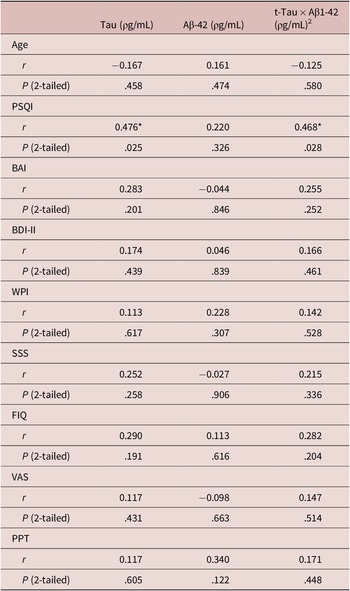
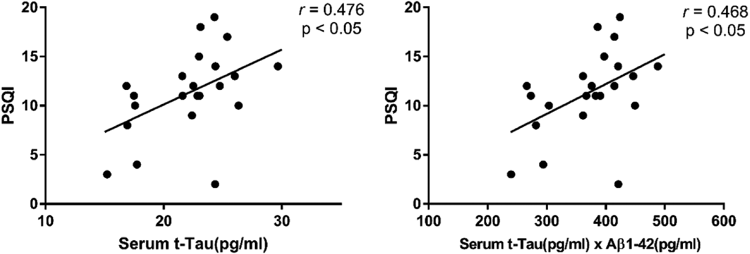
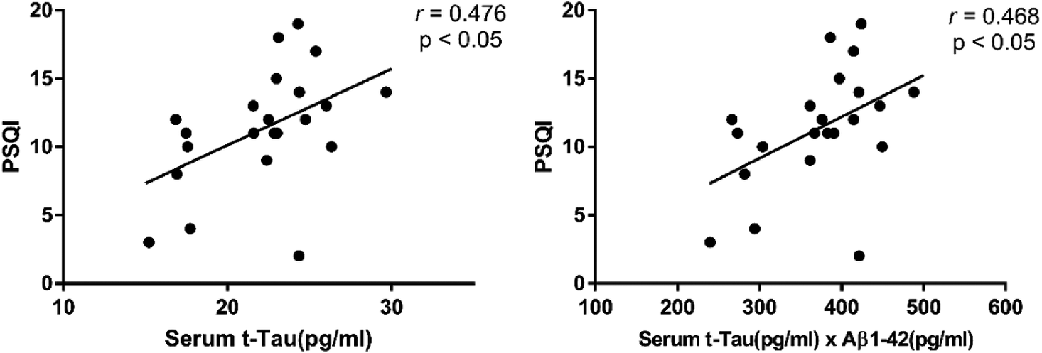
PSQI was significantly correlated with serum tau level (r = 0.476, P = .025, Figure 3A) and t-tau × Aβ1-42 (r = 0.468, P = .028, Figure 3B). However, no correlation between sleep quality scores and serum Aβ1-42 level was observed (P = .326). We did not observe any significant associations of serum levels of t-tau, Aβ1-42, and t-tau × Aβ1-42 values with other variables (age; WPI; SSS, FIQ, VAS, BDI-II, and BAI scores; and PPT, Table 3). The correlations were not statistically significant between the duration of FM and the levels of Tau/Aβ-42 (r = 0.154, P = .493; r = −0.087, P = .701, respectively).
Correlations between sleep quality and serum level of t-tau, product of t-tau and Aβ1-42 in FM group. In the FM group, PSQI scores had significant positive correlations with (A) levels of serum t-tau and (B) t-tau × Aβ1-42. t-Tau, total tau protein; Aβ1-42, 42-amino-acid sequence of beta-amyloid protein; FM, fibromyalgia; r, Pearson correlation coefficient.
Discussion
We observed that levels of serum tau and Aβ-42 proteins were significantly higher in patients with FM than in controls. Tau is a microtubule-associated protein which plays a main role in the stability of axonal microtubules.Reference Trojanowski, Schuck, Schmidt and Lee 24 High blood tau levels can predict poor neurological outcomes in patients with ischemic stroke, cardiac arrest, and neonatal hypoxia.Reference Lv, Wang, Chen, You, Ren and Li 25 Furthermore, the abnormal phosphorylation of tau leads to their disorganization and accumulation to form tangles called neurofibrillary tangles,Reference Alonso, Zaidi, Novak, Grundke-Iqbal and Iqbal 26 resulting in abnormal axonal transport and neurodegeneration.Reference Lue, Sabbagh and Chiu 27 Aβ is the major component of the destructive synaptic formation amyloid plaque, which can be found in the brains of patients with AD and Down syndrome.Reference O’Brien and Wong 28 Aβ is generated from amyloid precursor protein (APP), a modulator of synaptic formation, neurons plasticity, and synaptic transmission.Reference Turner, O’Connor, Tate and Abraham 29 APP can be split into types of Aβ based on the number of amino acids. Aβ-42 is considered the main substance that forms plaques in patients with AD.Reference Fleming and Espay 30 The positive associations among blood tau level, cerebrospinal fluid (CSF) tau level, and brain tau deposition have been demonstrated.Reference Park, Han and Yi 31 In addition, the transport of Aβ from the CNS into the peripheral blood has been proven.Reference Roberts, Elbert and Kasten 32 Therefore, high serum tau and Aβ concentrations might represent the amount of tau and Aβ in the brain. To our knowledge, the present study is the first to investigate levels of serum tau and Aβ-42 in patients with FM. This finding demonstrates that patients with FM could have a higher burden of neuronal damage and a higher risk of subsequent neurodegeneration.
Although accelerated neurodegeneration in the brain has been reported in patients with FMReference Kuchinad, Schweinhardt, Seminowicz, Wood, Chizh and Bushnell 4 the pathomechanism remains unclear. We investigated the relationship between tau and Aβ-42 serum levels with symptom profiles in patients with FM. Among all symptoms profiles, we only observed a significantly positive correlation between serum tau levels and PSQI scores. This finding supports the hypothesis that sleep disturbance has a key role in the neurodegeneration in people with FM. Sleep disturbance, including reduced total sleep time, sleep onset difficulty, increased wake after sleep onset and difficulty in falling asleep after waking, light sleep, decreased sleep efficiency, and non-refreshing sleep, is a common problem for patients with FM. Sleep plays a vital role in conserving brain energy and facilitating memoryReference Steriade 33. Sleep fragmentation and reduction of slow-wave sleep can be found in patients with FM, and alpha-delta wave activity is correlated with pain level in these patientsReference Perlis, Giles and Bootzin 34. The decline in attention, memory, and verbal knowledge were found in people with poor sleep quality in the elderlyReference Yaffe, Falvey and Hoang 35. An association of poor sleep with increased tau levels in CSF and tau deposition in the brain has been observed as wellReference Holth, Fritschi and Wang 36. During sleep deprivation, interstitial fluid tau levels in mice increased by 100%, and CSF tau levels increased by 50% in sleep-deprived humans compared with those well-rested.Reference Holth, Fritschi and Wang 36 These findings imply that sleep disturbance could be a crucial modulator in one of pathomechanisms of neurodegeneration in patients with FM.
It has been reported that pain duration, intensity, and sensitivity are related to gray matter depletion in specific brain regions in patients with FM.Reference Jensen, Srinivasan and Spaeth 37 Furthermore, depression and anxiety disorders are common in people with FM. Some studies have indicated a relationship between psychological symptoms and neurocognitive impairmentReference Gelonch, Garolera and Valls 38 and altered brain structure in patients with FM.Reference Diaz-Piedra, Guzman, Buela-Casal and Catena 7 An association was demonstrated between depression severity and tau accumulation in specific brain regions, including the entorhinal cortex and inferior temporal area.Reference Gatchel, Donovan and Locascio 39 However, the relationship of pain, depression, and anxiety with serum tau and Aβ-42 levels in patients with FM was not observed in the present study. The contribution of pain and psychological disorders to altered brain structure in FM could involve different pathomechanism.
Studies have noted the association of impaired sleep with CSF Aβ levels and brain Aβ deposition.Reference Shokri-Kojori, Wang and Wiers 12 The accumulation of Aβ after one night of sleep deprivation was observed in the hippocampus and thalamus regions. However, increased plasma Aβ-42 was observed but did not reach significance in young people after 24 hours of sleep deprivation.Reference Wei, Zhao and Huo 40 In the current study, serum Aβ-42 levels were significantly higher in FM patients than in controls. However, no significant association between serum Aβ-42 levels and sleep in patients with FM was noted. Patients with obstructive sleep apnea have higher plasma Aβ levels than do other people, and Aβ levels are correlated with hypoxia level in these patients.Reference Salminen, Kauppinen and Kaarniranta 41 Patients with FM demonstrate nocturnal hypoxia during sleep.Reference Alvarez Lario, Alonso Valdivielso, Alegre Lopez, Martel Soteres, Viejo Banuelos and Maranon Cabello 42 Therefore, we conjecture blood Aβ-42 levels may be associated with hypoxia during sleep, rather than with sleep quality, in patients with FM. This hypothesis warrants additional studies.
Dysregulated neuroimmune activation is considered to play a potential role in the central abnormalities of FM patients. Recent study demonstrates that increased microglia but not astrocyte activation in the specific brain areas can be noted by positron emission tomography in FM patients.Reference Albrecht, Forsberg and Sandstrom 43 Astrocytic phagocytosis of synaptic elements is upregulated during acute sleep deprivation and shows a significant increase after prolonged sleep loss which may reflect the physiological response to the increase in synaptic activity associated with prolonged wake. By contrast, chronic sleep loss leads to a sustained low-level microglia activation which may make the brain more susceptible to damages.Reference Bellesi, de Vivo, Chini, Gilli, Tononi and Cirelli 44 These findings can serve as a foundation for understanding the linkage between sleep disturbance and neuroinflammation in FM patients. The complex inflammation process on neuron–glia-tau relations have been highlighted in Alzheimer’s disease. Further exploration of the interaction of glial cells and tauopathy in sleep disturbance would open a potential therapeutic strategy in the treatment for FM.
In a study, blood t-tau × Aβ1-42 could aid in identifying probable AD in patients without cognitive impairments with 92% accuracy; the cutoff value of t-tau × Aβ1-42, its sensitivity, and its specificity were 382.68, 0.96, and 0.90, respectively.Reference Lue, Sabbagh and Chiu 27 We calculated the values of t-tau × Aβ1-42 in both FM and healthy groups in the present study. On the basis of the aforementioned cutoff value of 382.68, 12 patients in the FM group and two persons in the control group had t-tau × Aβ1-42 values exceeding the cutoff. This is consistent with previous epidemiological observations that noted that patients with FM had a higher risk of developing dementia.Reference Tzeng, Chung and Liu 6 Our results indicated that a significant ratio of patients with FM may have high AD risk on the basis of the observation of these biomarkers.
Our study has several crucial clinical implications. First, compared with determining the concentrations of tau and Aβ in CSF and the brain, assessing these proteins in peripheral blood is more convenient. Tau and Aβ may be considered as potential biomarkers in the blood for assessing neurodegenerative disease risk in patients with FM. Furthermore, sleep disturbance evaluation is currently mainly based on subjective reports from patients with FM. We recommend that serum tau protein level be used as a surrogate biomarker to objectively evaluate sleep disturbance in patients with FM. Moreover, interventions to manage sleep disturbance in patients with FM to minimize further neurodegeneration risk, particularly in those with higher tau or Aβ-42 levels, should be considered. A pilot study demonstrated that cognitive behavioral therapy for insomnia might slow or reverse gray matter loss in patients with sleep disturbance.Reference McCrae, Mundt and Curtis 45 Finally, future studies should investigate whether recently developed immune-based tau-targeting therapies can reduce neurodegenerative disease risk in patients with FM.
Some limitations should be addressed in the present study. First, the sample size was relatively small. Second, some reports have suggested that lifestyle habits, such as exercise, smoking, alcohol consumption, and diet, might also affect tau and Aβ levels.Reference Gratuze, Julien, Morin, Marette and Planel 46 These uncontrolled factors may have affected the results of the current study. Third, although we excluded patients who received a diagnosis of dementia clinically, detailed evaluations of neurocognitive function were not performed in the present study. Fourth, we did not analyze the specific sleep disorders in the present study. Although alpha-wave intrusion sleep with non-refreshing sleep is recognized as a major feature of sleep abnormality in FM patients, sleep-disordered breathing and restless leg syndrome are reported to be prevalent in FM population.Reference Viola-Saltzman, Watson, Bogart, Goldberg and Buchwald 47 , Reference Meresh, Artin and Joyce 48 Different sleep disorders may have different impacts on the brain. Further study should be considered to explore the effects of co-existing sleep disorders on tau/Aβ associated abnormality in FM patients. Finally, the causal relationship cannot be determined in a cross-sectional study, and thus, a longitudinal study is warranted to confirm the association we observed in patients with FM.
Conclusion
We found significantly increased serum tau and Aβ-42 levels in patients with FM than in controls. Thus, patients with FM could have an increased subsequent neurodegeneration risk. Moreover, sleep disturbance had a significantly positive association with higher concentrations of serum tau but not serum Aβ-42. These findings imply that sleep disturbance may be important in the pathomechanism of neurodegeneration in patients with FM.
Abbreviations
Glossary
- Aβ
-
beta-amyloid
- ACR
-
The American College of Rheumatology
- AD
-
Alzheimer’s disease
- BAI
-
Beck Anxiety Inventory
- BDI
-
Beck Depression Inventory
- BMI
-
body mass index
- CNS
-
central nervous system
- CSF
-
cerebrospinal fluid
- FIQ
-
Fibromyalgia Impact Questionnaire
- FM
-
fibromyalgia
- IMR
-
immunomagnetic reduction
- ISF
-
interstitial fluid
- MRI
-
magnetic resonance images
- NREM
-
non-rapid eye movement
- PSQI
-
Pittsburgh Sleep Quality Index
- p-tau
-
phosphorylated tau protein
- SSS
-
symptom severity scale
- t-tau
-
total tau protein
- t-tau × Aβ1-42
-
products of serum t-tau and Aβ1-42
- VAS
-
visual analog scale
- VBM
-
voxel-based morphometry
- WPI
-
widespread pain index
Data Availability Statement
The data and analytical methods of this study are available from the corresponding author upon reasonable request.
Funding
This research is financially supported by the Ministry of Education, Taiwan (Higher Education Sprout Project, DP2-108-21121-01-A-01-01) and Ministry of Science and Technology, Taiwan (MOST108-2314-B-038-101-MY2). This work was also supported by the Imaging Center for Integrated Body, Mind, and Culture Research at National Taiwan University.
Disclosures
Bich-Hanh Tai Nguy and Yu-Tai Chang have nothing to be disclosed based on the ICMJE requirements. Wen-Te Liu reports, in past 12 months, receiving honoraria for lectures delivered for CME-related conferences hosted by professional academic societies. He has been a principal investigator or consultant on studies sponsored by G Innings Medical, Ltd., APEX Medical, Clinico, ResMed, Boehringer Ingelheim, and AmCad Biomed, Ltd. Beyond 12 months, He has been a consultant of Teleplus HeathCare, Rooti, and AngelCare Biomedical companies. Chih-Peng Lin reports, in the past 12 months, receiving honoraria for lectures delivered for CME-related conferences hosted by professional academic societies including Taiwan Pain Society and Taiwan Society of Anesthesiologists. No other financial issues related to pharmaceutical company. Jiunn-Horng Kang reports, in the past 12 months, receiving honoraria for lectures delivered for CME-related activities and plenaries and lectures at professional academic conferences and receiving remuneration from academic books published by Ho-Chi Publisher. He has been a principal or co-investigator on studies sponsored by Bayer Pharmaceutical Company and received grant support from LongGood Company. Beyond 12 months, JHK reports having received honoraria for CME-related lectures and for lectures delivered at professional academic conferences.