Introduction
Subtle motor and sensory deficits, or neurological soft signs (NSS), are frequently found in schizophrenia (Heinrichs & Buchanan, Reference Heinrichs and Buchanan1988). NSS have been consistently described in patients with schizophrenia, in prodromal subjects and even in healthy subjects with an increased liability towards the disease (Schröder et al. Reference Schröder, Niethammer, Geider, Reitz, Binkert, Jauss and Sauer1992; Niethammer et al. Reference Niethammer, Weisbrod, Schiesser, Grothe, Maier, Peter, Kaufmann, Schröder and Sauer2000; Lawrie et al. Reference Lawrie, Byrne, Miller, Hodges, Clafferty, Cunningham Owens and Johnstone2001). Although NSS can be reliably demonstrated by clinical examination, it is generally accepted that the respective signs cannot be used for topological diagnosis, as emphasized by the suffix ‘soft’.
In fact, NSS mainly involve complex rather than simple tasks, necessitating the integration of different cerebral sites and systems. Recent longitudinal studies found NSS to decrease with clinical stabilization from the acute to the post-acute phase of the disorder. This effect is less pronounced in patients with an unfavourable rather than a more benign course of the disease (Schröder et al. Reference Schröder, Niethammer, Geider, Reitz, Binkert, Jauss and Sauer1992; Whitty et al. Reference Whitty, Clarke, Browne, McTigue, Kamali, Feeney, Lane, Kinsella, Waddington, Larkin and O'Callaghan2003; Bachmann et al. Reference Bachmann, Bottmer and Schroder2005), a finding that may be related to a persisting ‘process activity’ of the underlying neurobiological disease in the former group. However, even in patients with a more favourable course, NSS scores significantly exceed those obtained in healthy controls. From a clinical standpoint these fluctuations may refer to functional deficits involving pre-formatted morphological deficits of the respective cerebral sites.
Previous studies identified slightly decreased volumes of the basal ganglia, the thalamus (as indicated by an increased width of the third ventricle) and the cerebellum to be associated with increased NSS scores (see Dazzan & Murray, Reference Dazzan and Murray2002; Bottmer et al. Reference Bottmer, Bachmann, Pantel, Essig, Amann, Schad, Magnotta and Schröder2005 for review). Similar associations were found with decreased activation of the sensorimotor cortices and the supplementary motor area using functional neuroimaging techniques (Schröder et al. Reference Schröder, Wenz, Schad, Baudendistel and Knopp1995, Reference Schröder, Essig, Baudendistel, Jahn, Gerdsen, Stockert, Schad and Knopp1999). Although these findings include all parts of the cortico-cerebellar-thalamic-cortical circuitry as conceptualized in the model of ‘cognitive dysmetria’ (Andreasen et al. Reference Andreasen, Paradiso and O'Leary1998), the hypothesis that NSS may serve as an indicator of the respective deficits has not yet been addressed in a clinical study. Moreover, because of methodological constraints the above-mentioned structural imaging studies were restricted to those areas that can be segmented manually or by means of semi-automatic algorithms.
Voxel-based morphometry (VBM) allows examination of the entire brain for structural correlates of clinical signs and symptoms or for group differences (Ashburner & Friston, Reference Ashburner and Friston2000). In a first VBM analysis with respect to NSS, Dazzan et al. (Reference Dazzan, Morgan, Orr, Hutchinson, Chitnis, Suckling, Fearon, Salvo, McGuire, Mallett, Jones, Leff and Murray2004) found subcortical structures (thalamus, putamen and globus pallidus), the pre-central, temporal and lingual gyrus to be important sites for NSS in patients with newly diagnosed schizophrenia. However, potential cerebellar changes were not addressed.
Low levels of NSS can also be demonstrated in healthy individuals, in whom they relate to peristatic factors such as age or less developed skills and their potential cerebral correlates. As the latter factors may occur randomly in any individual, it seems unlikely that they correspond to the same pattern of cerebral changes that is observed in schizophrenia patients. This hypothesis is supported by the longitudinal studies cited above that indicate that NSS in schizophrenia not only refer to pre-morbid deficits but also may involve changes inherent to psychosis itself.
In the present study, we therefore investigated the morphological correlates of NSS in first-episode patients with schizophrenia and in healthy controls by using optimized VBM under the following hypotheses: (i) in patients with schizophrenia NSS correspond to changes in the cortico-cerebellar-thalamic-cortical circuit including the sensorimotor cortex, basal ganglia, thalamus and the cerebellum; and (ii) these associations do not apply to healthy controls, in whom NSS are not systematically related to any particular cerebral site.
Method
Subjects
Forty-two subjects with first-episode schizophrenia and 22 healthy controls matched for age, gender, ethnicity, education and handedness were enrolled. Patients were consecutively admitted to the in-patient unit of the Department of Psychiatry, University of Heidelberg, and diagnosed as suffering from schizophrenia or schizophreniform disorder according to DSM-IV criteria. The patient group consisted of 21 women and 21 men, all Caucasians with a mean age of 27.7 years (s.d.=5.8) and a mean of 12.1±1.5 years of education. Patients were treated with an atypical antipsychotic according to their psychiatrists' choice (mean dose of 542.9±172.2 mg chlorpromazine equivalents) (Woods, Reference Woods2003). The mean duration of total neuroleptic treatment was 34.5±10.8 days. Twenty-two healthy controls were recruited through advertisements and screened for major psychiatric disorders before being included. The group comprised 11 male and 11 female Caucasians with a mean age of 27.6±3.5 years and a mean of 11.9±1.5 years of education. Clinical evaluation included ascertainment of personal and family history and detailed physical and neurological examination. None of the participants had a lifetime history of neurological or medical illness, head injury or substance abuse. All subjects were dominantly right-handed (Oldfield, Reference Oldfield1971).
The investigations were approved by the ethics committee of the Medical Faculty, Heidelberg University. Written informed consent was obtained from all participants after the procedures of the study had been fully explained.
Clinical evaluation
After remission of acute symptoms, NSS were examined with the Heidelberg Scale (Schröder et al. Reference Schröder, Niethammer, Geider, Reitz, Binkert, Jauss and Sauer1992), which consists of five items assessing motor coordination (Ozeretzki's test, diadochokinesia, pronation/supination, finger-to-thumb opposition, speech articulation), three items assessing integrative functions (station and gait, tandem walking, two-point discrimination), two items assessing complex motor tasks (finger-to-nose test, fist-edge-palm test), four items assessing right/left and spatial orientation (right/left orientation, graphesthesia, face-hand test, stereognosis), and two items assessing hard signs (arm holding test, mirror movements). Ratings are given on a 0 (no prevalence) to 3 (marked prevalence) point scale. All neurolological examinations were carried out by the same rater (S.B.). A sufficient internal reliability (Cronbach's α=0.85/0.89 for patients with schizophrenia/healthy subjects), inter-rater (0.88, p<0.005) and test–retest reliability (r tt=0.80, p<0.001) had been established previously (Schröder et al. Reference Schröder, Niethammer, Geider, Reitz, Binkert, Jauss and Sauer1992; Bachmann et al. Reference Bachmann, Bottmer and Schroder2005).
Diagnoses were established using the German version of the Structured Clinical Interview for DSM-IV (SCID; Wittchen et al. Reference Wittchen, Wunderlich, Gruschwitz and Zaudig1997). Psychopathological symptoms and predictors of outcome were rated on the Positive and Negative Syndrome Scale (PANSS; Kay et al. Reference Kay, Fiszbein and Opler1987) and on the Strauss–Carpenter Scale (SCS; Strauss & Carpenter, Reference Strauss and Carpenter1974). Extrapyramidal side-effects were assessed with the scales by Simpson & Angus (Reference Simpson and Angus1970), the Barnes Rating Scale for Drug-Induced Akathisia (Barnes, Reference Barnes1989) and the Abnormal Involuntary Movement Scale (AIMS; Guy, Reference Guy1976).
Magnetic resonance imaging (MRI) data acquisition
MRI data were obtained at the German Cancer Research Centre with a 1.5-T Magnetom Vision MR scanner (Siemens Medical Solutions, Erlangen, Germany) using a T1-weighted 3D magnetization prepared rapid gradient echo sequence (MP-RAGE, 126 coronal slices, image matrix=256×256, voxel size=0.98×0.98×1.8 mm3, TR=10 ms, TE=4 ms, flip angle=12°).
Optimized VBM
The VBM protocol proposed by Good et al. (Reference Good, Johnsrude, Ashburner, Henson, Friston and Frackowiak2001) was applied for preprocessing of imaging data (P.A.T.). This method minimizes the probability of misclassifications within the tissue segmentation by the introduction of additional preprocessing steps prior to normalization and subsequent segmentation. Before preprocessing, all structural images were checked for artefacts, the origin was placed on the anterior commissure and images were reoriented manually to approximate the anterior and posterior commissure to the horizontal plane.
(1) Initially, a group-specific template was generated by normalizing each MR image to the template provided by the Montreal Neurological Institute, using linear (ordinary least square, 12 parameter affine transformation) and nonlinear (modelled by the linear combination of a set of harmonic basis functions, cut-off=25 mm) algorithms (Friston et al. Reference Friston, Ashburner, Frith, Poline, Heather and Frackowiak1995; Ashburner & Friston, Reference Ashburner and Friston1999). The normalized images were segmented into grey matter, white matter, and cerebrospinal fluid, smoothed with an isotropic Gaussian kernel of 10-mm full-width at half-maximum (FWHM) and averaged for grey and white matter separately. Thus, two group- and tissue-specific templates were created with a voxel size of 1×1×1 mm3.
(2) MR images (in native space) were segmented into tissue classes, followed by a series of additional automated morphological operations to remove unconnected non-brain voxels from the segments. The grey- and white-matter images were normalized to the group-specific templates as described in (1).
(3) The calculated normalization parameters were then reapplied to the structural images and these images were resliced to a voxel size of 1×1×1 mm3. This approach led to improved normalization for the full brain images, and also guarantees optimal final segmentation because of the probability maps, used as Bayesian priors, being in stereotactic space.
(4) The normalized structural images were segmented again. Because of local volume effects (growing or shrinking) in nonlinear spatial normalization, a voxel-wise multiplication with the Jacobian determinant (derived from normalization parameters) was performed to preserve the volume of a particular voxel.
(5) Finally, the tissue segments were smoothed with a 10-mm FWHM Gaussian kernel (Ashburner & Friston, Reference Ashburner and Friston2000).
Statistical analysis
Using SPM2 software (www.fil.ion.ucl.ac.uk/spm) we tested for correlations between local grey and white-matter density and NSS levels within the two groups separately. To avoid false-positive findings caused by effects other than group affiliation or NSS level, an additional multiple correlation analysis was performed. Effects of ‘no interest’ were modelled by regressors for age, gender, treatment duration and neuroleptic dose. The F-map computed for the full model was thresholded for an α error probability of p<0.001, uncorrected. No spatial threshold was applied. The regions formed by the surviving voxels contained nuisance effects; we created mask images that allowed their exclusion in further analysis.
Regional correlations between local density values and NSS levels were assessed by a voxel-wise regression analysis containing the NSS subscales and the total score as explanatory variables. Linear contrasts were computed for each main effect, resulting in six T-maps thresholded for a significance level of p<0.001 uncorrected and a spatial extent of 200 voxels. Finally, voxels associated with nuisance variables were excluded by using the mask images mentioned above. In a second statistically more stringent step, we repeated the aforementioned analysis by applying a family-wise error correction for multiple comparisons over the whole brain (p<0.05).
Selected results were overlaid onto the inflated cortical surface of a standard brain, provided by the anatomical toolbox of Eickhoff et al. (Reference Eickhoff, Stephan, Mohlberg, Grefkes, Fink, Amunts and Zilles2005) using the Computerized Anatomical Reconstruction and Editing Toolkit (CARET; Van Essen et al. Reference Van Essen, Drury, Dickson, Harwell, Hanlon and Anderson2001). Significant results in subcortical regions were visualized by superimposing them onto a high-resolution T1-weighted standard brain.
Results
Demographic and clinical data
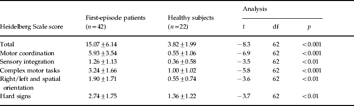
The two groups did not differ significantly in age, gender, educational extent and handedness. DSM-IV assessment revealed diagnoses of schizophrenia in 26 patients and diagnoses of schizophreniform disorder in 16 patients. Patients' total NSS scores and single item scores on the Heidelberg Scale were significantly increased when compared to controls (Table 1). Extrapyramidal side-effects were low according to the Simpson and Angus Rating Scale (12.1±1.9), the Barnes Scale (0.9±1.6) and the AIMS (0.8±2.1). Mean scores on the PANSS were 53.2±10.4 and 57.7±7.6 on the SCS. On the latter, duration of illness was estimated according to the period since first onset of hallucinations or delusions (4: <1 week; 3: 1 week to 6 months; 2: 6 months to 2 years; 1: 2–5 years; 0, >5 years). The distribution was as follows: 4: 21.4%, 3: 59.5%, and 2: 19.1%.
Levels of neurological soft signs (NSS) in patients with first-episode schizophrenia and in healthy comparison subjects

NSS and grey-/white-matter density
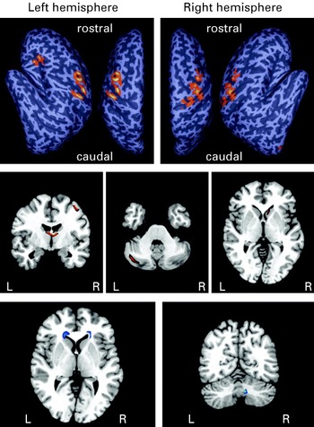
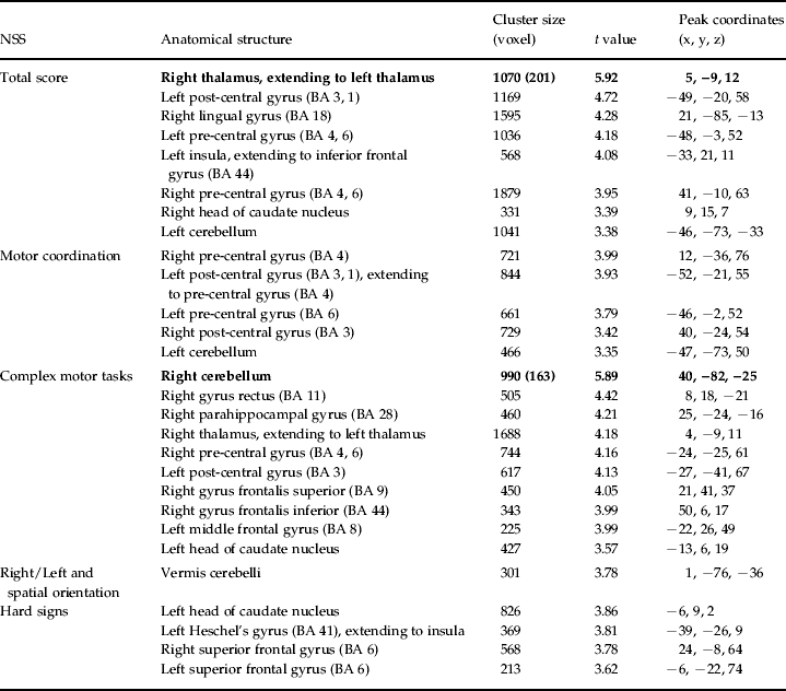
In the patient group, higher NSS scores were associated with a decreased grey-matter density of the thalamus bilaterally, the left post-central gyrus, the right lingual gyrus, the left insula (extending to the inferior frontal gyrus), the right pre-central gyrus, the head of the right caudate nucleus, and the left hemisphere of the cerebellum (Table 2; Fig. 1).
Brain regions showing a significant inverse correlation between grey-matter (top, middle) and white-matter (bottom) density and neurological soft signs (NSS) total score in patients with first-episode schizophrenia superimposed onto an inflated standard brain surface (top) and onto slices of a T1-weighted standard brain (middle, bottom): height threshold p<0.001, uncorrected; extent threshold=200 voxels.

Anatomical structures showing significant inverse correlation between grey-matter density and levels of neurological soft signs (NSS) in patients with first-episode schizophrenia

BA, Brodmann area.
Height threshold p<0.001, uncorrected; extent threshold=200 voxels. Regions surviving a threshold of p<0.05, corrected for multiple comparisons, in bold with corresponding cluster size in parentheses.
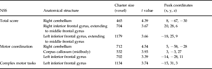
Similar associations applied to the NSS subscales of motor coordination, complex motor tasks and hard signs (Table 2). Increased scores on the subscale right/left and spatial orientation were also correlated with grey-matter loss in the cerebellar vermis (Table 2). No significant association was found for the subscale sensory integration. The associations between NSS total score and the thalamus bilaterally and between the NSS subscale complex motor tasks and the right cerebellar hemisphere (Table 2) remained significant after a stringent family-wise error correction for multiple comparisons over the whole brain (p<0.05). In addition, higher NSS levels were associated with a reduced white-matter density in the right inferior frontal gyrus (NSS total score), the left inferior frontal gyrus (NSS total score, motor coordination, complex motor tasks), the right cerebellum (NSS total score, motor coordination) and the corpus callosum (motor coordination) (Table 3, Fig. 1).
Anatomical structures showing significant inverse correlation between white-matter density and levels of neurological soft signs (NSS) in patients with first-episode schizophrenia

Height threshold p<0.001, uncorrected; extent threshold=200 voxels.
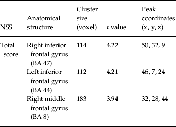
In the control group, VBM revealed that a higher NSS total score was significantly correlated with reduced grey-matter density in the right middle frontal gyrus and the inferior frontal gyrus bilaterally (Table 4). No significant association between NSS scores and white-matter density was detected.
Anatomical structures showing significant inverse correlation between grey-matter density and levels of neurological soft signs (NSS) in healthy control subjects

BA, Brodmann area.
Height threshold p<0.001, uncorrected; extent threshold=100 voxels.
Discussion
Our study yielded two major findings: (i) increased NSS scores were associated with a decreased density in the pre- and post-central gyrus, pre-motor areas, inferior and middle frontal gyri, caudate nucleus, thalamus and cerebellum; and (ii) this pattern only applied to patients with schizophrenia but not to healthy controls.
The putative relationship between NSS levels and cerebral alterations in schizophrenia has been addressed by independent research groups (Dazzan & Murray, Reference Dazzan and Murray2002; Bottmer et al. Reference Bottmer, Bachmann, Pantel, Essig, Amann, Schad, Magnotta and Schröder2005 for review). Structural imaging studies reported higher NSS levels to be associated with sulcal or ventricular enlargement, smaller hemispheres and frontal areas, cerebellar atrophy, lower volumes of the basal ganglia, reduced thalamic size and cortical changes in sensorimotor areas. Studies using functional MRI demonstrated an association between NSS and reduced activation of sensorimotor and supplementary motor areas (Schröder et al. Reference Schröder, Wenz, Schad, Baudendistel and Knopp1995, Reference Schröder, Essig, Baudendistel, Jahn, Gerdsen, Stockert, Schad and Knopp1999).
Except for the study conducted by Dazzan et al. (Reference Dazzan, Morgan, Orr, Hutchinson, Chitnis, Suckling, Fearon, Salvo, McGuire, Mallett, Jones, Leff and Murray2004), the aforementioned structural imaging studies analysed data using the region-of-interest approach, which is based on a priori choices and can therefore only be applied to a certain set of brain structures. Newer imaging tools such as VBM allow for the detection of structural alterations throughout the entire brain. Whole-brain analysis seemed to be particularly suitable for our purpose as hitherto existing research indicated that NSS are not attributable to changes in discrete brain structures but rather to cerebral alterations affecting multiple areas. Particular care was taken to include the cerebellum because of its potential association with NSS in schizophrenia (Keshavan et al. Reference Keshavan, Sanders, Sweeney, Diwadkar, Goldstein, Pettegrew and Schooler2003; Bottmer et al. Reference Bottmer, Bachmann, Pantel, Essig, Amann, Schad, Magnotta and Schröder2005). Overall, our results strongly support the findings of both structural and functional neuroimaging studies, as VBM analysis revealed a statistically significant association between NSS level and reduced density values in the pre- and post-central gyrus, pre-motor areas, inferior and middle frontal gyri, caudate nucleus, thalamus and cerebellum. After family-wise error correction for multiple comparisons over the whole brain, regional associations remained significant for the NSS total score and thalamus bilaterally as well as for the NSS subscale complex motor tasks and right cerebellar hemisphere. In their voxel-based MRI investigation of 77 first-episode patients with schizophrenia, Dazzan et al. (Reference Dazzan, Morgan, Orr, Hutchinson, Chitnis, Suckling, Fearon, Salvo, McGuire, Mallett, Jones, Leff and Murray2004) found left and right thalamus grey-matter reductions in patients who scored high on sensory integration signs. This finding corresponds with the association between NSS and an increased width of the third ventricle as a rough approximation of thalamic size described in a computed tomography (CT) study (Schröder et al. Reference Schröder, Niethammer, Geider, Reitz, Binkert, Jauss and Sauer1992). Involvement of the thalamus in the development of NSS seems plausible as it is generally considered as a relay centre in coordinating motor activity through basal ganglia and cerebellar pathways (Schell & Strick, Reference Schell and Strick1984) and mediates sensory perception and gating (Jones & Friedman, Reference Jones and Friedman1982).
Our finding of cerebellar involvement in the performance of complex motor tasks is supported by the studies cited above (Keshavan et al. Reference Keshavan, Sanders, Sweeney, Diwadkar, Goldstein, Pettegrew and Schooler2003; Bottmer et al. Reference Bottmer, Bachmann, Pantel, Essig, Amann, Schad, Magnotta and Schröder2005). That this association applied primarily to complex motor task parallels the findings of Keshavan et al. (Reference Keshavan, Sanders, Sweeney, Diwadkar, Goldstein, Pettegrew and Schooler2003) and underlines its important role in motor coordination (Ramnani & Miall, Reference Ramnani and Miall2001).
On the cortical level, decreased grey-matter density values in the primary sensory cortex, the primary motor area, and the pre-motor area were associated with higher NSS levels. These findings support the hypothesis that changes in the sensorimotor cortex may be involved in the development of NSS, as indicated by functional MRI studies (Schröder et al. Reference Schröder, Wenz, Schad, Baudendistel and Knopp1995, Reference Schröder, Essig, Baudendistel, Jahn, Gerdsen, Stockert, Schad and Knopp1999).
In an earlier CT study (Schröder et al. Reference Schröder, Niethammer, Geider, Reitz, Binkert, Jauss and Sauer1992) we found the size of the rostral part of the caudate nucleus (estimated by the ratio of the frontal horn distance to the distance between the heads of the caudate nuclei) to be significantly correlated with NSS in 50 schizophrenia patients. Accordingly, the present investigation revealed an association between increased NSS levels (total score, complex motor tasks, and hard signs) and decreased densities of the heads of the caudate nuclei. Volume losses in the basal ganglia in patients with higher NSS scores were also reported by Dazzan et al. (Reference Dazzan, Morgan, Orr, Hutchinson, Chitnis, Suckling, Fearon, Salvo, McGuire, Mallett, Jones, Leff and Murray2004). The basal ganglia constitute a major centre in the extrapyramidal motor system and are involved in the automatic execution of learned motor plans and in the preparation of movements (Marsden, Reference Marsden1982). Studies examining the effects of neuroleptic treatment on brain structure (Scherk & Falkai, Reference Scherk and Falkai2006 for review) found the basal ganglia to be particularly sensitive to morphological changes, especially under treatment with typical antipsychotics. However, an influence of medication in our study is unlikely as all patients were treated with atypical neuroleptics and duration of treatment was relatively short.
In our sample of healthy comparison subjects, significant associations between NSS levels and regional changes in grey-matter density (p<0.001, uncorrected) were restricted to three circumscribed frontal areas. This finding is partly convergent with the VBM study of Dazzan et al. (Reference Dazzan, Morgan, Chitnis, Suckling, Morgan, Fearon, McGuire, Jones, Leff and Murray2006) that found a reduced grey-matter density in frontal cortical fields in subjects with increased scores for integrative signs. Additionally, and in contrast to our findings, the group reported a significant inverse association of NSS with volumes of the middle and superior temporal gyri. This discrepancy might be partly related to the relatively small sample size of healthy individuals in our study. Furthermore, the present sample of healthy controls was composed only of Caucasians, whereas in the study by Dazzan et al. (Reference Dazzan, Morgan, Chitnis, Suckling, Morgan, Fearon, McGuire, Jones, Leff and Murray2006) other ethnic groups were over-represented. As Caucasians have been described as presenting with lower NSS scores than other ethnic groups (Heinrichs & Buchanan, Reference Heinrichs and Buchanan1988), this difference is of potential importance.
In both schizophrenia patients and healthy controls, increased levels of NSS were related to frontal grey-matter reductions. By contrast, associations between NSS and other brain regions were restricted to the patient group. This dissociation leads to the hypothesis that some neurological signs share a common anatomical substrate in patients with schizophrenia and in healthy controls, whereas others are more specifically related to the pathogenesis of schizophrenia. Levels of NSS demonstrated in healthy controls are generally rather low and are stable over time (Bachmann et al. Reference Bachmann, Bottmer and Schroder2005). It is generally assumed that NSS in healthy controls refer to peristatic factors such as age or less developed skills and their potential cerebral correlates. By contrast, the associations found within the patient group between NSS and cerebral changes correspond to the cortico-cerebellar-thalamic-cortical circuitry as proposed by Andreasen et al. (Reference Andreasen, Paradiso and O'Leary1998). Additional evidence that NSS in healthy subjects and patients with schizophrenia are based on a different pathogenesis comes from the northern Finland 1966 general population birth cohort study, where ratings for infant motor development at age 1 year were related to cerebral changes assessed by MRI at age 33–35 years (Ridler et al. Reference Ridler, Veijola, Tanskanen, Miettunen, Chitnis, Suckling, Murray, Haapea, Jones, Isohanni and Bullmore2006). In healthy subjects, but not in the patients, delay of infant motor development was associated with cerebral changes in areas important for NSS. According to this dissociation, NSS in schizophrenia only partly depend on delays of infant motor development. The corresponding hypothesis that the cerebral changes underlying NSS are not entirely pre-formatted or static but may also increase as psychosis develops is supported by longitudinal studies indicating progressive changes of the sensorimotor cortices and the supplementary motor area (Thompson et al. Reference Thompson, Vidal, Giedd, Gochman, Blumenthal, Nicolson, Toga and Rapoport2001), of the thalamus (Rapoport et al. Reference Rapoport, Giedd, Kumra, Jacobsen, Smith, Lee, Nelson and Hamburger1997), and the cerebellum (Pantelis et al. Reference Pantelis, Velakoulis, McGorry, Wood, Suckling, Phillips, Yung, Bullmore, Brewer, Soulsby, Desmond and McGuire2003).
Patients' disease state, their medication, and the mode of recruitment have to be considered as potential confounding factors. All patients were first hospitalized (first-episode patients) and examined after remission of acute psychotic symptoms before discharge; the NSS scores obtained were in the range that was expected from previous studies of our group. This also applied to the NSS scores measured in the healthy controls. Patients received atypical neuroleptics; significant extrapyramidal side-effects did not occur. In this regard, NSS did not relate to extrapyramidal side-effects or to changes in the D2-dopamine receptor systems even in patients who underwent their first neuroleptic therapy (Schröder et al. Reference Schröder, Silvestri, Bubeck, Karr, Demisch, Scherrer, Geider and Sauer1998). The patient sample was recruited consecutively among referrals from a large university department in an urban area. According to this, the study carried out in South London by Dazzan et al. (Reference Dazzan, Morgan, Orr, Hutchinson, Chitnis, Suckling, Fearon, Salvo, McGuire, Mallett, Jones, Leff and Murray2004) yielded comparable findings.
In conclusion, we found grey and white-matter density of distinct brain regions to be significantly correlated with severity of NSS in first-episode schizophrenia patients. These regions predominantly involved the inferior and middle frontal gyri, sensorimotor and pre-motor cortices, the thalamus, basal ganglia and the cerebellum. Our findings emphasize the disruption of the cortico-cerebellar-thalamic-cortical circuitry in schizophrenia as conceptualized in the model of ‘cognitive dysmetria’ (Andreasen et al. Reference Andreasen, Paradiso and O'Leary1998) and strongly suggest that the resulting disconnection syndrome underlies both psychopathological symptoms and neurological abnormalities.
Acknowledgements
This study was supported by the Stanley Medical Research Institute. We are grateful to Dr K. J. Brooks, Oxford University, UK, for his assistance in the preparation and proofreading of the manuscript.
Declaration of Interest
None.