I. INTRODUCTION
The self-assembly of amphiphilic molecules in aqueous environments is a bottom-up approach to the construction of nanoscale artifacts observed in nature that can be readily translated into workable synthetic nanostructures.Reference Philp and Stoddart1–Reference Barauskas, Johnsson and Tiberg7 In nature it can be seen that the shape of lipid-based assemblies is driven by the molecular structure of the lipid, and the shape of the assembly has an effect on the function of the aggregate.Reference Kunitake3, Reference Zhang5, Reference Schnur6, Reference Kunitake, Okahata, Shimomura, Yasunami and Takarabe8–Reference Reichel, Roelofsen, Geurts, Hämäläinen, Feiters and Boons11 Likewise, by making rational alterations to the structure of a precursor amphiphile, the shape and function of synthetic aggregates can be tuned.Reference Shimizu, Masuda and Minamikawa12
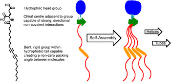
One such useful aggregated structure is straight, high axial ratio nanotubes, which form by the tight, helical turning of a bilayer ribbon.Reference Kunitake3, Reference Schnur6, Reference Shimizu, Masuda and Minamikawa12–Reference Spector, Selinger, Schnur, Green, Nolte and Meijer15 In order to design a precursor amphiphile capable of assembling in this manner, several molecular characteristics have to be exploited. At the most basic level, the molecular geometry of the hydrophilic head and hydrophobic tail can be used to predict the likelihood of either the bilayers forming helically based tubes or forming a micellar assembly.Reference Israelachvili, Mitchell and Ninham10 Molecular motifs specific to ribbon and tube assembly include a rigid segment, often provided by unsaturation, within an otherwise flexible hydrophobic tail. This rigidity encourages bilayer assembly, and if the rigid segment causes angular displacement between neighboring hydrophobic tails, then the formation of elongated ribbon structures are possible.Reference Kunitake, Okahata, Shimomura, Yasunami and Takarabe8 In order to facilitate the twisting of long ribbons into helices, groups that provide strong, directional interactions between neighboring molecules, such as amide and aromatic groups, are desirable.Reference Kunitake3, Reference Schnur6, Reference Shimizu, Masuda and Minamikawa12 These groups are best positioned at the interface between a polar head group and a nonpolar tail, and close to a chiral center.Reference Kunitake, Okahata, Shimomura, Yasunami and Takarabe8, Reference Helfrich and Prost16, Reference Selinger, Spector and Schnur17 Chirality imparts a specific direction to the nonzero packing angle between nearest neighbor molecules that is generated by the bent rigid segment within the hydrophobic tail (Fig. 1). It is this concerted molecular packing that drives the assembly of amphiphiles into helical ribbons that can become so tightly wound that they can form straight nanotubes with high axial ratios.Reference Helfrich and Prost16–Reference Thomas, Lindeman, Corcoran, Cotant, Kirsch and Persichini18
Generic molecular structure for amphiphiles that self-assemble via helical intermediates. Important molecular characteristics for tubular self-assembly include the polar head group, a group able to take part in strong directional interactions close to a chiral center and the flexible hydrophobic tail with a bent rigid group.

As distinct from other nanostructures with similar aspect ratios, amphiphile-based nanotubes can be synthesized with internal diameters ranging from 10–1000 nm.Reference Shimizu, Masuda and Minamikawa12 In addition, these nanotubes can be designed to include hydrophilic surfaces with a range of available surface chemistries.Reference Frusawa, Fukagawa, Ikeda, Araki, Ito, John and Shimizu19 This flexibility in design makes amphiphile-based nanotubes suitable for a diverse range of applications including their use as a platform to elucidate the 3D structure of proteinsReference Wilson-Kubalek, Brown, Celia and Milligan20, Reference Ringler, Müller, Ringsdorf and Brisson21 and their employment as drug delivery vehicles.Reference Rudolph, Stilwell, Cliff, Kahn, Spargo, Rollwagen and Monroy22–Reference Schnur, Price and Rudolph25 Lipid nanotubes are also ideal as templates to synthesize useful structures from materials that do not lend themselves to self-assembly,Reference Yang, Kamiya, Shimizu, Koshizaki and Shimizu26–Reference Kogiso, Zhou and Shimizu30 or in combination with other materials as nanocomposites.Reference Archibald and Mann31–Reference Zhou, Kogiso, He, Shimizu, Koshizaki and Shimizu34
Amphiphilic molecules that have been shown to self-assemble into helically based tubes include diacetylenic phosphatidylcholines,Reference Yager and Schoen35–Reference Mahajan, Zhao, Du and Fang37 a range of glycolipids,Reference Archibald and Mann31, Reference Polidori, Pucci, Zarif, Lacombe, Riess and Pavia38–Reference Yunis and Lee40 cholesterol,Reference Zastavker, Asherie, Lomakin, Pande, Donovan, Schnur and Benedek41, Reference Chung, Benedek, Konikoff and Donovan42 and ammonium-Reference Nakashima, Asakuma, Kim and Kunitake43–Reference Wang and Hollingsworth45 and amino acid-Reference Yamada, Ihara, Ide, Fukumoto and Hirayama46–Reference Lee, Kim, Seo, Do, Lee, Jung, Kogiso and Shimizu48 headed surfactants. Shimizu et al.Reference Shimizu, Masuda and Minamikawa12 have conducted significant research into such self-assembling structures. One direction they have taken investigates the assembly of glycolipids with glucopyranosylamine head groups connected by an amide bond to a C18 hydrocarbon tail.Reference Kamiya, Minamikawa, Jung, Yang, Masuda and Shimizu39 Here they reiterated the morphological importance of the kinked rigidity in the hydrophobic tail to the self-assembly of helically based tubes. In this case they optimized tubular assembly by varying the position and number of double bonds in the hydrophobic tail, finding that the inclusion of a single cis-double bond at C11 provided the best results. This current work investigates the thermal properties of the nanotubes self-assembled from this lipid, N-(11-cis-octadecenoyl)-β-D-glucopyranosylamine, illustrating the highly ordered nature of the supramolecular structure. We also have constructed a novel hybrid structure of a glycolipid nanotube decorated with silver nanoparticles.
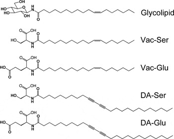
In order to contribute to the understanding of the rational design and construction of these self-assembled systems, we report the design, synthesis, and characterization of four new precursor amphiphiles and their subsequent aggregated structures. These amphiphiles are composed of the vaccenic acid based hydrophobic tail of the glycolipid and the diacetylenic tail used extensively in other nanotube systemsReference Yager and Schoen35, Reference Lee, Koepsel, Stolz, Warriner and Russell44, Reference Wang and Hollingsworth45, Reference Lee, Kim, Seo, Do, Lee, Jung, Kogiso and Shimizu48, Reference Thomas, Corcoran, Cotant, Lindeman, Kirsch and Persichini49 with hydrophilic amino acid head groups (Fig. 2). Aqueous dispersions of these amphipiles produced a range of aggregated structures including nanotubes and the related helical and twisted bilayer ribbons.
Chart of molecules synthesized to investigate amphiphilic self-assembly.

II. EXPERIMENTAL PROCEDURES
A. Materials
All materials, except methanol, were used as received. 11-cis-octadecenoic acid was purchased from Nu-Chek Prep, Inc. (Nu Chek Prep, Elysian, NM) and O-(7-azabenzotriazol-1-yl)-N,N,N′,N′-tetramethyluronium hexafluorophosphate (HATU) and 1-hydroxy-7-azabenzotriazole (HOAt) were purchased from GL Biochem (Shanghai, China). All other reactants and reagents were purchased from Sigma-Aldrich Pty. Ltd. (Aldrich Australia, Sydney, NSW, Australia), as were Toyopearl TSKgel HW-40S size-exclusion media and dimethyl sulfoxide. Other solvents were AR grade obtained from Ajax Finechem Pty. Ltd. (Ajax, Adelaide, SA, Australia). Methanol was degassed for 2 h before size exclusion chromatography (SEC) and refluxed over magnesium turnings before distillation (65 °C) for use in nanotube preparation. All water used for self-assembly was purified using a LABCONCO Water Pro PS deionizer operating at a resistance of 18.2 MΩ.
B. Instrumentation
13C and 1H NMR spectra of glycolipids and precursors were recorded on a Varian Mercury (200 MHz) spectrometer (Novachem Pty Ltd., Melbourne, Australia) and conducted in dimethyl sulfoxide-d6 or D2O with chemical shifts reported in parts per million downfield from tetramethylsilane. 13C and 1H NMR spectra of amino acid lipids and precursors were recorded on a Bruker (400 MHz) spectrometer (Novachem Pty Ltd., Melbourne, Australia) and conducted in dimethyl sulfoxide-d6 or deuterated chloroform with chemical shifts reported in parts per million downfield from tetramethylsilane.
FTIR was carried out with a Thermo Electron Corporation Nicolet Nexus 870 spectrophotometer (Thermo Fisher Scientific, Cheshire, UK) using a Smart Collector attachment (Thermo Fisher Scientific, Cheshire, UK) for high temperature (HT) DRIFT analysis, and an MCT/A detector, on samples ground with anhydrous KBr powder. An environmental chamber enabled heating of the samples in 25° increments from 100–200 °C in a nitrogen atmosphere.
Electron microscopy was used to image nanotube samples. Transmission electron microscopy (TEM) was conducted on air-dried aqueous solutions using a JEOL JEM-1200EX instrument (JEOL Australia, Sydney, NSW, Australia) operated at 80 kV, a Philips CM100 instrument (Philips Research, Eindhoven, The Netherlands) operated at 80 kV, or a Philips CM200 instrument operated at 200 kV.
Thermal analysis was carried out on crushed, lyophilized samples of glycolipid nanotubes. Modulated temperature differential scanning calorimetry (MT-DSC) was performed using a TA Instruments MDSC 2920 (TA Instruments c/-Waters Australia, Rydalmere, NSW, Australia) calibrated for heat capacity using a sapphire standard, and for temperature and cell constant with indium. Scans were performed from 50–180 °C under a high purity nitrogen atmosphere (flow rate 50 mL·min−1) with a modulation of ±1 °C every 60 s. Thermal gravimetric analysis (TGA) was performed on a TA Instruments Hi-Res modulated TGA 2950HR under an atmosphere of nitrogen (flow rate 50 mL·min−1) from room temperature to 400 °C at a rate of 2 °C min−1 and from 400 °C to 900 °C at 10 °C min−1.
C. Synthesis of β-D-glucopyranosylamine
D-(+)-glucose monohydrate (1.00 g, 5.05 mmol) and ammonium hydrogen carbonate (0.399 g, 5.05 mmol) were added to ammonia solution, 28% (25 mL) and stirred at 42 °C for 48 h. The resultant solution was then reduced to half its volume and water (15 mL) was added before reducing the solvent again to half its volume. This cycle was repeated several times until the ammonia odor disappeared, at which point the solution (15 mL) was cooled overnight to –20 °C, and then to –196 °C just prior to lyophilization (DYNAVAC Freeze Drying Unit, Model FD1, Dynavac, Sydney, NSW, Australia; temperature at –42 °C; Vacuum at 1.3 × 10⁻¹ mbar) for at least 24 h. The resulting light brown highly hydrophilic solid was produced in essentially quantitative yields. 13C NMR (200 MHz, D2O): δ 62.0 (C-6); 70.9 (C-4), 73.8 (C-2), 77.8 (C-3) and 77.8 (C-5); 88.0 (C-1[β]).
D. Synthesis of N-(11-cis-octadecenoyl)-β-D-glucopyranosylamine and the self-assembly of glycolipid nanotubes
The glycolipid, N-(11-cis-octadecenoyl)-β-D-glucopyranosylamine, was synthesized and subsequently self-assembled as described previouslyReference Kamiya, Minamikawa, Jung, Yang, Masuda and Shimizu39 with the exception of an additional purification step, that being recrystallization from methanol. This produced a waxy white solid (0.1826 g, 33%), for which spectral data matched those in the literatureReference Kamiya, Minamikawa, Jung, Yang, Masuda and Shimizu39 and the morphology of the self-assembled nanotubes, incubated for 72 h, was confirmed by TEM. 1H NMR (200 MHz, DMSO-d 6): δ 0.85 (t, 3H); 1.24 (m, 20H); 1.47 (m, 2H); 1.98 (m, 2H); 2.08 (m, 4H); 3.01–3.67 (m, 6H); 4.45–4.98 (m, 4H); 4.69 (d, J = 9 Hz, 1H); 5.32 (m, 2H); 8.25 (d, J = 9.2 Hz, 1H). 13C NMR (200 MHz, DMSO-d 6): δ 14.0-35.5 (16C), 61.0, 70.0, 72.5, 77.6, 78.5, 79.4, 129.7, 172.6.
Anal. Calcd for C24H45O6N: C, 64.98; H, 10.22; N, 3.16. Found: C, 64.94; H, 9.80; N, 3.17.
E. Photoreduction of silver nitrate in aqueous dispersions of glycolipid nanotubes
Degassed 20% ethanolic aqueous solution of silver nitrate (3 mL, 10 mmol) were internalized into lyophilized nanotubes by capillary action using the procedure established by Yang et al.Reference Yang, Kamiya, Shimizu, Koshizaki and Shimizu26 Glycolipid nanotubes with internalized 20% ethanolic aqueous solutions of silver nitrate were suspended in pure water (3 mL) and placed in a 10 mm quartz cell. This cell was irradiated by ultraviolet light (λmax = 254 nm) emitted by sixteen 8 W fluorescent tubes contained in an Oliphant Ultra Violet Lamp cabinet with temperatures between 20–25 °C maintained by fan-forced airflow. A sample of nanotubes not removed from the ethanolic aqueous solution of silver nitrate was also exposed to UV irradiation using the same method.
F. L-glutamic acid dimethyl ester hydrochloride (L-GluDME·HCl)
Thionyl chloride (30 mL, 150 mmol) was added dropwise to methanol (120 mL) that was rapidly stirring at –10 °C. To this solution was added L-glutamic acid (7.36, 50 mmol) and then the reaction mixture was allowed to warm to room temperature and left to stir overnight. The solvent was removed in vacuo and the residue was redissolved in methanol and then reduced in vacuo on two further occasions to remove residual thionyl chloride. Purification was via dissolution in a minimal quantity of methanol, followed by precipitation with diethyl ether and then filtration. This purification procedure was repeated revealing a white solid in 90% yield. 1H NMR (400 MHz, DMSO-d 6): δ 2.08 (quartet, J = 7.00, 2H), 2.46–2.62 (m, 2H), 3.61 (s, 3H), 3.73 (s, 3H), 4.02 (t, J = 6.64, 1H), 8.78 (s, 3H); 13C NMR (400 MHz, DMSO-d 6): δ 25.1, 28.9, 51.1, 51.5, 52.5, 169.5, 172.1.
G. L-serine methyl ester hydrochloride (L-SerME.HCl)
A synthetic procedure based on the one described for L-GluDME·HCl was followed. Starting with the variant amino acid, L-serine (10.51 g, 100 mmol), all other quantities were adjusted to maintain the molar ratio to the amino acid. A white solid was isolated in 98% yield. 1H NMR (400 MHz, DMSO-d 6): δ 3.74 (s, 3H), 3.82 (s, 2H), 4.08 (t, J = 3.58, 1H), 5.61 (s, 1H), 8.59 (s, 3H); 13C NMR (400 MHz, DMSO-d 6): δ 52.7, 54.4, 59.4, 168.4.
H. N-(11-cis-octadecenoyl) glutamic acid dimethyl ester (Vac-GluDME)
11-cis-octadecenoic acid (0.572, 2.0 mmol) was stirred under nitrogen in the minimum quantity of ethyl acetate/tetrahydrofuran (1:1 v/v) to dissolve the carboxylic acid at room temperature. Once dissolved, N,N-disopropylethylamine (DIEA; 0.85 mL, 5 mmol), 1-hydroxy-7-azabenzotriazole (HOAt; 0.27 g, 2 mmol), and O-(7-azabenzotriazol-1-yl)-N,N,N′,N′-tetramethyluronium hexafluorophosphate (HATU; 0.95 g, 2.5 mmol) were added and this mixture was stirred for a further hour. Then L-GluDME·HCl (0.70 g, 4 mmol) was introduced and stirring under nitrogen continued at room temperature overnight. The resultant solution was diluted in ethyl acetate/tetrahydrofuran (1:1 v/v, 100 mL) and washed with aqueous solutions of sodium hydrogen sulfate (5% w/v, 2 × 50 mL), sodium hydrogen carbonate (5% w/v, 2 × 50 mL), and brine (2 × 50 mL). The volume of the organic phase was maintained during the washing process by including additional tetrahydrofuran. After washing, the organic phase was then dried on magnesium sulfate and filtered before the solvent was removed by evaporation. Purification was by recrystallization from acetonitrile revealing a white solid (mp 29.0 °C) in 75% yield. 1H NMR (400 MHz, CDCl3): δ 1.28 (t, J = 7.58, 3H), 1.54 (m, 2H), 1.74 (quintet, J = 7.61, 2H), 1.84 (quintet, J = 7.00, 2H), 2.00 (m, 1H), 2.21, (m, 1H), 2.27 (t, J = 7.48, 2H), 2.40 (m, 2H), 2.72 (quartet, J = 7.59, 2H), 3.67 (s, 3H), 3.75 (s, 3H), 4.04 (t, J = 6.38, 2H), 4.64 (dt, J = 7.80, 5.20, 1H), 6.19 (d, J = 7.60, 1H), 6.98 (d, J = 8.96, 2H), 7.31 (d, J = 8.36, 2H), 7.80 (d, J = 8.32, 2H), 7.88 (d, J = 8.92, 2H) ppm; 13C NMR (400 MHz, CDCl3): δ 15.5, 25.3, 25.8, 27.5, 28.9, 29.1, 30.2, 36.5, 51.7, 52.0, 52.7, 68.1, 114.8, 122.7, 124.7, 128.6, 147.2, 151.2, 161.5, 172.6, 172.9, 173.5.
I. N-(11-cis-octadecenoyl) serine methyl ester (Vac-SerME)
A synthetic procedure based on the one described for Vac-GluDME was followed. In this case the variant methyl ester, L-SerME·HCl (0.62 g, 4.2 mmol), was used and all other quantities were adjusted to maintain the molar ratio to the ester. A white solid was isolated in 53% yield. 1H NMR (400 MHz, DMSO-d 6): δ 0.85 (t, J = 6.80, 3H), 1.24–1.31 (s, 20H), 1.47 (m, 2H), 1.98 (m, 4H), 2.13 (t, J = 7.40, 2H), 3.57–3.69 (m, 2H), 3.61 (s, 3H), 4.33, (dt, J = 7.74, 5.07, 1H), 4.96 (t, J = 5.72, 1H), 5.32 (m, 2H), 8.02 (d, J = 7.68, 1H) ppm; 13C NMR (400 MHz, DMSO-d 6): δ 13.9, 22.0, 25.1, 26.6, 27.5, 28.2, 28.6, 28.6, 28.8, 28.8, 28.9, 29.0, 29.1, 31.1, 34.9, 51.6, 54.5, 61.3, 129.6, 171.2, 172.4.
J. N-(10,12-pentacosadiynoyl) glutamic acid dimethyl ester (12-diyne-9-GluDME)
A synthetic procedure based on the one described for Vac-GluDME was followed. Starting with the variant carboxylic acid, 10,12-pentacosadiynoic acid (0.749 g, 2.0 mmol), all other quantities were adjusted to maintain the molar ratio to the carboxylic acid. A white solid (mp 47.6 °C) was isolated in 70% yield. 1H NMR (400 MHz, CDCl3): δ 0.87 (t, J = 6.54, 3H), 1.25–1.29 (s, 24H), 1.36 (m, 2H), 1.50 (m, 4H), 1.61 (m, 2H), 2.00 (m, 1H), 2.21 (m, 1H), 2.23 (m, 6H), 2.39 (m, 2H), 3.67 (s, 3H), 3.74 (s, 3H), 4.63 (m, 1H), 6.15 (d, J = 7.52, 1H) ppm; 13C NMR (400 MHz, CDCl3): δ 14.2, 19.2, 22.8, 25.6, 27.5, 28.5, 28.9, 29.0, 29.0, 29.2, 29.3, 29.3, 29.5, 29.6, 29.7, 29.8, 29.8, 30.2, 32.1, 36.6, 51.7, 52.0, 52.6, 77.6, 77.7, 172.6, 173.2, 173.5.
K. N-(10,12-Pentacosadiynoyl) serine methyl ester (12-diyne-9-SerME)
A synthetic procedure based on the one described for Vac-SerME was followed. Starting with the variant carboxylic acid, 10,12-Pentacosadiynoic acid (0.832 g, 2.2 mmol), all other quantities were adjusted to maintain the molar ratio to the carboxylic acid. A white solid was isolated in 89% yield. 1H NMR (400 MHz, CDCl3): δ 0.88 (t, J = 6.82, 3H), 1.26–1.39 (s, 26H), 1.51 (m, 4H), 1.65 (m, 2H), 2.22–2.28 (m, 6H), 2.46 (m, 1H), 3.80 (s, 3H), 3.90–4.01 (m, 2H), 4.69 (dt, J = 3.65, 7.30, 1H), 6.36 (d, J = 6.28, 1H) ppm; 13C NMR (400 MHz, CDCl3): δ 14.2, 19.4, 22.8, 25.6, 28.5, 28.5, 28.9, 29.0, 29.0, 29.3, 29.3, 29.5, 29.6, 29.8, 29.8, 29.8, 32.1, 36.6, 52.9, 54.9, 63.9, 65.4, 65.5, 77.6, 77.7, 171.2, 173.8.
L. N-(11-cis-octadecenoyl) glutamic acid (Vac-Glu)
Vac-GluDME (0.851 g, 1.9 mmol) was dissolved in tetrahydrofuran (3.5 mL) and stirred under nitrogen. To this was added aqueous potassium hydroxide (5.5 M, 3.5 mL) and the mixture refluxed overnight. Ethyl acetate/tetrahydrofuran (1:1 v/v, 100 mL) was added and the mixture was acidified with hydrochloric acid (1M, 2 × 100 mL). The organic phase was then washed once with water and then once with brine before drying on magnesium sulfate, filtration, and subsequent evaporation of the solvents. The isolated product was dissolved in the minimum quantity of ethyl acetate and then precipitated with hexane. The precipitate was then filtered under vacuum revealing a white solid (mp 94.3 °C) in 57% yield. 1H NMR (400 MHz, DMSO-d 6): δ 0.85 (t, J = 6.80, 3H), 1.24–1.29 (s, 20H), 1.47 (m, 2H), 1.75 (m, 2H), 1.94 (m, 2H), 1.97 (m, 4H), 2.09 (m, 2H), 2.26 (m, 2H), 4.19 (dt, J = 5.06, 8.53, 1H), 5.32 (m, 2H), 7.99 (d, J = 7.88, 1H), 12.30 (s, 2H) ppm; 13C NMR (400 MHz, DMSO-d 6): δ 13.9, 22.0, 25.2, 26.3, 26.6, 28.2, 28.6, 28.6, 28.8, 28.8, 28.9, 29.1, 29.1, 30.1, 31.1, 35.0, 51.0, 129.6, 129.6, 172.3, 173.4, 173.6.
M. N-(10,12-pentacosadiynoyl) glutamic acid (12-diyne-9-Glu)
A synthetic procedure based on the one described for Vac-Glu was followed. Starting with the variant ester, 12-diyne-9-GluDME (0.548 g, 1.0 mmol), all other quantities were adjusted to maintain the molar ratio to the ester. A white solid (mp 102.2 °C) was isolated in 87% yield. 1H NMR (400 MHz, DMSO-d 6): δ 0.85 (t, J = 6.84, 3H), 1.24 (s, 24H), 1.31 (m, 2H), 1.44 (m, 6H), 1.75 (m, 1H), 1.95 (m, 1H), 2.10 (m, 2H), 2.26 (m, 6H), 4.19 (dt, J = 5.08, 8.52, 1H), 8.00 (d, J = 7.88, 1H), 12.31 (s, 2H) ppm; 13C NMR (400 MHz, DMSO-d 6): δ 13.9, 18.3, 22.1, 25.2, 26.3, 27.7, 28.2, 28.2, 28.4, 28.5, 28.6, 28.7, 28.8, 28.9, 29.0, 30.1, 31.3, 35.0, 51.0, 65.3, 65.3, 77.9, 172.3, 173.4, 173.7.
N. N-(11-cis-octadecenoyl) serine (Vac-Ser)
Vac-SerME (0.394 g, 1.0 mmol) was dissolved in methanol (15 mL) and stirred under nitrogen. To this was added aqueous sodium hydroxide (1 M, 3 mL) and the mixture was refluxed for 4 h before the addition of aqueous hydrochloric acid (1 M, 7.5 mL). The mixture was then extracted with ethyl acetate/tetrahydrofuran (1:1 v/v, 100 mL) and washed twice with aqueous hydrochloric acid (1 M, 50 mL), once with water (50 mL) and then once with brine (50 mL). The organic phase was then dried on magnesium sulfate, filtered, and then the solvents removed by evaporation. The isolated product was dissolved in the minimum quantity of ethyl acetate and then precipitated with hexane. The solid was then filtered under vacuum revealing a white solid (mp 62.0 °C) in 82% yield. 1H NMR (400 MHz, DMSO-d 6): δ 0.85 (t, J = 6.80, 3H), 1.24–1.29 (s, 20H), 1.47 (m, 2H), 1.98 (m, 4H), 2.13 (t, J = 7.42, 2H), 3.58–3.68 (m, 2H), 4.26 (dt, J = 7.98, 4.83, 1H), 5.32 (m, 2H), 7.85 (d, J = 7.92, 1H) ppm; 13C NMR (400 MHz, DMSO-d 6): δ 13.9, 22.0, 25.2, 26.6, 28.2, 28.6, 28.6, 28.8, 28.9, 28.9, 29.1, 29.1, 31.1, 35.0, 54.5, 61.4, 129.6, 129.6, 172.1, 172.3.
O. N-(10,12-pentacosadynoyl) serine (12-diyne-9-Ser)
A synthetic procedure based on the one described for Vac-Ser was followed. Starting with the variant ester, 12-diyne-9-SerME (0.915 g, 1.9 mmol), all other quantities were adjusted to maintain the molar ratio to the ester. A white solid (mp 97.5 °C) was isolated in 85% yield. 1H NMR (400 MHz, DMSO-d 6): δ 0.86 (t, J = 6.82, 3H), 1.26–1.33 (s, 26H), 1.44 (m, 6H), 2.13 (t, J = 7.42, 2H), 2.27 (t, J = 6.88, 4H), 3.58–3.68 (m, 2H), 4.26 (dt, J = 7.94, 4.85, 1H), 7.85 (d, J = 7.96, 1H) ppm; 13C NMR (400 MHz, DMSO-d 6): δ 13.9, 18.2, 18.3, 22.1, 25.2, 27.7, 27.7, 28.1, 28.2, 28.3, 28.6, 28.7, 28.8, 28.9, 29.0, 31.3, 35.0, 54.5, 61.4, 65.3, 65.3, 77.9, 172.1, 172.2.
P. Self-assembly of amino acid based lipids
The lipid (1 mg) was dissolved in methanol (2.5 mL) by sonication at 63 °C. To this was added pure water (7.5 mL; 63 °C) and sonication at 63 °C continued for 1 h at which point sonication was ceased, but heating continued for a further 5 h. The solution was then slowly cooled to room temperature.
III. RESULTS AND DISCUSSION
A. Self-assembly of N-(11-cis-octadecenoyl)-β-D-glucopyranosylamine
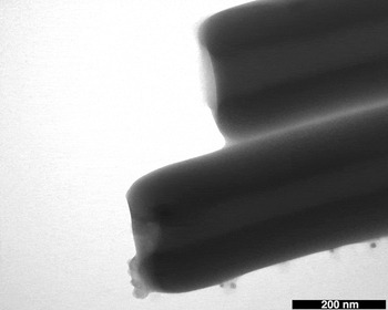
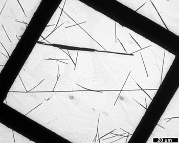
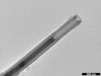
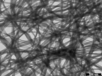
The self-assembly of the glycolipid occurred in a facile manner. After being dispersed in water by heating and sonicating, the mixture is slowly cooled through the transition temperature of the lipid, occurring at 71 °C.Reference Kamiya, Minamikawa, Jung, Yang, Masuda and Shimizu39 At this point, the transparent dispersion assembles into a white suspension of aggregates. Transmission electron microscopy (TEM) revealed the aggregates to be almost exclusively nanotube structures with open ends (see Figs. 3 and 4). The lengths of these nanotubes falls in a range on the order of 10 mm to an order of 100 mm, and the mean internal diameter of the tubes is 64 nm (SD 22 nm), which is in close agreement with the literatureReference Kamiya, Minamikawa, Jung, Yang, Masuda and Shimizu39 (61 nm, SD 19 nm). The mean external diameter is 285 nm (SD 82 nm), which is consistent with tube walls of 112 nm made up of approximately 25 bilayers of 4.5 nm each, the bilayer spacing being established previously by Kamiya et al.Reference Philp and Stoddart1 using x-ray diffraction.
TEM close-up image of glycolipid nanotubes showing open ends.

TEM image of nanotubes self-assembled from the glycolipid.

The external diameter is relatively large and varied compared to the most consistent external diameters achieved by Shimizu et al.Reference Kamiya, Minamikawa, Jung, Yang, Masuda and Shimizu39 of 200 nm (SD 23 nm). This is a reflection of the incubation conditions, which have been shown to affect this parameter.Reference Kamiya, Minamikawa, Jung, Yang, Masuda and Shimizu39 Why the external diameter of these tubes is more variable than the internal diameter has not been entirely resolved. One explanation is that tube formation will physically set the pitch and width of the helical ribbon. It may be that the point where constraints of molecular packing that halt further assembly are actually reached for the internal diameter. In contrast, the external diameter reaches no such intrinsic molecular packing constraint and the number of bilayers that can add to the external tube wall is then determined by the thermodynamic and kinetic factors of the incubation process.
B. Thermal analysis
The thermal properties of the glycolipid nanotubes were analyzed using thermal diffuse reflectance infrared Fourier transform (DRIFT) spectroscopy, temperature modulated differential scanning calorimetry (TMDSC) and thermogravimetric analysis (TGA).
C. Thermal analysis: Thermal DRIFT
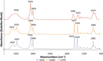
Firstly, the supramolecular integrity of the nanotube structure was evaluated using thermal DRIFT spectroscopy. Figure 5 shows the spectra for lyophilized nanotubes taken at 22 °C, 150 °C, and 175 °C. While there is little difference between the spectra taken at 22 °C and 150 °C, there are major variations between these spectra and the spectrum taken at 175 °C. The broadening and shifting of peaks in the spectrum at 175 °C relative to the others is indicative of a lowering of the order of hydrogen bonding interactions.
Thermal DRIFT spectra of glycolipid nanotubes taken at 22 °C, 150 °C, and 175 °C.

The spectrum taken at 150 °C has sharp OH stretching patterns (3452 cm−1 and 3332 cm−1) that are attributed to hydrogen bonded OH groups, which absorb at lower frequencies than free OH groups due to extended oxygen to hydrogen bonds.Reference Crews, Rodriguez and Jaspars50, Reference Williams and Fleming51 Hydrogen bonded OH groups, however, usually exhibit broader absorbance bands due to the variation in the extent of hydrogen bonding and, hence, the length of oxygen to hydrogen bonds in amorphous solids.Reference Williams and Fleming51 Hydrogen bonded OH groups that have been reported to exhibit sharp absorbance bands include intramolecular hydrogen bonds and the hydrogen bonds exhibited by dimers where the variation in hydrogen bonding is limited.Reference Williams and Fleming51 This implies that the interactions between the head groups of the glycolipids in the nanotubes are highly organized and consistent, such that variation in hydrogen bonding is limited.Reference John, Masuda, Okada, Yase and Shimizu52 Indeed, there is organization to the extent that it is possible to resolve two distinct types of hydrogen bonding, with the band at 3332 cm−1 similar to those seen for strong hydrogen bonds found in crystalline materials.Reference Williams and Fleming51 At 175 °C the absorbance due to hydrogen bonded OH groups is typical of amorphous solid materials. This suggests that the highly ordered structure of the nanotubes has been destroyed.
Further evidence for the crystalline-like structure, or more specifically the anisotropic nature of the lipid organization within the nanotubes, is illustrated by the change in character of the bands due to the amide group (amide I, C=O; 1637 cm−1, amide II, N-H; 1543 cm−1 at 150 °C)Reference Crews, Rodriguez and Jaspars50, Reference Williams and Fleming51 upon heating. The substantial broadening and reduction in intensity of these peaks as the material is heated from 150 °C to 175 °C is indicative of a loss of organization of the hydrogen bonding, and hence the organization of the nanotube structure. The shift of the amide I (C=O) band from 1637 cm−1 to 1668 cm−1, and the amide II (N-H) band from 1543 cm−1 to 1533 cm−1 is also indicative that the hydrogen bonding has become weaker at 175 °C.Reference Williams and Fleming51, Reference Jung, John, Yoshida and Shimizu53–Reference Jung, Do, Lee and Shimizu55
D. Thermal analysis: TMDSC
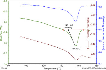
Confirmation that there is indeed a breakdown of the highly ordered supramolecular structure of the nanotubes upon heating, suggested by the thermal DRIFT analysis, was obtained by TMDSC. Figure 6 shows an endothermic process analogous to melting. The “melting point” occurs at 156.7 °C with an onset at 148.2 °C. This melting onset is in good agreement with the value of 148 °C reported by Kamiya et al.,Reference Kamiya, Minamikawa, Jung, Yang, Masuda and Shimizu39 using an optical microscope with a heating stage. The sensitivity of TMDSC is able to clearly identify this supramolecular transition and this method also provides the samples’ response to temperature modulation. Certain effects, such as a glass transition, can follow the modulating heating rate, whereas others, such as crystallization, are slower in responding.
TMDSC thermogram of glycolipid nanotubes heated at 2 °C min−1 from 50–180 °C under inert atmosphere with a modulation of ±1 °C every 60 s. (Only the numbers for the total heat flow are included in the figure for clarity. Reversing and nonreversing heat flows are described in the text.)

The topmost thermogram in Fig. 6 shows the reversing heat flow (9.11 J·g−1) and the portion of total heat flow (41.06 J·g−1) that is due to reversible effects, while the lowest thermogram shows the portion of total heat flow due to nonreversible effects (31.95 J·g−1; the center thermogram illustrates the total combined heat flow). Therefore, the nonreversing effects are responsible for 78% of the total heat flow. This level is usually associated with highly ordered structures, such as crystalline structures. This contribution to heat flow of nanotube melting is likely due to the breakdown of the strong hydrogen bonds between the sugar head groups while the reversing heat flow can be attributed to the breaking of van der Waals forces between the interdigitatedReference Jung, John, Yoshida and Shimizu53, Reference Guo, Yui, Minamikawa, Masuda, Kamiya, Sawada, Ito and Shimizu56 alkyl chains. Given that hydrogen bonds are approximately 10 times stronger than van der Waals forces,Reference Atkins and de Paula57 it is reasonable to say that hydrogen bonds are responsible for the bulk of the energy involved in the supramolecular association of the glycolipids, and hence the bulk of the heat flow upon dissociation. Thermal DRIFT analysis also showed the character of the groups involved in hydrogen bonding is significantly altered during supramolecular decomposition, while the peaks associated with alkyl chains in the FTIR spectra (Fig. 5) are much less influenced. The differential in various bond strengths suggests that at temperatures close to the “melting point,” van der Waals attraction between excited alkyl chains may break, but then easily reform with the modulation of temperature, as the hydrogen bonding between head groups keeps the alkyl chains in close association. Consequently, the resulting heat flow is reversible. However, once the hydrogen bonding is broken, the van der Waals forces are insufficient to sustain this highly ordered structure and consequently it collapses. As the lipids are no longer in an aqueous environment during the TMDSC measurements, there is no longer an amphiphilic drive to self-organize, so there is no way for the specific hydrogen bonds that are part of the highly ordered nanotube to reform. Consequently, the heat flow due to the breaking of the hydrogen bonds between head groups appears in the nonreversing signal. These types of results are typical of a melt of a highly organized anisotropic structure.
The heat loss for the 0.65 mg sample is calculated at 18.2 kJ·mol–1 given a molar mass of 443.62 g·mol–1 for the glycolipid. This seems low for a strong supramolecular association between molecules, until one realizes that supramolecular associations continue after the melting point, and 18.2 kJ·mol–1 is simply the reduction in free energy created by the organization of glycolipids into nanotubes. This analysis is supported by the continued absorbance due to hydrogen bonded OH groups (3340 cm−1) in the thermal DRIFT spectrum at 175 °C (see Fig. 5). This low energy differential also explains why careful incubation or annealing is required to form the nanotubes.
E. Thermal analysis: TGA
TGA confirms that the mass change at the proposed melting point of 156.7 °C was insignificant, and decomposition of the glycolipid occurred at 245.2 °C with an onset of 234.9 °C; appreciably higher than the proposed supramolecular decomposition. TGA therefore confirms that the effects noted in the thermal DRIFT and TMDSC analyses, and attributed to the breakdown of the organized structure of the nanotubes, was not in fact due to the decomposition of the molecular structure of the glycolipid.
F. Templated and hybrid structures
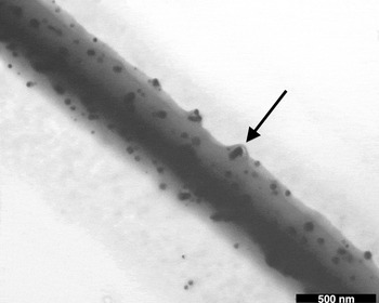
The great utility of nanotubes self-assembled from N-(11-cis-octadecenoyl)-β-D-glucopyranosylamine is that they are robust enough to withstand the removal of water from the lumen of the tube by freeze drying. Previously, Shimizu et al. reported that the nanotubes could be filled with gold and silver nanoparticles,Reference Yang, Kamiya, Shimizu, Koshizaki and Shimizu26 CdS quantum dots,Reference Zhou, Ji, Shimizu, Koshizaki and Shimizu58 and ferritinReference Yui, Shimizu, Kamiya, Yamashita, Masuda, Ito and Shimizu59 via capillary action. In this research, ethanolic solutions of silver nitrate were drawn into lyophilized tubes, the excess silver nitrate solution being washed away and the internalized silver nitrate being photoreduced to produce silver nanoparticles within the tubes. Of more novelty are the hybrid structures formed when an ethanolic silver nitrate solution was photoreduced as a bulk solution containing tubes. In this case, the outer surface of the nanotubes became decorated with silver nanoparticles (see Fig. 7), being incorporated into the outer bilayers of the tubes (the arrow in Fig. 7 indicates the membrane is raised around a nanoparticle). This incorporation could be caused either by diffusion of the silver ions into the outer layers of the tubes, or further assembly of dissolved lipid onto a tube surface already associated with silver ions during the photoreduction experiment. The latter reasoning has experimental support as the UV irradiation used to photoreduce the silver also leads to some degree of photoisomerization of the cis double bond in the hydrophilic tail. The highly ordered molecular packing of the tubes cannot tolerate such changes in molecular conformation and consequently isomerization leads to the disintegration of some tubular aggregates and an increase in solubilized lipid. This was observed by a decrease in the amount of tubes and a concomitant increase in amorphous and fibrous aggregates after UV irradiation. In contrast, nanotube populations remain constant when simply exposed to the ethanolic solutions of silver salts without irradiation, confirming the isomerization effect. It is also significant that no such incorporation of nanoparticles occurs when the tubes are dispersed in solution with previously formed nanoparticles. Organic-inorganic hybrid structures such as these can find utility in the construction of electrical and optical nanodevices,Reference Zhou and Shimizu29 and silver nanoparticle decorated nanotubes should provide an antibacterial effect,Reference Sondi and Salopek-Sondi60 valuable for applications such as drug delivery.Reference Zhou, Kogiso, Asakawa, Dong and Kiyama61
TEM image of a glycolipid nanotube decorated with silver nanoparticles. The arrow highlights incorporation of a nanoparticle into the nanotube wall.

G. Synthesis of amino acid lipids
Hydrophilic amino acids, serine and glutamic acid, were coupled with hydrophobic tails, vaccenic acid and 10,12-pentacosadiynoic acid, to create four new lipids (see Fig. 2) for which the self-assembling properties were investigated. These design modifications were made for two main reasons. The first reason was to eliminate the various synthetic issues, most notably low coupling yields, in working with the β-D-glucopyranosylamine head group. The second reason was to understand the contribution that variations in the molecular structure of the components can make to the rational design of supramolecular tubular assemblies.
In designing these amino acid based lipids, several molecular characteristics were taken into account such that tubular assemblies may be encouraged. Both of the hydrophobic tails selected have been used previously in the self-assembly of lipid nanotubes.Reference Shimizu, Masuda and Minamikawa12, Reference Yager and Schoen35, Reference Thomas, Safinya, Plano and Clark36, Reference Kamiya, Minamikawa, Jung, Yang, Masuda and Shimizu39, Reference Lee, Koepsel, Stolz, Warriner and Russell44, Reference Lee, Kim, Seo, Do, Lee, Jung, Kogiso and Shimizu48, Reference Lee, Koepsel and Russell62 Importantly, the unsaturation in both hydrophobic tails provides the rigidity and a kink in the tail that promotes packing between neighboring molecules at some nonzero angle. This angle is required to twist the bilayer ribbon into a tube.Reference Helfrich and Prost16–Reference Thomas, Lindeman, Corcoran, Cotant, Kirsch and Persichini18 In addition, both tails have acidic functional groups through which they can be attached to a head group by an amide bond, thus providing a means for generating strong and directional hydrogen bonding between molecules. The new amino acid head groups provide cheap and convenient access to a chiral center of high fidelity, close to an amino group (used to form an amide bond with the acidic tail group). The amino acids used here, glutamic acid and serine, were selected specifically for their highly hydrophilic character, with carboxylic acid and hydroxyl groups in the side chains providing significant hydrogen bonding opportunities between the head groups.
The synthesis of the amino acid based lipids was significantly more efficient (up to 89% yield) compared to the synthesis of the glycolipid (literature maximum yield of 33Reference Barclay, Matisons and Clarke63). Indeed, in the synthesis of a library of 67 amino acid based amphiphiles the average yield for the coupling reaction was 87%, with a maximum of 98%.Reference Barclay64 This is comparable to other similar reactions in the literature where the coupling of alanine esters to diacetylenic acids achieved up to 95% yield.Reference Lee, Kim, Seo, Do, Lee, Jung, Kogiso and Shimizu48
H. Self-assembly of amino acid lipids
The self-assembly of the four new amphiphiles were investigated. Initially, the self-assembly method previously developed for the glycolipid was used. However, for the most part, macroaggregates were formed, with only a small amount of loosely organized microaggregates appearing. Therefore, a new method for the self-assembly of these lipids was developed to better accommodate the chemistry of the amino acid head group and in this case the lipids were dispersed in a methanol/water mixture (1:3). Vaccenic acid-based lipids were relatively soluble in the aqueous methanolic solutions, which resulted in lower yields of organized aggregates. Nonetheless, the organized aggregates that did form using the new procedure had a higher order. The lipids with diacetylenic groups in the hydrophobic tails did not have the same solubility issues, having both greater bulk and reduced polarity for the hydrophobic tail in comparison to that of vaccenic acid tails.
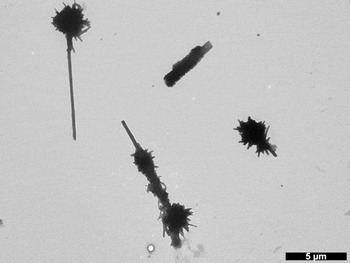
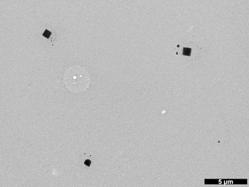
Serine combined with the vaccenic acid hydrophobic tail (Vac-Ser) self-assembled into two types of organized structures, as well as amorphous aggregates that were created by the rapid solidification of solubilized lipids upon sample preparation for TEM. The most prevalent of the organized structures were nanotubes (see Fig. 8) that aggregated together in masses, often one larger tube (mean external diameter of 292 nm) being associated with many other smaller tubelike aggregates (see Fig. 9). Also noticed were square structures that appear to be crystalline with a mean side length of 851 nm (see Fig. 10). Close observation of the tube aggregates in Fig. 9 shows squarelike shapes from which tubes seem to radiate and grow. Possibly, the square crystals act as seeds in the formation of the nanotubes, providing the requisite orientation of amphiphiles for a subsequent thermodynamically favored transition to helically based tubes. These structures may be related to those observed in the crystallization of cholesterol from supersaturated bile, where rectangular platelike crystals of cholesterol monohydrate can form from helically based tubular intermediates.Reference Zastavker, Asherie, Lomakin, Pande, Donovan, Schnur and Benedek41, Reference Chung, Benedek, Konikoff and Donovan42, Reference Konikoff, Chung, Donovan, Small and Carey65 In this case, the orientation of the cholesterol provided in the tube is reflected in the crystal, the unchecked formation of which leads to gallstones.Reference Konikoff, Chung, Donovan, Small and Carey65 If greater understanding of such transitions between crystals and tubes can be developed, benefits to the treatment of this ailment may ensue.
TEM image of a nanotube self-assembled from the Vac-Ser amphiphile.

TEM image of self-assembled tube clusters of Vac-Ser amphiphile.

TEM image of self-assembled Vac-Ser amphiphile showing square crystal-like structures.

The vaccenic acid tail combined with the glutamic acid head group (Vac-Glu) also suffers from high solubility, similar to the serine analogue. Here the aggregates show substantially more amorphous character, with only a few tubes being observed (see Fig. 11) and therefore an in depth analysis of these structures was not possible. Obviously the difference between the two vaccenic acid based lipids is limited to the amino acid head groups. The variations in self-assembly between the Vac-Ser and Vac-Glu lipids is not surprising as the amino acid side chains provide different chemistry and chain lengths, affecting the interactions between head groups. Similar behavior has been noted for a library of amino acid lipids with azobenzene moieties in the hydrophobic tails; these tails vary in proximal and distal chain lengths. In this case, the glutamic acid head groups provide a higher solubility than analogous serine based lipids and they have a tendency to form less ordered aggregates of ribbons (flat, twisted, and helical), rather than the tubes more often seen with serine head groups.Reference Barclay64
TEM image of nanotubes self-assembled from the Vac-Glu amphiphile.

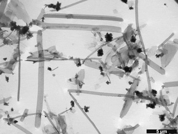
Using the alternate diacetylenic hydrophobic tail with the serine head group (DA-Ser) significantly changed the aggregated structures. Here there was a large range of ordered structures (see Fig. 12) including tubes, various plates and ribbons, and strange bundles that appear akin to an incomplete starting structure of a tubular based gel (see Fig. 13). The ribbons have a mean width of 438 nm (SD 185 nm) with a distribution that tails off toward larger widths, as illustrated by the lower median value of 393 nm. In contrast, the tubes have a normal distribution of external diameters, the mean being 77 nm (SD 16 nm). There is no evidence that the large flat ribbons are precursors for tubes, with no curling or twisting intermediate structures observed. Confirmation of the unrelated nature of the ribbon and tube aggregates is provided by the variation in apparent order of the molecular packing. The degree of order in each of the structures is indirectly evident by the apparent packing density provided by the relative opacity in the TEM images. Both the gel-like bundles and the tubes are dark and opaque, while the ribbons and plates have a lighter, transparent appearance. This indicates that for DA-Ser, either the self-assembly and incubation procedure was not optimized for the most ordered aggregates, or the difference in energy between highly ordered and less ordered aggregates is insufficient to be able to easily favor the desired highly ordered aggregates.
TEM image of nanotubes self-assembled from the DA-Ser amphiphile.

TEM images of self-assembled structures of DA-Ser amphiphile showing ribbons and plates. The denser aggregates in close-up images (not shown) are revealed to be nascent or damaged intersections from a tubular network.

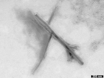
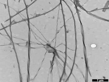
Changing the head group altered aggregate structures for the diacetylenic lipids, with DA-Glu efficiently forming high axial ratio ribbon structures (see Fig. 14), some of which displayed twisted and helical characteristics (see Fig. 15), matching closely the morphologies seen for azo-based lipids with glutamic acid head groups.Reference Barclay64 The flat ribbons have mean widths of 58 nm (SD 6.3 nm), whereas the mean ribbon width of twisted ribbons was 85 nm (SD 7 nm) with a twist pitch of 664 nm (SD 27 nm). The pitch for the helical ribbons was much smaller, at 121 nm (SD 21 nm) and the mean helical diameter was 62 nm (SD 7.7 nm). The types of self-assembled nanotubes sought in this investigation are formed from such helical ribbons, that in tubes are more tightly wound, and the curvature of these ribbons is described as mean. Twisted ribbons are a closely related structure with Gaussian curvature. There is a continuous transition between mean and Gaussian curvature in lipidic assembliesReference Mishra, Garrett and Thomas66 with the mean curvature of the helical ribbon being of greater chiral order.Reference Jung, John, Yoshida and Shimizu53, Reference John, Jung, H., Yoshida and Shimizu67, Reference Spector, Singh, Messersmith and Schnur68 Therefore, the presence of twisted ribbons is indicative that there is insufficient chiral order to the packing to create the desired tubular structures, despite the generation of high axial ratio nanostructures. Further support to this notion is found in the TEM image in Fig. 15. Here it can be seen that the helical ribbons self-assembled from DA-Glu are all left-handed, obviously an expression of the intrinsic molecular chirality, while the twisted ribbons have both left and right handed morphologies. This anomaly can be attributed to looser packing about the chiral center in the twisted ribbon. This allows the diacetylenic moiety to drive assembly via chiral symmetry breaking, which in the absence of assertive molecular chirality will form chiral aggregates of both handedness.Reference Thomas, Lindeman, Corcoran, Cotant, Kirsch and Persichini18, Reference Thomas, Lindemann and Clark69
TEM images of an interconnected network of ribbons self-assembled from the DA-Glu amphiphile.

TEM images of self-assembled structures of DA-Glu amphiphile showing twisted and helical ribbons.

It is of interest to compare the self-assembly of these diacetylenic species, DA-Ser and DA-Glu, with the assembly of similar single chain diacetylenic amphiphiles. Alanine was coupled to 10,12-heptadecadiynoic acid and to a saturated analogue, steric acid.Reference Lee, Kim, Seo, Do, Lee, Jung, Kogiso and Shimizu48 The diacetylenic amphiphile, with eight less carbons in the distal chain than the lipids investigated here, self-assembled in water into helices, upon cooling from 95 °C at 5 °C min−1, and into tubes when cooled at 0.5 °C min−1. The helices of the L-form were found to be left-handed, similar to the helices found for DA-Glu. In contrast, the saturated analogue self-assembled into a linear ribbon structure, as was seen in this investigation for the bulk of the organized structure for DA-Glu. These results suggest that the glutamic acid head group is unable to provide the level of chiral order provided by the alanine head group.
Another amphiphile found to self-assemble into tubes in water is based on the same diactetylenic tail used in this work, but with an achiral head group comprised of the hydrobromide salt of N-ethylethylenediamine.Reference Lee, Koepsel, Stolz, Warriner and Russell44, Reference Lee, Koepsel and Russell62 Here the authors believe the tubes assemble by the rolling of bilayer sheets and while the inducement of chirality in self-assembly was observed by circular dichrosim, the possibility that the tubes assemble from helical intermediates was not considered. Finally, aspartic acid was used as a head group coupled to alkyl acids with 12, 14, 16, and 18 carbon linear chains.Reference Imae, Takahashi and Muramatsu70 These amphiphiles formed gels made up of helical aggregates, but only for certain narrow pH ranges in which the head group was appropriately ionized.
The preceding examples show that self-assembly into helically based aggregates is possible for lipids either without chiralityReference Lee, Koepsel, Stolz, Warriner and Russell44, Reference Lee, Koepsel and Russell62 or without the kink in the hydrophobic tailReference Imae, Takahashi and Muramatsu70 provided by unsaturation. While it should be noted that helical assembly is rare when either of these features are missing, it does indicate that both diacetylenic hydrophobic groups and amino acid head groups are powerful drivers of this type of self-assembly. Consequently, such groups were chosen and combined in this research to try to maximize the possibility that the lipids would self-assemble into tubes based on helical intermediates.
IV. CONCLUSIONS
There is no doubt that the glycolipid developed by Shimizu et al.Reference Kamiya, Minamikawa, Jung, Yang, Masuda and Shimizu39 aggregates into robust tubes in high populations making it a uniquely valuable molecule for this type of assembly and in the creation of templated and hybrid structures. The current work provides synthetic alternatives with higher yields at relatively low cost while also investigating the rational design of such structures. It is clear that for either of the amino acid head groups, the vaccenic acid hydrophobic tail is better able to form tubes, with a large proportion of such organized structures being evident in TEM. However, with the chosen method of self-assembly, solubility becomes an issue as much of the observed aggregates are amorphous and actually created in sample preparation for TEM. Using the diacetylenic tail removes the issue of high solubility and enables large populations of organized structures to form. The relative populations of tubes are low using the serine head group, as other organized structures dominate. Combining a glutamic acid head group with a diacetylenic tail gave no tubes at all, though small populations of the precursor twisted and helical ribbons are found among the interconnected networks of linear ribbons that show no visible sign of chiral packing. Our work therefore continues in this area investigating both new tail and head groups.