Introduction
Arachnoid cysts arising from the internal auditory canal (IAC) or facial nerve canal are rare.Reference Brackmann and Bartels1–Reference Starkman, Brown and Linell3 They constitute only 0.5 per cent of all neoplasms found in these locations.Reference Schuknecht and Gao4, Reference Pappas and Brackmann5 Because of their location, such arachnoid cysts usually manifest with clinical symptoms similar to those of acoustic neuroma.Reference Pappas and Brackmann5, Reference Brooks, Mayer, Sataloff, Myers, Spiegel and Ruiz6 Previously, intracanalicular arachnoid cysts have been reported only in a small number of patients. Most of these reported lesions were large and presented with obvious clinical symptoms of nerve dysfunction.Reference Brooks, Mayer, Sataloff, Myers, Spiegel and Ruiz6–Reference Harenberg and Helms11 Arachnoid cysts confined to nerve canals have rarely been reported as asymptomatic.
We herein present four cases of intracanalicular arachnoid cyst, recently discovered during temporal bone examinations.
Materials and methods
All temporal bone samples were obtained from the temporal bone collection of the department of otolaryngology, Teikyo University, Japan.
These temporal bones had been removed during autopsy, and were prepared according to routine pathology procedures. The temporal bones were cut horizontally into 20-µm sections. Every 10th slice was selected, stained with haematoxylin and eosin (H&E) and observed under a light microscope.
The clinical records of the corresponding patients were reviewed and compared with the results of histological observation.
Results
Case one
A 74-year-old woman with breast cancer and acoustic neuroma died of cancer metastasis and sepsis.
At the age of 71 years, the patient had complained of left-sided hearing loss and tinnitus, two weeks prior to admission to hospital. She denied otalgia or otorrhoea in her left ear. No facial nerve palsy or vestibular problems could be detected on physical examination. Otoscopic examination demonstrated an intact tympanic membrane. The patient had previously undergone a mastectomy for breast cancer, aged 58 years, and radiotherapy (6000 rads) due to recurrence at 70 years. No relevant family history was recorded.
Pure tone audiography revealed moderate sensorineural hearing loss, with an average hearing threshold of 68.75 dB HL, in the patient's left ear. Click-evoked auditory brainstem response (ABR) disclosed no response in the same ear at 100 dB nHL. No response could be induced on caloric testing. Electronystagmography of the left ear produced no spontaneous, positional or gaze nystagmus.
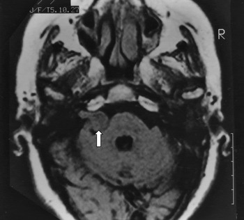
Computed tomography (CT) and magnetic response imaging (MRI) revealed a space-occupying, tumorous mass in the cerebellopontine angle and in the left IAC (Figure 1). A diagnosis of acoustic neuroma was made.
Magnetic resonance imaging scan for case one, demonstrating the enlarged acoustic tumour (arrow) in the left cerebellopontine angle.

The patient was recommended to have the tumour removed, but she refused. She died of breast cancer metastasis and sepsis at the age of 74 years. Both temporal bones were removed at autopsy. Routine horizontal serial sections were prepared for observation.
Histopathological findings
Two arachnoid cysts were detected in the left temporal bone. One was found in the facial nerve canal, the other in the IAC.
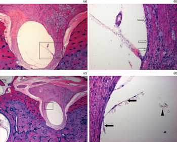
Light microscopy of serial temporal bone sections showed a cystic lesion in the left facial nerve canal which had compressed the facial nerve downward. The surrounding nerve fibres and ganglion cells had atrophied. The facial nerve canal was slightly enlarged at that position. The wall of the cyst was thin, comprising a single layer of epithelium in some places, and was collapsed in some areas. A small amount of acidophilic material and a few mononuclear inflammatory cells were seen inside the cystic cavity; this may have been due to rupture of the cyst during histological preparation (Figure 2).
Photomicrographs of the arachnoid cyst present within the geniculum of the left facial nerve canal, in case one. (a) The lesion is located within the geniculum of the facial nerve (×50). (b) Higher magnification of the boxed area in Figure 2(a), showing atrophy of the ganglion cells around the arachnoid cyst (arrows) (×50). (c) Another section, slightly inferior to that shown in Figure 2(a) (×25). (d) Higher magnification of the boxed area in Figure 2(c), showing a thin cyst wall, comprising a single epithelial layer in places, which has collapsed in some areas (arrows); the surrounding nerve fibres have degenerated, and a small amount of acidophilic material is present in the cystic cavity (arrowhead) (×50). (H&E)

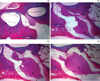
The other arachnoid cyst was seen in the middle part of the left IAC. The cyst very firmly compressed the facial nerve downward and forward within the IAC. The wall of this cyst was a little thicker than the cyst in the facial nerve canal, and contained several layers of cells and some matrix. The cyst was bi-lobed, and it was unclear whether the two parts communicated with each other. A great many deformed erythrocytes and inflamed cells were found around the cyst (Figure 3).
Photomicrographs of an arachnoid cyst and acoustic neuroma found within the left internal auditory canal (IAC) in case one. (a) Section near the top of the arachnoid cyst, showing that the cyst wall is composed of several layers of epithelium and matrix (arrows). (b) The arachnoid cyst compresses the facial nerve trunk (arrowhead), forming a niche-like indentation (arrows); note that the arachnoid cyst is lobulated from within. (c) The acoustic neuroma (arrowhead) appears to have developed within the middle part of the IAC, causing the meatus diameter to enlarge at this point (arrows indicate the arachnoid cyst). (d) View of the inferior part of the arachnoid cyst (arrows), between the acoustic neuroma (arrowhead) and the meatus. Many red blood cells and inflammatory cells are seen around the cyst in each section. (H&E; original magnification ×25)

An acoustic neuroma could be seen in the enlarged left auditory canal, which took up most of the meatal space. This schwannoma was composed of mixed Antoni A and B type cells. The main part of the cochlear nerve and the inferior vestibular nerve trunks had been replaced by the tumour. In the cochlea, the number of spiral ganglion cells and cochlear nerve fibres decreased in each turn. However, the outer and inner hair cells were well preserved. Some fluid and thickened mucosa could be defined in the ipsilateral mastoid cells.
Case two
A 43-year-old man with haemophilia A, acquired immune deficiency syndrome and long-term encephalitis died of intracranial haemorrhage.
The patient had been diagnosed with haemophilia A at 16 years of age, after suffering continuous bleeding from a thigh wound. In the following years, he had received several blood transfusions for blood coagulation factor VIII.
In August 1990, he suffered from recurrent oral cavity inflammation and chest wall folliculitis, of uncertain microbiological origin. He was later investigated and found to be positive for serous human immunodeficiency virus antibody.
Four months later, the patient reported sensory disturbance in his left hand (fourth and fifth fingers), and it was noted that his pupils differed in size. Encephalitis was suspected, and this diagnosis was later confirmed at autopsy.
Two weeks later, he was hospitalised with severe nasal bleeding. At the end of a long period of hospitalisation, he suffered a sudden and fatal intracranial haemorrhage (on 24 July 1991). Computed tomography revealed a huge haemorrhage in the left basal ganglion area, which extended into the left cerebral ventricle. Cerebral herniation advanced very rapidly, and the patient died within hours.
No hearing data, hearing problems or facial palsy manifestations were documented in the patient's medical record.
Histopathological findings
Many red blood cells and mononuclear inflammatory cells were found in the subarachnoid space. A cystic, space-occupying lesion was present in the left IAC. This cyst was located exactly in the middle of the nerve trunks, and displaced the surrounding nerves in various directions. Both the cochlear nerve and the inferior vestibular nerve were compressed into an arch shape. Slight atrophy of the posterior part of the left cochlear nerve trunk was detected. The wall of the cyst was composed of several epithelial layers and small amounts of matrix. No obvious enlargement of the left IAC was detected (Figure 4).
Photomicrograph of the arachnoid cyst in case two, located within the left internal auditory canal and displacing the surrounding nerves. Note slight atrophy of the posterior part of the cochlear nerve (arrows). (H&E; original magnification ×25)

A moderate amount of acidophilic material had accumulated in the scala tympani, scala vestibule, cochlear duct, and perilymphatic and endolymphatic spaces of the semicircular canals, on both sides. The organs of Corti (at each turn), neurosensory epithelia of the utricle and saccule, semicircular canal cristae, and ganglion cells of various nerves were all well preserved.
Case three
A 22-year-old man died of sudden, progressive tuberculous meningitis. The disease had progressed rapidly, with only 25 days elapsing between onset and death.
The patient had initially experienced the sudden onset of a severe headache. He visited his physician but returned home, as no urgent signs could be found. Five days later, he experienced nausea and began vomiting. The suspected diagnosis at the time was vascular headache; however, the next day he suddenly lost consciousness. An emergency lumbar puncture was performed, and the intracranial pressure was found to be 290 mmH2O.
An emergency cerebral ventricular drainage procedure was performed. The patient regained consciousness a few days later.
Unfortunately, his condition reversed on the fifth post-operative day: he experienced neck stiffness and was positive for Kernig's sign. On the seventh post-operative day, deafness and peripheral facial nerve palsy occurred on the right but not on the left. No hearing examination was conducted.
The patient died three days later.
His temporal bones were removed 80 minutes after death, and were prepared for observation using routine procedures.
Post-operative histopathological examination disclosed tuberculous meningitis as the final diagnosis.
Histopathological findings
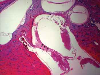
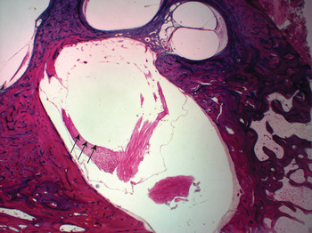
A clear, space-occupying cyst was located in the left IAC, among the nerve trunks. The facial nerve and the superior vestibular nerve were both displaced upward. The body of the cyst compressed the cochlear nerve and the inferior vestibular nerve forward and backward, respectively, with atrophy of the latter. The cystic wall was folded at the top of the cystic cavity; this may be because the arachnoid cyst had ruptured and collapsed during the autopsy procedure, or during histological preparation (Figure 5).
Photomicrograph of the arachnoid cyst in case three, located deep within the left internal auditory canal. Note compression and atrophy of the inferior vestibular nerve (arrows). (H&E; original magnification ×25)

A few mononuclear inflammatory cells had accumulated in the subarachnoid space of the IAC. Nerve fibres were infiltrated with mononuclear inflammatory cells, and some were seen to be swollen and degenerate. The neurosensory epithelia of the apical and middle turns of the cochlea was absent. The cristae of the semicircular canals and the maculae of the saccules and utricles were severely damaged on both sides.
Discussion
Theoretically, arachnoid cysts can occur at any site where arachnoid membrane exists.Reference Schuknecht and Gao4 In practice, these cysts commonly occur on the surface of the brain at the level of the main brain fissures, i.e. the sella turcica, the anterior cranial fossa and the middle cranial fossa.Reference Gelabert-González12, Reference Wester13
Neither the IAC nor the facial nerve canal is a common site for arachnoid cysts. To our knowledge, arachnoid cysts confined to the facial nerve canal have not previously been reported in the temporal bone.
Aetiology
Classification of arachnoid cysts into primary and secondary types is widely accepted. Although the aetiopathogenesis of primary arachnoid cysts is unknown, the secondary type occurs in association with trauma, inflammation and neoplasms of the meninges.Reference Pillai, Menon, Manjooran, Kariyattil, Pillai and Panikar14–Reference Cadoni, Agostino, Volante and Scipione17
According to this classification, all four arachnoid cysts in the presented series should be classified as the secondary type. Case one suffered from intracanalicular acoustic neuroma; case two had long-term intracranial inflammation; and case three had tuberculous meningitis. Histologically, each of these arachnoid cysts had inflammatory cells and red blood cells in spaces surrounding the cyst.
The coexistence of arachnoid cysts and neoplasms has been reported in several papers. Francis et al. reported the coexistence of heterotopic neuroglial tissue with an intracanalicular arachnoid cyst.Reference Francis, Nager, Holliday and Long7 Parnes et al. and Thijssen et al. both described a close relationship between arachnoid cysts and intracanalicular acoustic neuroma.Reference Parnes and Nelson8, Reference Thijssen, Marres and Slooff9 Surprisingly, in our first case we found one arachnoid cyst in the IAC and another in the facial nerve canal, together with an acoustic neuroma in the same temporal bone.
Morphology
The morphology of intracanalicular arachnoid cysts is unique. They develop within narrow spaces, forming distinct spherical or oval-shaped, space-occupying cysts with a sharp boundary with the surrounding tissue.Reference Schuknecht and Gao4, Reference Go, Houthoff, Blaauw, Stokroos and Blaauw18 In our series, we found largely transparent cyst contents. Each of our cases showed compression-induced nerve trunk atrophy, meeting the diagnostic criteria recommended by Schuknecht et al. Reference Schuknecht and Gao4 Each of our cysts had a very thin wall, histologically consisting of a single layer or countable layers of epithelial cells. Surprisingly, intracanalicular arachnoid cysts can heavily compress surrounding nerve trunks, even if their cyst wall is very thin. For example, in our first case, both cysts compressed the facial nerve trunk, leading to atrophy of nerve fibres and ganglion cells. This compression may have been due to the location of these intracanalicular arachnoid cysts, within hard, bony meatal walls, with extremely limited space.
• Arachnoid cysts confined to the internal auditory canal or facial nerve canal are rare
• We report three arachnoid cysts occurring in the internal auditory canal and one in the facial nerve canal
• The intracanalicular arachnoid cysts identified in this series, via temporal bone histology, were asymptomatic
In contrast to arachnoid cysts lying on the surface of the brain, the arachnoid cyst in case one, occurring within the IAC, was discovered to be lobulated from within. It has previously been assumed that arachnoid cysts are usually unilocular; however, within the IAC the situation may be different.Reference Go, Houthoff, Blaauw, Stokroos and Blaauw18 In case one, the intracanalicular space was deep, narrow and relatively static (being slightly distant from the cerebrospinal fluid circulation, and with an acoustic neuroma blocking the outer part of the IAC). Therefore, it is possible that the intracanalicular cystic wall on one side may have extended such that it touched another part of the wall, then adhered to it from within, thus displaying a lobulated appearance on histological examination.
Clinical manifestations
Many reports have described intracanalicular arachnoid cysts as aggressive lesions. These lesions are capable of destroying the surrounding bony canal walls and the cochlea, leading to florid symptoms of nerve dysfunction such as facial palsy, hearing loss, tinnitus and loss of balance.Reference Brooks, Mayer, Sataloff, Myers, Spiegel and Ruiz6–Reference Harenberg and Helms11 In contrast, the presented cases did not exhibit any detectable cranial nerve dysfunction, although severe nerve trunk compression was revealed on histological analysis. (Our first patient reported left ear hearing loss and tinnitus two weeks before hospitalisation; however, it is more likely that these symptoms were induced by this patient's acoustic neuroma rather than her arachnoid cysts.) This distinct clinical difference may have occurred because these patients' arachnoid cysts were at an early stage, in contrast to the relatively advanced-stage lesions which generally present to the specialist clinic. On the other hand, it might also indicate that meatal nerves have a powerful self-adjustment capacity when compressed within nerve canals.
Arachnoid cysts are not invasive in every case. Previous reports have described spontaneous resolution and long-term stability without growth.Reference Takagi, Sasaki and Basugi19–Reference Alaani, Hogg, Siddiq, Chavda and Irving21 In the present study, each of the four identified cysts differed from the others. At present, it is not possible to distinguish between those arachnoid cysts that are strong enough to destroy surrounding structures and to induce severe complications, and those that are not. It is possible that an unknown mechanism may prevent some intracanalicular arachnoid cysts from growing large enough to induce severe symptoms. Additionally, it may be the case that some occult intracanalicular arachnoid cysts do not grow large, but do induce unremarkable meatal nerve dysfunction. Since so many patients present to specialist clinics suffering from hearing loss, tinnitus, vertigo, facial palsy or spasm of unknown cause, we propose the hypothesis that some of these symptoms may be due to occult arachnoid cysts within the IAC or facial nerve canal. In light of the present pathological study, we suggest that dynamic observation, together with high resolution, cross-sectional imaging, may be helpful in detecting such occult lesions at an early stage.
Conclusion
Arachnoid cysts can develop within the IAC and facial nerve canal, and may compress adjacent nerves. In the present, post-mortem study, the identified arachnoid cysts were asymptomatic, in contrast to most of the intracanalicular arachnoid cysts encountered in clinical practice.
Acknowledgements
The authors are grateful to Ms Kayoko Sekiguchi for her help in observational work. We also thank Ms Dongtao Lin for copy-editing this manuscript. This work was supported in part by a research grant from the Society for the Promotion of International Otorhinolaryngology (Japan), by a scholarship from the Takeda Science Foundation (Japan), and by a school president grant from Xi'an Jiaotong University (xjj 2008019, China).