Introduction
Environmental enteropathy (EE), also referred to as ‘environmental enteric disorder’ (EED), is a syndrome of altered small intestine structure and function characterized by partial villus atrophy and crypt hyperplasia.Reference Kelly, Menzies and Crane 1 It is believed to be ubiquitous among individuals with high exposure to enteric pathogens,Reference Salazar-Lindo, Allen and Brewster 2 which, by recent WHO/UNICEF estimates, comprise a third of the world’s population. 3 As the gold standard for EE diagnosis is via small bowel biopsy,Reference Mckay, Gaudier, Campbell, Prentice and Albers 4 prevalence studies of EE in children are non-existent. For the same reason, investigations of the causes and consequences of EE have primarily relied upon non-invasive measures of intestinal function including primarily the lactulose:mannitol (L:M) urine test of intestinal permeabilityReference Lunn, Northrop-Clewes and Downes 5 but also increasingly fecal and plasma or serum biomarkers of intestinal inflammation,Reference Kosek, Haque and Lima 6 , Reference Campbell, McPhail, Lunn, Elia and Jeffries 7 intestinal repair,Reference Peterson, Buss and Easley 8 and bacterial translocation,Reference Mondal, Minak and Alam 9 systemic immune activation and altered host metabolism.Reference Kosek, Mduma and Kosek 10
Recently, it has been suggested that EE/EED in early life may also be a risk factor for later cardiometabolic diseases.Reference Guerrant, Deboer, Moore, Scharf and Lima 11 Current evidence to support this comes from epidemiological findings that EE is associated with poorer growth in infancy and early childhood,Reference Lunn, Northrop-Clewes and Downes 5 , Reference Kosek, Haque and Lima 6 , Reference Campbell, Elia and Lunn 12 , Reference Goto, Mascie-Taylor and Lunn 13 which is in turn associated with an increased risk of prediabetes or cardiovascular disease (CVD) in adulthood.Reference Barker, Osmond, Forsén, Kajantie and Eriksson 14 – Reference Deboer, Lima and Oría 16 Symptomatic childhood diarrhea, which has been characterized as the ‘tip of the iceberg’ atop an underlying base of EE,Reference Smets, Spears and Vyas 17 has also been associated with adult CVDReference Buck and Simpson 18 and with facets of metabolic syndrome in adulthood including dyslipidemia, elevated fasting glucose and abdominal adiposity.Reference DeBoer, Chen and Burt 19 , Reference Margolis 20
We hypothesize two biological pathways, one inflammatory and one non-inflammatory, by which EE may increase the risk of developing later the cardiometabolic disease There is evidence that EE is associated with alterations to the microbiome and bacterial overgrowth, as well as intestinal permeability, which result in bacterial translocation,Reference Mondal, Minak and Alam 9 , Reference Campbell, Elia and Lunn 12 , Reference Lin, Arnold and Afreen 21 resulting in chronic, low-level, systemic inflammation.Reference Kelly, Shawa and Mwanamakondo 22 Endotoxemia, potentially resulting from these alterations, is also associated with inflammation and with an increased risk of developing obesity and type 2 diabetes in humans.Reference Creely, Mcternan and Kusminski 23 , Reference Pussinen, Havulinna, Lehto, Sundvall and Salomaa 24 Further, in animal models, metabolic syndrome can be both induced and reversed by controlling endotoxin absorption.Reference Teixeira, Souza and Chiarello 25 , Reference Kaliannan, Hamarneh and Economopoulos 26
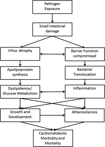
A second mechanism by which EE may impact the cardiometabolic profile is through alterations to the production of intestinal enterocyte-derived apolipoprotein concentrations. Apolipoproteins are major components of lipoprotein molecules, with the various subtypes being related to specific classes of lipoprotein molecules. Apolipoprotein A (Apo-A) is a major component of high-density lipoprotein cholesterol (HDL-c), whereas apolipoprotein-B (Apo-B) is associated with lipoproteins of lower density. Patients with untreated celiac disease (a condition that is considered to bear some histopathological similarities to EE) show depressed apolipoprotein-AI (Apo-AI) synthesis,Reference Florén and Alm 27 which is suspected to be the cause of decreased HDL-c concentrations among these patientsReference Brar, Kwon and Holleran 28 , Reference Lewis, Sanders, Logan, Fleming, Hubbard and West 29 Apo-AI has also been implicated in glucose metabolism,Reference Siebel, Heywood and Kingwell 30 and has been found to predict CVD risk among adultsReference Thompson and Danesh 31 and to precede gross markers of metabolic syndrome in pre-pubertal children.Reference Rodríguez-Moran, Aradillas-García and Guerrero-Romero 32 Childhood undernutrition has also been associated with alterations in the Apo-B/Apo-AI ratio,Reference Idohou-Dossou, Wade and Guiro 33 as well as to dyslipidemia and specifically lowered HDL-c.Reference Veiga, Ferreira, Sawaya, Calado and Florêncio 34 Together, these findings suggest that this chronic enteric syndrome could have long-term implications for the cardiometabolic risk of the individual. In Fig. 1, we present a conceptual framework of the hypothesized pathways investigated in this report. This model adapts and combines a previously published framework linking EE to growth faltering,Reference Lunn 35 with a framework linking infections in childhood to adult chronic disease.Reference Crimmins and Finch 36
This framework adapts and combines the previously published framework of Lunn linking environmental enteropathy to growth faltering, with that of Crimmins and Fitch linking infectious and chronic disease in order to describe the two pathways (dyslipidemia induced by depressed apolipoprotein production and inflammation resulting from bacterial translocation) investigated in this report.

Method
Study design
This report drew upon the Peru cohort of the ‘Etiology, Risk Factors and Interactions of Enteric Infections and Malnutrition and the Consequences for Child Health and Development (MAL-ED)’ study, a prospective, longitudinal birth cohort to evaluate the relationship between enteric and nutritional exposures and child physical growth, cognitive development, and immune response among infants exposed to repeated bouts of overt enteric illness and chronic subclinical enteropathogen exposure in the first 2 years of life. 37 , Reference Yori, Lee and Olórtegui 38 This MAL-ED study protocol included detailed assessments of intestinal permeability and markers of intestinal and systemic immune activation.Reference Yori, Lee and Olórtegui 38 – Reference Caulfield, Bose and Chandyo 40
When children were 3–5 years of age, we conducted a follow-up to evaluate whether EE in infancy and childhood is associated with systemic metabolic changes that may influence the likelihood of adult cardiometabolic disease. The a priori specific aims of this sub-study were (i) to determine whether measures of intestinal permeability (L:M) and chronic bacterial translocation [anti-endotoxin-core antibodies (EndoCAb)] in early childhood are associated with altered components of the cardiometabolic profile; (ii) to evaluate whether a history of EE in infancy is associated with the cardiometabolic profile in early childhood; (iii) to test whether any observed associations between EE biomarkers and the cardiometabolic profile (specifically, HDL-c) appeared to be mediated by altered apolipoprotein profiles (Apo-AI and the Apo-B/Apo-AI ratio).
Data collection: MAL-ED study
The Peruvian birth cohort of the MAL-ED study followed ~300 Peruvian infants over the first 0–60 months of life. 37 , Reference Yori, Lee and Olórtegui 38 All infants born between December 2009 and February 2012 who met the following criteria were eligible to enroll: healthy singleton newborns weighing >1500 g at birth, who were aged <17 days at recruitment, did not already have a sibling participating in the study, and whose mothers were 16 years of age or older, able to give informed consent and whose families were planning to remain in the community for the next 6 months. Infants were not also eligible if they had any indications of a serious disease, including hospitalization for something other than a typical healthy birth, or any severe or chronic condition diagnosed by a medical doctor (such as a congenital condition). 37
During the first 24 months of age, data related to infant infection and nutrition were collected as part of the core MAL-ED protocol. Twice-weekly house visits were conducted to characterize illness symptoms and identify symptomatic enteric disease, and infant feeding patterns.Reference Richard, Barrett and Guerrant 41 At 7, 15 and 24 months, blood samples were collected to characterize plasma α-1-glycoprotein (AGP), C-reactive protein (CRP), as well as EndoCAb, which is a measure of bacterial lipopolysaccharide exposure.Reference Mondal, Minak and Alam 9 , Reference Barclay 42 , Reference Prendergast, Rukobo and Chasekwa 43 At 3, 6, 9 and 15 months, 5-h L:M tests were administered to characterize intestinal permeability.Reference Kosek, Guerrant and Kang 39 Measures of length, weight were taken at enrollment, and then on each child’s monthly birth day by trained personnel.Reference Caulfield, Bose and Chandyo 40
Follow-up evaluation
For the sub-study described in this report, all children who participated in the original MAL-ED cohort until a minimum of 18 months of age and were still living in the community or in Iquitos city were eligible to participate, if they were between 3–5 years of age at the time of the follow-up, which took place from February to April, 2015. Power calculations were based on the expected number of cohort participants to enroll (N=160), which provided 80% power to detect differences of 0.1 mmol/l for plasma HDL-c, as well as differences of 0.1 mmol/l for triglycerides (TG), 0.2 mmol/l for fasting glucose and 3.0 mmHg for systolic blood pressure between children with elevated v. normal intestinal permeability. Institutional review board approval was obtained from Johns Hopkins University and A.B. Prisma, and written, informed consent was obtained from each participating family and it was made clear that participation or non-participation in this study would not affect their continued participation in the MAL-ED cohort.
Parents were asked to fast their children overnight before the visit, which were conducted in early morning (between 5 am to 9 am). At the study visit, a fasting blood sample was taken. After blood was drawn, a L:M test was administered, with every child receiving a 20 ml dose of L:M solution. After 30 min, a standard breakfast containing no dairy or sugars (a ‘juane’, which is a regional dish consisting of steamed rice with chicken and egg) was provided to the child. Urine was collected for 2 h following the test or in some cases longer when the child did not urinate immediately. At the same visit, during the urine collection at a time when the child was calm, blood pressure was measured three times for each participant, using a manual sphygmometer with an appropriately sized child cuff and waiting at least 1 min between each measurement as is recommended.Reference Pickering, Hall and Appel 44 Anthropometric measurements were taken by trained personnel, including height, weight, waist circumference, and subscapular, bicep, tricep and calf skinfolds.Reference Lohman, Roche and Martorell 45 In addition, a stool sample was collected within ±7 days of the visit. These samples were collected without fixative and frozen at −70°C before testing.Reference Kosek, Guerrant and Kang 39
Cardiometabolic factors
Blood was collected for the determination of fasting plasma glucose, insulin and lipid profiles. Glucose, TG, total cholesterol (TC), HDL-c, low-density lipoprotein cholesterol and very low-density lipoprotein cholesterol (VLDL-c) concentrations were determined using an enzymatic colorimetric method on an automated Cobas c 311 clinical chemistry analyzer, and insulin was determined using an electrochemiluminescence immunoassay on a Cobas E411 immunoassay analyzer. Apolipoprotein profiles were determined using a multiplex Luminex panel (Milliplex™ MAP human apolipoprotein magnetic bead panel, Millipore Corporation, MA, USA).
EE biomarkers
EndoCAb were determined via a commercial ELISA (EndoCab ELISA Kit; Hycult Biotech, Uden, The Netherlands)/stool samples were assayed for fecal myeloperoxidase (Alpco, Salem, NH, USA), fecal neopterin (GenWay Biotech, San Diego, CA, USA) and fecal α-1-antitrypsin (Biovendor, Candler, NC, USA) using commercial ELISA assays and subsequently dehydrated in order to estimate dry fecal weight concentrations.
Urine samples from the L:M test were analyzed by liquid chromatography–mass spectrometry, conducted by the same laboratory (Oregon Analytics) that had previously conducted the testing on 0–24 month L:M samples,Reference Lee, Kosek and Lima 46 in order to obtain the percent excretion of lactulose (%Lac), mannitol (%Man) and their ratio (L:M ratio).
Definitions
We used modified criteria of the National Cholesterol Education Program (NCEP) guidelines to define cut-offs for each of the cardiometabolic risk factors present,Reference Stewart, Christian and Schulze 47 , 48 where low HDL-c was defined as <0.93 mmol/l for girls and <0.98 mmol/dl for boys;Reference Daniels and Greer 49 high TG as ⩾1.35 mmol/l for girls and 0.98 mmol/l for boys;Reference Daniels and Greer 49 high glucose as >5.55 mmol/l for boys and girls. 50 High blood pressure was taken as the average of the three measurements, and defined as ⩾90th percentile for a given sex, age and height,Reference Falkner, Daniels and Flynn 51 high waist circumference defined as ⩾90th percentile based on an age- and sex-specific Mexican-American reference population (⩾54.2, ⩾57.6, and ⩾61.0 for boys 3, 4, and 5 years of age; and ⩾55.3, ⩾58.3 and ⩾61.4 for girls aged 3, 4 and 5, respectively). Insulin resistance (IR) was estimated by using the homeostasis model assessment (HOMA)=(Insulin×Glucose)/22.5, where fasting plasma insulin concentration is reported as milliunits per liter and fasting plasma glucose is reported as millimoles per liter.Reference Matthews, Hosker and Rudenski 52
L:M was defined as low (<0.07 or elevated⩾0.07) based on previously reported cut-offs in the literature.Reference van der Merwe, Moore and Fulford 53 , Reference Wessells, Hess and Rouamba 54 As cut-offs for normal v. abnormal values have not been established for percent lactulose excretion, percent mannitol excretion and EndoCab, these were each defined as less than, or greater than, the median (cut-offs=0.08% lactulose excretion, 0.86% mannitol excretion, 75 GMu/ml EndoCAb). Extant MAL-ED and biomarker data were used to build profiles of EE exposure during the first 18 months of life, including the cumulative total number of symptomatic disease events reported [the incidence and prevalence of diarrheal disease, acute lower respiratory tract infections and non-enteric fevers (i.e. maternally reported fever in the absence of maternally reported loose stools) from 0–7, 7–15 and 15–18 months of age, respectively].
Child anthropometry [weight-for-height Z-score (WHZ)] were calculated using the WHO child growth standards. 55 Each child’s breastfeeding history was summarized as the total prevalence of full (exclusive or predominant) breastfeeding from 0 to 6 months of age and from 0 to 18 months of age. Socio-economic status (SES) was summarized by the improved Water/sanitation, Assets, Maternal education, and Income (WAMI) index, a socio-economic score that was developed and validated by Psaki et al.Reference Psaki, Seidman and Miller 56 for the MAL-ED study and includes indicators of water sanitation and hygiene, assets, household size, dwelling size, maternal education and income. This score ranges from 0 to 1 and, therefore its coefficient in each model can be interpreted as the association of SES on the outcome of interest comparing the poorest possible SES score with the greatest possible SES score.
Statistical analysis
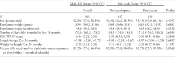
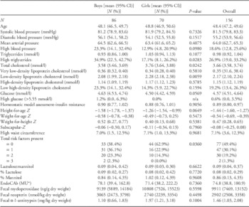
Characteristics of participants included in this report were compared with those who did not participate using t-tests and χ2 tests, as appropriate. For variables with an approximately normal distribution, and t-tests and χ2 tests of log-transformed values for variables with a non-normal distribution (Table 1). Among participating study children, characteristics were similarly compared by gender (Table 2).
Characteristics of sub-study participants

MAL-ED, the Etiology, Risk Factors, and Interactions of Enteric Infections and Malnutrition and the Consequences for Child Health Study; CI, confidence interval.
Characteristics of study children

EndoCAb, anti-endotoxin-core antibodies; CI, confidence interval.
t-test and 95% intervals for non-normal distributions were calculated based on their log-transformed values. Cut-offs for high blood pressure, high triglycerides, low high-density lipoprotein cholesterol, high glucose and high waist circumference are described in the Methods section.
*MU are EndoCAB standard median-units, defined by the manufacturer based on medians of ranges for 1000 healthy adults in a particular locality.
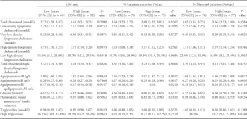
Aim i: To evaluate the association between child intestinal permeability and concurrent cardiometabolic profile, two-sided t-tests were used to compare cardiometabolic results between children with low v. high L:M, percent mannitol excretion and percent lactulose excretion, respectively. We next fitted generalized linear models regressing each cardiometabolic risk factor on each measure of intestinal permeability. In these models, percent lactulose excretion, percent mannitol excretion and L:M were presented as standard deviations [e.g. natural log (L:M)/standard deviation natural log (L:M)]. Potential confounders considered included child age, child sex, breastfeeding history, WHZ and SES (WAMI) score. Backwards elimination was used to determine variable inclusion, where the initial model was fitted with all variables, then all variables with P-values>0.200 were removed (except for the gut function biomarker in each model, which was retained regardless of P-value), and then the model was re-run and the procedure repeated until all P-values remaining were ⩽0.200 (Table 3).
Associations between concurrent lactulose:mannitol (L:M) test results and cholesterol and apolipoprotein profiles

CI, confidence interval.
To compare inflammatory and cardiometabolic risk factors, EndoCAb, prior inflammation and symptomatic disease histories were compared using two-sided t-tests between children with low v. high blood pressure, TG, HDL-c and waist circumference, based on the definitions noted above. As with the permeability measures, generalized linear models regressing each cardiometabolic risk factor on each of these inflammatory factors were constructed and the same potential confounders were evaluated.
Aim ii: To evaluate the association between EE in infancy and the child cardiometabolic profile, a similar approach was used. First, two-sided t-tests were used compare factors measured in infancy (L:M, mannitol, lactulose, inflammatory biomarkers and symptomatic disease histories) between children with low v. high blood pressure, TG, HDL-c and waist circumference, based on the definitions noted above, followed by the construction of generalized linear models regressing each cardiometabolic risk factor on each of these inflammatory factors and evaluation of potential confounders.
Aim iii: To test whether the relationship between percent mannitol excretion and HDL-c was mediated by Apo-AI a mediation analysis was conducted. For ease of interpretation, Apo-AI concentration, and HDL-c were expressed per unit standard deviation, and percent mannitol excretion was expressed as binary (low v. high mannitol). An interaction term between the exposure (percent mannitol excretion) and the mediator (Apo-AI) was also included, and child age was also considered.
A second mediation analysis was also conducted in to test whether WHZ mediated the relationship between factors measured in infancy, and later blood pressure. In this analysis, an interaction term between the exposure (inflammation in infancy) and the mediator (WHZ) was also considered, and child age was also adjusted for. Both mediation analyses were conducted using the ‘medeff’ command in Stata.Reference Imai, Keele and Tingley 57 , Reference Hicks and Tingley 58 All statistical analysis was conducted using Stata 14.2 (Stata Corporation, College Station, TX, USA).
Results
Of 303 children initially enrolled in the MAL-ED cohort and during the sub-study enrollment period from February to April 2015, three had previously withdrawn from the MAL-ED cohort; 15 had aged out of active surveillance (>5 years old); and three had died. Among those children remaining, 76 were ineligible for the sub-study because they had withdrawn from the MAL-ED cohort before 18 months of age; and 42 were eligible but were not living in the study community at the time of sub-study enrollment. A total of 164 children were invited to participate in the sub-study and 156 (95%) accepted. Of these, 150 had complete surveillance data from 0 to 18 months of life and six had withdrawn from the MAL-ED cohort before 18 months of age but later returned (i.e. they were technically ineligible for the sub-study but were invited to participate due either to clerical error or because the family expressed an active interest in participation). Therefore, the final sample size was 156 children, four less than our estimated sample size (n=160).
One child reported plasma glucose concentrations >5.55 mmol/l (5.61 mmollL). One child was not given the L:M test, because parents declined the urine test. Two children had mannitol absorption results below the lower limit of the assay, and therefore the L:M ratio could not be calculated. Finally, due to insufficient sample volumes, we were unable to complete apolipoprotein profiles in one child (final N=155) and unable to complete EndoCab testing in two children (final N=154). Characteristics of children participating in the sub-study compared with those of the overall cohort are shown in Table 1. Anthropometry and biochemical results are shown in Table 2. Overall, by the cut-offs defined about, 23.3% of children had blood pressure above the 90th percentile for their height, sex and age; 34.9% had high plasma TG; 1.2% had high glucose; 23.3% had low HDL-c; and 7.0% had an elevated waist circumference.
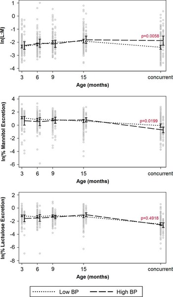
Aim i: Percent mannitol excretion was positively associated with HDL-c, Apo-AI and Apo-B, and negatively associated with VLDL-c, the TC/aHDL-c ratio, the Apo-B/Apo-AI ratio, HOMA-IR, high TG and high blood pressure (Table 3 and Fig. 2). In general, the magnitude of the associations between gut function biomarkers and cardiometabolic biomarkers were similar between unadjusted models, and models adjusting for child age, sex, anthropometry, breastfeeding history and socio-economic status. Therefore, only simple (bivariate t-test) comparisons are shown in Table 3 and Figs 2 and 3 and unadjusted and adjusted generalized linear models are reported in Supplemental Tables 1a–1c.
Estimates of the mean lactulose:mannitol (L:M) ratio, percent mannitol excretion and percent lactulose excretion at tests conducted at 3, 6, 9 and 15 months (5-h L:M tests) as well as concurrent with the cardiometabolic testing (2-h L:M test). Two-sided t-tests were conducted separately for each time point. Children with higher blood pressure (BP) at 3–5 years had lower percent mannitol excretions and higher L:M ratios at the time of the testing (P=0.006 and P=0.020). Related models adjusting for child age, sex, weight-for-height, breastfeeding history and socio-economic status were also considered, and no major differences in the magnitude or statistical significant of associations were observed. Therefore, only bivariate comparisons are reported here.

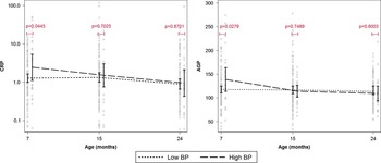
Estimates of the mean α-1-glycoprotein (AGP) and C-reactive protein (CRP) as measured at 7, 15 and 24 months of age. Two-sided t-tests were conducted separately for each time point. Two-sided t-tests were conducted separately for each time point. Children with higher blood pressure (BP) at 3–5 years had higher levels of inflammation at 7 months of age as measured by both biomarkers. However, there was no association between inflammation as measured at 15 or 24 months, and BP at 3–5 years. A 1 s.d. in CRP or AGP at 7 months corresponded to a 58% increased odds of higher BP at 3–5 years (odds ratio=1.58 for CRP and 1.57 for AGP, simple logistic regression models not shown). Related models adjusting for child age, sex, weight-for-height, breastfeeding history and socio-economic status were also considered, and no major differences in the magnitude or statistical significant of associations were observed. Therefore, only bivariate comparisons are reported here.

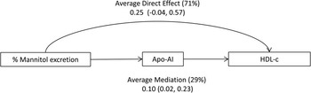
β Coefficients resulting from a mediation analysis testing the hypothesis that the association between mannitol excretion and high-density lipoprotein cholesterol (HDL-c). No other covariates were adjusted for in this analysis. Percent mannitol excretion, apolipoprotein-AI (Apo-AI) concentration and HDL-c are shown here per unit standard deviation (e.g. a 1 s.d. in percent mannitol excretion was associated with a 0.24 s.d. in Apo-AI concentration). The association between percent mannitol excretion and HDL-c was found to be mediated by Apo-AI. Percent mannitol recovery is considered to be a biomarker of small intestinal absorption and particularly villous tip area. Apo-AI is produced on the villous tip. Therefore, our results support the hypothesis that the intestinal damage associated with EE results in decreased Apo-AI concentrations, leading in turn to lower plasma HDL-c concentrations.

Aim ii: There were no consistent associations between the L:M ratio measured at 3, 6, 9 and 15 months of age and the cardiometabolic profile at 3–5 years of age (Fig. 2 and Supplemental Table 2). Among the inflammatory factors considered, there was no association between EndoCAb measured at 7, 15 or 24 of age; maternally reported diarrheal disease and fever from 0 to 18 months of age, and the cardiometabolic profile at 3–5 years of age. There was, however, an association between inflammation at 7 months of age (Fig. 3), as a 1 s.d. in CRP or AGP at 7 months corresponded to an ~58% increased odds of higher blood pressure at 3–5 years for both biomarkers (odds ratio=1.58 for CRP and 1.57 for AGP; Supplemental Table 3). There was no association between inflammation as measured at 15 or 24 months, and blood pressure at 3–5 years. The magnitude of these associations was similar when adjusting for potential confounders (Supplemental Table 3).
Aim iii: Mediation analysis suggested that Apo-AI mediated the relationship between percent mannitol excretion and HDL-c. In addition, a significant positive interaction between % mannitol excretion and Apo-AI was observed. When mannitol excretion was low (below the median), Apo-AI mediated 8% of the relationship between mannitol excretion and HDL-c (direct effect=0.17, indirect effect=0.03); and when mannitol excretion was high (above the median), Apo-AI mediated 51% of the relationship between mannitol excretion and HDL-c (direct effect=0.33, indirect effect=0.18). The overall average effect (no interaction) is shown in Fig. 4, and mediation analysis including the interaction term is shown in Supplemental Table 4.
In contrast, although both inflammatory biomarkers measured at 7 months of age and concurrent WHZ were positively associated with blood pressure, there was no evidence that the relationship between inflammation at 7 months and blood pressure was mediated by child WHZ (Supplemental Fig. 1).
Discussion
This is the first study, to our knowledge, to investigate whether alterations to the gut, that have also been reported to impact infant growth and oral vaccine response,Reference Lunn, Northrop-Clewes and Downes 5 , Reference Naylor, Lu and Haque 59 are also associated with alterations in the cardiometabolic profile of young children. As EE is believed to be a chronic condition affecting both children and adults, these alterations, if maintained over the long term, might result in an increased risk of chronic disease in adulthood. If so, EE in infancy and early childhood may have economic and health ramifications over the lifespan for individuals living in low and middle income countries (LMICs) where the rates of chronic CVDs in adulthood have rapidly increased.
We set out to test two pathways by which EE may be associated with components of the cardiometabolic profile, one inflammatory, represented by the biomarker EndoCAb, and one non-inflammatory, represented by the urinary L:M test. Our results suggest greater support for the non-inflammatory pathway, as percent mannitol excretion, a biomarker of small intestinal surface area, was associated with multiple cardiometabolic factors, as well as with the apolipoprotein profile. Although we did not find evidence that chronic bacterial translocation, as measured by EndoCAb, was related to these same factors, we did observe that systemic inflammation early in life was associated with to later blood pressure. A limitation of our study was that we did not measure concurrent systemic inflammation.
Our results suggest that the population experienced significant EE in infancy: by 15 months of age, 46% had L:M ratios above the 90th percentile for a healthy tropical reference population,Reference Kosek, Lee and Guerrant 60 70% had subclinical inflammation as defined by AGP>1 g/l and fecal inflammatory markers were 1.5–6 times higher than published non-tropical reference values.Reference Kosek, Haque and Lima 6 Children in this community also faced high rates of infection by enteropathogens and frequent diarrheal disease.Reference Platts-Mills, Babji and Bodhidatta 61 These indicators were comparable with those seven other diverse populations in the MAL-ED study. Nevertheless, the burden of enteropathogen exposures faced by these children may be relatively greater than that facing the majority of children living in LMIC settings.
Two components of the cardiometabolic profile where a relatively greater proportion of children in the cohort were above or below the NCEP criteria we considered were blood pressure, and HDL-c. We found a high proportion of children with relatively elevated blood pressure (>90th percentile for a US reference population) blood pressure. As the measurement of blood pressure in children is challenging, it is possible that our measurements were biased. Therefore, although our results suggest that children with relatively higher blood pressure had greater intestinal permeability, they should not be used to infer that this population has a high prevalence of clinically relevant elevated blood pressure. Nevertheless, our estimates of blood pressure were very similar to what has been reported in an urban Peruvian cohort of similar age.Reference Mispireta, Caulfield and Zavaleta 62
As has been reported by other studies of both childrenReference Buitrago-Lopez, van den Hooven and Rueda-Clausen 63 , Reference Kuzawa, Adair, Avila, Cadungog and Le 64 and adultsReference Lazo-Porras, Bernabe-Ortiz and Málaga 65 in Latin American populations, we found a proportion of children with relatively low HDL-c in the absence of other risk factors. It has previously been reported that this low HDL-c is inversely associated with SES in Colombian children.Reference Buitrago-Lopez, van den Hooven and Rueda-Clausen 63 Our results suggest that lower percent mannitol excretions (greater mannitol being an indicator of villous surface area and absorptive capacity) were associated with lowered HDL-c and that this relationship was partially mediated by Apo-AI. When percent mannitol excretion was high, there were both direct and indirection (mediated by Apo-AI) relationships between mannitol and HDL-c relationship, but when mannitol excretion was low, the strength of the indirect relationship was reduced. These findings support our hypothesis that a loss of intestinal surface area results in lowered Apo-AI production on the villous tip, and, thus, lowered HDL-c. We also observed associations between percent lactulose excretion (an indicator of intestinal permeability) and apolipoprotein profiles but not with other aspects of the cardiometabolic profile; notably, lactulose and percent mannitol excretions were strongly positively correlated with one another. Recently, it has been demonstrated by Kelly et al.Reference Kelly, Besa and Zyambo 66 that percent lactulose excretion is influenced by intestinal surface area in addition to permeability, which may help to explain this result and may also explain why we observed association between mannitol excretion and HDL-c, for instance, but not between HDL-c and L:M.
Only a percentage of the relationship between percent mannitol excretion and HDL-c was mediated by Apo-AI. Neither bacterial translocation (EndoCAb) or WHZ appeared to be an additional mediator of the mannitol–HDL-c relationship. Furthermore, although concurrent WHZ was positively associated with blood pressure and HOMA-IR, we failed to find evidence that WHZ mediated the relationship between EE and cardiometabolic biomarkers. Therefore, other potential mechanisms to explain the association between intestinal permeability and the cardiometabolic profile should also be investigated. For example, although the effects of constant exposure to fecal contamination on commensal flora are still not well understood, studies have found important difference in microbial populations between well- and malnourished infants,Reference Blanton, Charbonneau and Salih 67 , Reference Brown, Wlodarska and Willing 68 which, given the emerging associations between the gut microbiota and the risk of obesity and metabolic syndrome,Reference Cani 69 merit further investigation.
The strengths of this study include the very high-resolution data collected, through the MAL-ED cohort, on enteric infections and intestinal function in the first 18 months of life. The limitations include the relatively small sample size, as well as the multiple comparisons considered (five EE biomarkers compared with multiple components of the cardiometabolic profile). To minimize the risk of improper inference based on multiple comparisons, we were careful to define our primary hypothesis, that L:M and EndoCAb would be associated with lowered Apo-AI and HDL-c, a priori. An additional limitation is the early age of follow-up: metabolic syndrome cannot be defined at 3–5 years of age, although there is evidence that cardiometabolic risk factors track from childhood into adulthood.Reference Camhi and Katzmarzyk 70
An additional limitation is that, although L:M is currently the most common test for EE, and EndoCAb has demonstrated promise as a potential additional biomarker,Reference Mondal, Minak and Alam 9 , Reference Lin, Arnold and Afreen 21 , Reference Naylor, Lu and Haque 59 there are technical limitations of both assays, including difficulties in standardizing L:M between studies and laboratory procedures.Reference Lee, Kosek and Lima 46 To minimize these issues, L:M was run on an liquid chromatography tandem mass spectrometry (LC-MSMS) system; and all EndoCAb assays were taken from a single lot.
In conclusion, this study supports the potential for a relationship between EE and the cardiometabolic profile through both inflammatory and non-inflammatory mechanisms. Furthermore, infancy may be a critical period during which to define the impact of infectious exposures on long-term, non-communicable disease risk.
Acknowledgments
The authors thank the staff and participants of the MAL-ED Network for their important contributions. In addition, the authors would like to thank Lleny Amasifuen Llerena (A.B. Prisma) and Reyna Camus Chufandama for their help in data collection, and Dr Ramya Ambikapathi, Dr Benjamin JJ McCormick, and Dr Zeba Rasmussen (Fogarty International Center/National Institutes of Health, Bethesda, MD, USA) for their helpful insights into the project.
Financial Support
This project was supported as an Early Career Award to G.O.L. by the Thrasher Foundation, as well as the projects ‘Improved Biomarkers for Environmental Enteropathy’ (to M.N.K.) and ‘the Etiology, Risk Factors and Interactions of Enteric Infections and Malnutrition and the Consequences for Child Health and Development’ (MAL-ED) Project (to the Foundation of the National Institutes of Health) both funded by the Bill and Melinda Gates Foundation.
Conflicts of Interest
None.
Ethical Standards
Ethics permission was granted by the Institutional review boards of Johns Hopkins University and A.B. Prisma.
Supplementary Material
To view supplementary material for this article, please visit https://doi.org/10.1017/S2040174417000071