





Non-technical Summary
Several echinoderm groups are rare in the Silurian fossil record, especially when compared to the Ordovician and Devonian. Therefore, new discoveries of Silurian echinoderms have the potential to significantly contribute to our understanding of echinoderm evolutionary dynamics throughout this period. In this study, we describe a new species, Eucystis charlotteae n. sp., from the Silurian of Missouri, USA. This new species expands our understanding of the biogeographic pathways of echinoderms in the early Paleozoic.
Introduction
Localities with well-preserved echinoderms are uncommon globally in the Silurian (Donovan and Harper, Reference Donovan and Harper2003; Fearnhead and Donovan, Reference Fearnhead and Donovan2007; Sheffield et al., Reference Sheffield, Ausich and Sumrall2018, Reference Sheffield, Lam, Phillips and Deline2022a; Thompson et al., Reference Thompson, Ausich and Cournoyer2022), although there are rare exceptions with more well-preserved fossil deposits (e.g., Meléndez, Reference Meléndez1951; Paul, Reference Paul1971). Echinoderm preservation is variable in the fossil record, resulting from taphonomic bias related to their multielemental construction; without rapid burial, echinoderms can disarticulate rapidly, typically within hours or days (Brett et al., Reference Brett, Moffat and Taylor1997). However, the disappearance of several echinoderm groups in the Silurian and their return in the Devonian (e.g., solutes and pleurocystitid rhombiferans) could indicate the underrepresentation of some environments in the Silurian record (Lefebvre et al., Reference Lefebvre, Eble, Navarro and David2006).
The paucity of Silurian specimens in the fossil record limits our ability to understand echinoderm dynamics throughout this time (e.g., phylogenetic relationships and biogeography) (Deline and Thomka, Reference Deline and Thomka2017). Other groups, too, have fewer fossils throughout this time; arthropods (xiphosurans), for example, have a nearly 80 Myr interval between Ordovician and Devonian-aged specimens, with only a very recent discovery of a Silurian species from Indiana, USA, filling this gap (Lamsdell, Reference Lamsdell2025). Other authors have noted, too, that there are fewer Silurian Lagerstätten (Mikulic et al., Reference Mikulic, Briggs and Kluessendorf1985; von Bitter et al., Reference von Bitter, Purnell, Tetreault and Stott2007), and many Laurentian Lagerstätten yield fewer fossils than expected (Kluessendorf, Reference Kluessendorf1994). Therefore, new fossil discoveries from the Silurian have the potential to add considerably to the evolutionary dynamics of extinct organisms.
Herein, we describe a new species of diploporan blastozoan, Eucystis charlotteae new species, from the Moccasin Springs Formation in the Silurian of Missouri, USA. This occurrence of Eucystis Angelin, Reference Angelin1878, is the first representative of this genus known from Laurentia. Eucystis has previously been described from a comparatively broad geographic range (Kesling, Reference Kesling and Moore1968) and this new occurrence provides further insight into how its biogeographic range changed across the Ordovician and Silurian. This latest occurrence also adds important context to previously published studies (e.g., Lam et al., Reference Lam, Stigall and Matzke2018, Reference Lam, Sheffield and Matzke2021; Sheffield et al., Reference Sheffield, Lam, Phillips and Deline2022a) of dispersal and speciation pathways of Echinodermata and other marine fauna.
Background
Geologic setting
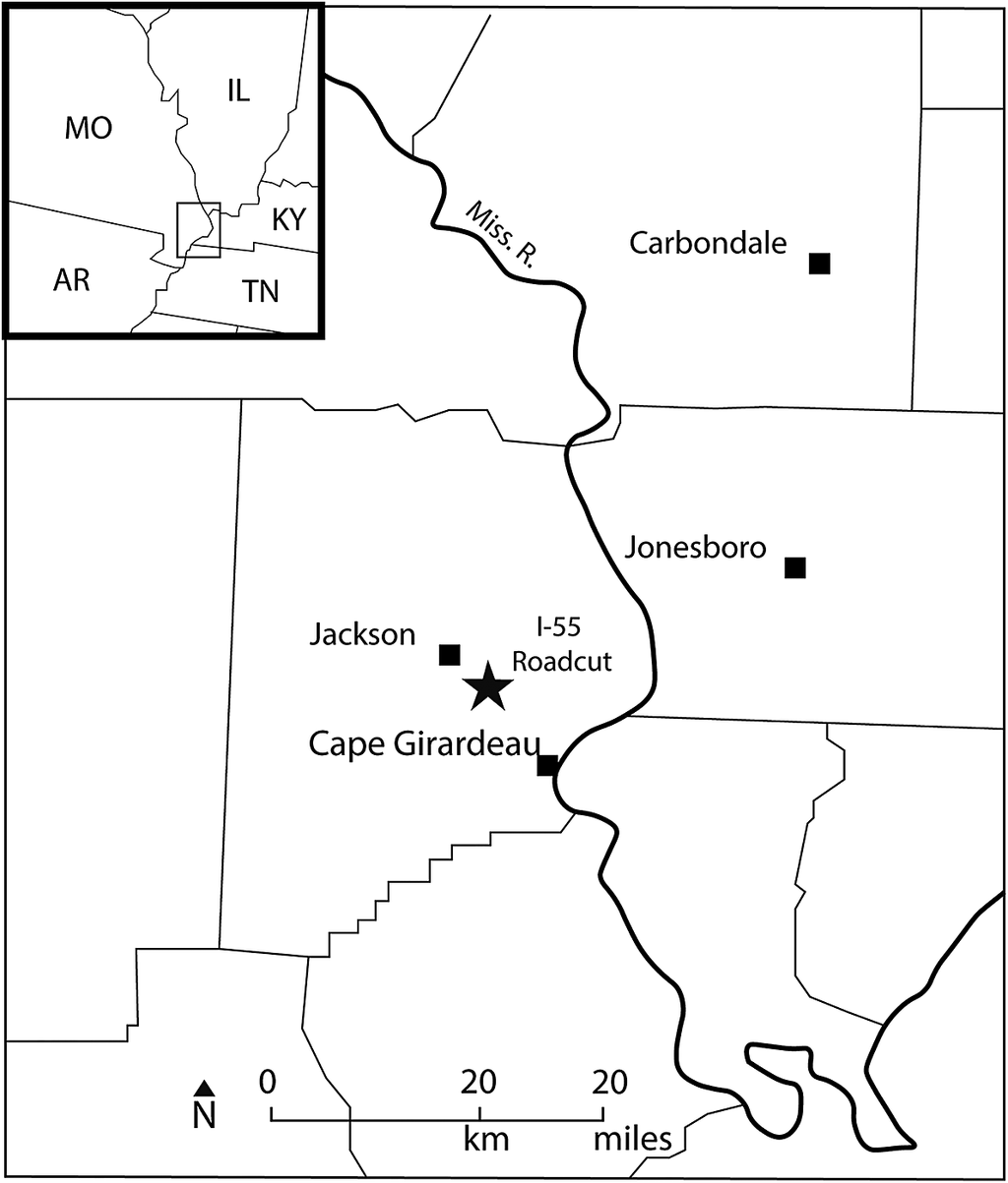
Eucystis charlotteae n. sp. is known from two relatively complete specimens, embedded together in the same matrix, and preserved as original calcite. The specimens were collected from a road cut exposure of the upper Silurian (Pridoli Series) Moccasin Springs Formation (as defined by McAdams et al., Reference McAdams, Cramer, Bancroft, Melchin, Devera and Day2019) in Cape Girardeau, Missouri, USA. The outcrop is found on the eastern side of the northbound lane of Interstate I-55, ~2.5 km north of the Missouri State Road 34 exit (Fig. 1). The outcrop was accessed from County Road 618, which parallels I-55 (37.37111°N, 89.61194°W). In the Silurian, this area of Missouri would have been located ~25° south of the equator and covered by the epicontinental sea of the southern Illinois Basin (McAdams et al., Reference McAdams, Cramer, Bancroft, Melchin, Devera and Day2019). This subtropical geography and epicontinental sea setting would have been warm, calm, and relatively shallow. The outcrop consists of steeply dipping beds of calcareous shales and mudstones that were likely tectonically altered due to their proximity to the St. Genevieve fault zone (McAdams et al., Reference McAdams, Cramer, Bancroft, Melchin, Devera and Day2019). The locality contains various echinoderm skeletal fragments (i.e., stems and disarticulated thecal plates) primarily belonging to unidentified crinoids. The presence of the graptolite index fossil Neocolonograptus parultimus Jaeger, Reference Jaeger1975, in Moccasin Springs Formation refines the age to the Pridoli Epoch, uppermost Silurian (McAdams et al., Reference McAdams, Cramer, Bancroft, Melchin, Devera and Day2019).
The Eucystis charlotteae n. sp. locality. The black star on the map represents the approximate location of the road cut exposure of the Moccasin Springs Formation outside of Cape Girardeau, Missouri, USA (modified from McAdams et al., Reference McAdams, Cramer, Bancroft, Melchin, Devera and Day2019). AR = Arkansas; IL = Illinois; KY = Kentucky; Miss. R. = Mississippi River; MO = Missouri; TN = Tennessee.

Figure 1. Long description
A two-part map layout.
* Inset Map: Located in the top-left corner, showing state boundaries. M O is in the Northwest, I L in the Northeast, A R in the Southwest, and K Y and T N in the Southeast. A small rectangle highlights the border region where these states meet.
* Main Map: A detailed view of the highlighted region. The Mississippi River, labeled Miss. R., flows from the North down through the center-east of the map.
* West of the river: The town of Jackson is marked with a square. To its Southeast is a large black star labeled I-55 Roadcut. Further South along the riverbank is Cape Girardeau, marked with a square.
* East of the river: The town of Jonesboro is marked with a square in the center-east, and Carbondale is marked with a square in the Northeast.
* Bottom: A scale bar indicates 0 to 20 kilometers and 0 to 20 miles. A North arrow points upward.
Diploporan morphology and biogeography
Diploporita Müller, Reference Müller1854 is a polyphyletic assemblage of blastozoan echinoderms (Ordovician–Middle Devonian) that was originally grouped by the presence of diplopore respiratory structures (Sumrall et al., Reference Sumrall, Brett, Dexter and Bartholomew2009; Lefebvre et al., Reference Lefebvre, Sumrall, Shroat-Lewis, Reich and Webster2013; Sheffield and Sumrall, Reference Sheffield and Sumrall2019, Sheffield et al., Reference Sheffield, Limbeck, Bauer, Hill and Nohejlová2022b). Diploporan blastozoans experienced both widespread biogeographic distribution and biodiversity through the Ordovician, diversifying into nearly 200 species across multiple paleocontinents (Lefebvre et al., Reference Lefebvre, Sumrall, Shroat-Lewis, Reich and Webster2013). The vast majority of diploporans went extinct during the Late Ordovician Mass Extinction (LOME), with few species surviving into the Silurian and Devonian (Sheffield et al., Reference Sheffield, Lam, Phillips and Deline2022a). Of those taxa that survived into the Silurian and Devonian globally, the overwhelming majority belong to the diploporan clade Sphaeronitida Neumayr, Reference Neumayr1889, with the vast majority of all occurrences either belonging to the Holocystites Fauna of Laurentia (Paul, Reference Paul1971; Frest et al., Reference Frest, Strimple and Paul2011) or the genus Eucystis (Jaekel, Reference Jaekel1899; Prokop, Reference Prokop1964). A few exceptions of glyptosphaeritids, e.g., the Silurian Gomphocystites Bockelie, Reference Bockelie1979 and the Devonian Tristomiacystis Sumrall et al., Reference Sumrall, Brett, Dexter and Bartholomew2009, both from Laurentia, also have ranges that extend past the LOME. Phylogenetic analyses have supported Sphaeronitida as a monophyletic grouping, whereas Glyptosphaeronitida has not been supported as a clade (Paul, Reference Paul, Paul and Smith1988; Sheffield and Sumrall, Reference Sheffield and Sumrall2019).
Sphaeronitid morphology
Sphaeronitids possess a spherical to ovoid-shaped theca, which constricts distally to a small or absent column or holdfast, and bear a moderate number of plates, with short ambulacra that are largely restricted to the oral area (Kesling, Reference Kesling and Moore1968; Sheffield and Sumrall, Reference Sheffield and Sumrall2019). Within sphaeronitids, there are two subgroups, as based on recent phylogenetic analyses (Sheffield and Sumrall, Reference Sheffield and Sumrall2019; Lam et al., Reference Lam, Sheffield and Matzke2021). One includes the sphaeronitids, whose ambulacra branch and each branch ends in a single brachiole facet (the group that includes Eucystis and Haplosphaeronis Jaekel, Reference Jaekel1926). The other group includes those whose ambulacra do not branch and instead end in a single, large brachiole facet; this group is informally known as the Holocystites Fauna (Sheffield and Sumrall, Reference Sheffield and Sumrall2019; Sheffield et al., Reference Sheffield, Limbeck, Bauer, Hill and Nohejlová2022b).
Eucystis is characterized by a combination of morphological features that set it apart from other sphaeronitids: (1) a 36° rotation of the oral plates in relation to the ambulacra, and (2) recumbent ambulacra ending in multiple terminal brachiole facets, with varying lengths of the ambulacral branches (Sheffield and Sumrall, Reference Sheffield and Sumrall2019) (Fig. 2). Because of the 36° rotation, the ambulacral food grooves overlay the oral plates, as opposed to the plesiomorphic condition of lying on the sutures of the oral plates of diploporan echinoderms (Sumrall, Reference Sumrall2017). This rotational feature is hypothesized to be homoplastic, re-evolving multiple times in diploporans, as seen in Glyptosphaerites Müller, Reference Müller1854 and in Holocystites Hall, Reference Hall1864 (Sheffield and Sumrall, Reference Sheffield and Sumrall2019; Sumrall et al., Reference Sumrall, Sheffield, Bauer, Thompson and Waters2023). Beyond the shared characters, there is also a clear morphological disparity among named species, highlighted in the differences in feeding structures, theca, and respiratory structures, especially. Prokop (Reference Prokop1964) considered the extreme variability in Eucystis to likely represent more than one genus, but a species-level phylogenetic analysis has not yet been done to test this hypothesis.
Oral area of Eucystis charlotteae n. sp. (CMCIP 53630) overlain on the holotype specimen. The ambulacral grooves are rotated ~36° so that the grooves do not follow the plesiomorphic condition of lying on the oral plate sutures. Heavy dashed lines (A–E) represent ambulacral grooves and facetal scars. Solid lines represent identified plate boundaries and the fine dashed lines represent unconfirmed plate boundaries due to poor preservation. GP? = suspected gonopore; HP = hydropore;; O1–O7 = oral plates; P = periproct.

Figure 2. Long description
At the center of the fossil is a dark, oval region labeled M. Radiating outward from this center are several solid black lines defining seven oral plates, labeled O 1 through O 7.
* Plate O 3 is at the top center.
* Moving clockwise, O 4 is at the top right, O 5 is at the bottom right, O 6 and O 7 are at the bottom center, O 1 is at the bottom left, and O 2 is at the far left.
Heavy dashed lines representing ambulacral grooves and facetal scars radiate from the center across these plates:
* Groove A extends upward through O 3.
* Groove B extends to the right through O 4.
* Groove C extends downward through O 5.
* Groove D extends downward through O 1.
* Groove E extends to the left through O 2.
Additional anatomical features include:
* H P, the hydropore, indicated by an arrow pointing to a small dark structure on the boundary of O 6.
* G P question mark, the suspected gonopore, located near the bottom edge below O 7.
* P, the periproct, marked by a solid black oval at the bottom right edge.
* Fine dashed lines indicate unconfirmed plate boundaries.
* A white scale bar is located in the bottom left corner.
Sphaeronitid biogeography
Sphaeronitids had an expansive biogeographic range, being found across several major paleo-oceanic basins; there is a concentration of Ordovician sphaeronitid fossils in central and southern Europe, Baltica, and eastern and western Asia (Kesling, Reference Kesling and Moore1968; Bockelie, Reference Bockelie1984; Lefebvre et al., Reference Lefebvre, Ghobadipour and Nardin2005), and there is a limited number of known Laurentian Ordovician sphaeronitids (e.g., Holocystites salmoensis Sheffield, Ausich, and Sumrall, Reference Sheffield, Ausich and Sumrall2018). In the Silurian and Devonian, outside of the Holocystites Fauna, which is concentrated in the Laurentian midcontinent and some limited finds in Australian Gondwana (Jell, Reference Jell2010), most sphaeronitid localities are far more limited, with few known occurrences, primarily in Baltica and peri-Gondwana terranes of southern Europe (Kesling, Reference Kesling and Moore1968).
Eucystis has the longest known temporal range of any diploporan taxon, with occurrences ranging from the Middle Ordovician to the Early Devonian (Kesling, Reference Kesling and Moore1968). As noted by Paul (Reference Paul1973), Eucystis is largely absent from Silurian strata; this is not extremely unusual, though, because Silurian-age echinoderms are less commonly preserved overall (Gladwell, Reference Gladwell2018; Sheffield et al., Reference Sheffield, Lam, Phillips and Deline2022a). In addition to its unusually broad temporal range compared to other diploporans, Eucystis has an equally broad geographic distribution, with occurrences in both subpolar and subtropical margins, which stands out even among the already wide distribution of the sphaeronitids. Eucystis has been found in Australia (Brown, Reference Brown1963; Jell, Reference Jell2010), Czech Republic (Prokop, Reference Prokop1964), France (Ferronnière, Reference Ferronnière1922; Le Maître, Reference Le Maître1958a; Chauvel and Le Menn, Reference Chauvel and Le Menn1972), Algeria (Le Maître, Reference Le Maître1958b), Morocco (Termier and Termier, Reference Termier and Termier1950; Klug and Korn, Reference Klug and Korn2001; Klug et al., Reference Klug, Samankassou, Pohle, De Baets, Franchi and Korn2018), Spain (Chauvel and Le Menn, Reference Chauvel and Le Menn1979), Germany (Jaekel, Reference Jaekel1899), Sweden (Regnéll, Reference Regnéll1945), Norway (Bockelie, Reference Bockelie1984), and Wales and Great Britain (Paul, Reference Paul1973). There is also a potential record of Eucystis in South China (Yunnan, China) (Reed, Reference Reed1917), which is listed in the Treatise of Invertebrate Paleontology (Kesling, Reference Kesling and Moore1968). However, the description and imagery provided by Reed (Reference Reed1917) are not definitive. The geographic range of Eucystis is now expanded by the findings presented in this study, with the addition of Eucystis charlotteae n. sp. in Laurentia.
Recent biogeographic studies (Lam et al., Reference Lam, Sheffield and Matzke2021; Sheffield et al., Reference Sheffield, Lam, Phillips and Deline2022a) indicate a strong likelihood that Baltica served as an origination point for diploporan dispersal events to other paleocontinental basins. Data from Lam et al. (Reference Lam, Sheffield and Matzke2021) revealed a distinct diploporan dispersal pathway between Baltica and Laurentia in the Late and Middle Ordovician, although other pathways, e.g., Baltica to the Western Midcontinent, were also uncovered as likely dispersal pathways for diploporans.
Morphological trends across the Ordovician-Silurian boundary
The LOME has been documented as a transition phase for crinoids from early Paleozoic faunas to those of the middle Paleozoic, during which crinoids with simple body plans and more stout arms became more common (Ausich et al., Reference Ausich, Kammer and Baumiller1994; Ausich and Deline, Reference Ausich and Deline2012; Cole and Wright, Reference Cole and Wright2022). Although this turnover in crinoids is well documented, similar patterns have been found in the fossil record of other suspension-feeding echinoderms, and in particular, diploporans. Among other morphological shifts, a trend toward simplified body plans, larger and stouter feeding appendages, and overall larger body sizes was noted when compared with their Baltic ancestors (Sheffield et al., Reference Sheffield, Lam, Phillips and Deline2022a), similar to shifts seen in middle Paleozoic crinoids.
Materials and methods
Materials
Two specimens of Eucystis charlotteae n. sp. (both contained in the same matrix) were collected from the upper Silurian Moccasin Springs Formation (Missouri, USA).
Repositories and institutional abbreviations
All specimens are reposited in the Cincinnati Museum Center, Cincinnati, Ohio, USA (CMCIP).
Systematic paleontology
Phylum Echinodermata Bruguière, Reference Bruguière1791
Subphylum Blastozoa Sprinkle, Reference Sprinkle1973
Class Diploporita Müller, Reference Müller1854
Superfamily Sphaeronitida Neumayr, Reference Neumayr1889
Family Holocystitidae Miller, Reference Miller1889
Genus Eucystis Angelin, Reference Angelin1878
Type species
Eucystis raripunctata Angelin, Reference Angelin1878
Eucystis charlotteae new species
Eucystis charlotteae n. sp., holotype (CMCIP 53630) and paratype (CMCIP 53630.1). (1) Oral view of holotype (larger specimen) and lateral view of paratype (smaller specimen) embedded in the same matrix. (2) Lateral view of holotype and oral view of paratype. The oral surface of the paratype is partially buried in matrix, obscuring some of the ambulacra. (3) Detail of the oral view of the holotype. Five ambulacra surround the oval peristome (mouth) and branch thrice; each branch ends in a large, raised brachiole facet. Diplopores pierce the oral plates proximal to the peristome; the peripore rim around the diplopores is not pronounced. The periproct (anus) is contained within the CD interrary below the mouth, separated by what is assumed to be O7 (the plate sutures are not well preserved). The hydropore is visible in the CD interray as a small pit between O1 and O6. The gonopore is likely preserved in the CD interray as a small, circular pit but is difficult to identify because of the presence of other small pits from weathering or preparation. (4) Lateral view of holotype; a crack, likely caused by lateral compression, runs along the specimen and extends distally down the theca into the holdfast. Diplopores are visible on the proximal side of theca, but most are weathered away in the distal portion of the theca. The holdfast widens distally and curves slightly. (5) Lateral view of paratype. Theca narrows distally into a holdfast, much more so than in the larger specimen. The large, raised brachiole scars that mark the end of each ambulacral branch are easily seen in this view; the majority of the branches are contained within the oral surface and do not extend past it. Diplopores are numerous and the peripore rims are shallow. (6) Oral view of paratype. Half of the oral area is embedded within the matrix; ambulacrum A is clearly visible, pointing up from the oval peristome with three clear ambulacral branches that each end in a large facet. Ambulacra D and E are also visible and follow the same branching patterns. The periproct is not visible. Specimens whitened with ammonium chloride sublimate. Scale bars = 5 mm.

Figure 3. Long description
A collection of six grayscale photographs labeled 1 through 6, each containing a white 5 mm scale bar.
Panel 1: Top-left. Shows two specimens in a textured matrix. The larger holotype is seen from an oral view at the top, while the smaller paratype is seen from a lateral view below it.
Panel 2: Top-right. Reverses the orientation, showing the holotype in lateral view above the paratype in oral view. The paratype’s oral surface is partially obscured by the surrounding rock.
Panel 3: Middle-left. A close-up oral view of the holotype. At the center is an oval peristome. Radiating outward are five branching ambulacra. Small diplopore pits are visible near the center. The C D interray area below the mouth contains the periproct and a small hydropore pit.
Panel 4: Middle-right. A lateral view of the holotype. A vertical crack runs from the top down toward the base. The surface is covered in small diplopore pits, which are more distinct near the top than the weathered bottom. The base widens into a slightly curved holdfast.
Panel 5: Bottom-left. A lateral view of the paratype. The body narrows significantly toward the bottom holdfast. Large, raised brachiole scars are prominent at the top edge. The surface is densely covered with small diplopores.
Panel 6: Bottom-right. A close-up oral view of the paratype partially embedded in matrix. Ambulacrum A is centered and points upward, branching into three parts that end in distinct facets. Ambulacra D and E are also visible.
Type specimens
Holotype, CMCIP 53630; paratype, CMCIP 53630.1.
Diagnosis
Five ambulacral grooves that branch at most three times, with unequal lengths; tumid, round facet scars that protrude above the surface of the body at the end of each branch. Ovate and relatively shallow peripores that encase the diplopore respiratory structures.
Occurrence
Eucystis charlotteae n. sp. is only known thus far from a roadcut exposure of the Moccasin Springs Formation in Cape Girardeau, Missouri, USA. Location is ~2.5 km north of the Missouri State Road 34 exit on Interstate I-55 (37.37111°N, 89.61194°W).
Description
Holotype
Theca is 16.28 mm in height and 16.20 mm in width as measured in a straight line across the A ambulacrum to the C-D interambulacrum; the specimen measures 21.45 mm in width, as measured in a straight line across the B-C and D-E ambulacra due to compaction. Theca is pyriform and relatively squat in shape; plates appear to be arranged semiregularly but not clearly organized into distinct circlets; there are ~40–50 thecal plates (some sutures are not well preserved or not exposed due to matrix); thecal plates are smooth and appear to lack ornamentation and granularity (see remarks for further discussion); six oral plates (O1–O6) border the peristome (mouth), and a possible O7 mght be present, although the sutures are poorly preserved; oral plates O1, O6, and O7(?) are contained between the C and D ambulacra (Figs. 2, 3.3); oral cover plates are not preserved; hydropore, lying between O1 and O6, is semitriangulate and poorly preserved; gonopore is possibly preserved, but due to artifacts of preservation, there are a few circular pits on O1, O6, and O7, where the gonopore would likely be, and it is not possible to confidently identify it here (Fig. 2); periproct is circular to subcircular, although the subcircular shape is likely a result of light compaction of the specimen, visible by a crack running down the theca (Fig. 3.4); periproct cover plates not preserved; peristome is relatively large and ovate; five ambulacral grooves lead from the mouth and cross the center O1–O5; ambulacral grooves are narrow and extend epithecally down the theca without underlying floor plates. The A, C, D, and E ambulacral grooves irregularly branch into three grooves of unequal lengths, whereas the B ambulacra branches twice—the longest of the grooves extend one-third to one-half way down the theca (see Remarks for further discussion); ambulacral grooves terminate in raised brachiole facet scars; facetal scars are of moderate size and relatively round, noticeably tumid, and raised above the surface of the oral area; on all ambulacra, the first branch occurs to the left; the first branch off each ambulacrum, except for the A and C ambulacrum, is notably shorter than the other two, with the branch ending in a facet that is on an oral plate, instead of extending farther down the theca. Thecal plates are mostly five-sided with equal suture lengths; plates are relatively flat and not tumid; sutures are not sunken or raised, but flush against the plates; simple diplopore respiratory structures present; diplopores appear to be oriented roughly parallel to the anteroposterior (AP) axis; diplopores are contained within the thecal plates and do not appear to cross plate sutures; diplopores are present across the entire theca, including the oral plates; diplopores are slightly more concentrated in number proximally as compared with distally; peripores are ovate in shape; theca flares out to holdfast distally that is curved to conform to the substratum (Fig. 3.4).
Paratype
Specimen conforms well to the holotype. Theca is incompletely preserved and embedded in the matrix (Fig. 3.2) and measures 14.37 mm in height and 13.12 mm in width. Theca is pyriform and relatively squat in shape; most plate sutures are not clearly preserved; the number of plates is not clear because of poor preservation; thecal plates appear smooth and do not appear to have ornamentation or granularity (see Remarks for further discussion); at the summit, the A, D, and E ambulacra border the peristome; the B and C ambulacra are not visible, although the very beginning of the B ambulacral groove can be seen leading from the mouth and is then obscured by matrix (Fig. 3.6); the A, D, and E ambulacral grooves branch into three grooves of unequal length—the longest extends just past the summit of the oral surface; all visible ambulacral grooves terminate in a raised brachiole facet scar, except for the rightmost branch in ambulacrum D, which does not appear to have a facet (likely due to poor preservation); facetal scars are of moderate size and relatively round; facet scars are noticeably tumid and raised above the surface of the oral area; on all visible ambulacra, the first branch occurs to the left; thecal plate sutures largely not visible, but some preserved detail shows five-sided equant plates; sutures do not appear tumid or sunken, but flush against the surface of the theca. Simple diplopore respiratory structures present; diplopores appear to be roughly aligned to the AP axis; diplopores are contained within the thecal plates and do not appear to cross plate boundaries; diplopores present across the entire theca, including proximal to the peristome on the oral plates (Fig. 3.5); diplopores are slightly more concentrated in number proximally as compared with distally; peripores are ovate in shape; theca constricts slightly into an elongated holdfast distally (Fig. 3.6) that appears to be complete.
Etymology
The proposed name was given in memory of Charlotte Sue Gibson, an avocational paleontologist who was active in the Dry Dredgers Fossil Club. Her interest in fossils began on her honeymoon in 1966 and lasted throughout her life. She volunteered weekly for more than 16 yrs at the Cincinnati Museum Center, concentrating her efforts on enhancing the Invertebrate Paleontology Collection.
Remarks
Holotype
The holotype specimen shows ambulacral grooves lying across the oral plates (Figs. 2, 3.3) as opposed to along their sutures, which is a synapomorphy of Eucystis (Sheffield and Sumrall, Reference Sheffield and Sumrall2019). Artifacts from the preparation of the specimen give the appearance of further branching on the D and E ambulacra. The theca shows evidence of lateral compaction, as evidenced by a vertical crack below the periproct, which slightly distorts the periproct and the thecal shape (Fig. 3.4). A seventh oral plate (O7) cannot be fully confirmed because of poor preservation of the sutures. However, we would expect it to be present because most sphaeronitids possess seven oral plates. The surface of the thecal plates appears to be smooth, lacking ornamentation; it is unclear if this is an artifact from preparation or a true morphological feature, although the plates are largely uniformly smooth. The plates of this specimen appear to be at least semiregular in appearance, but we cannot confirm whether or not they are arranged in circlets, because a statement such as this would require multiple specimens to confirm if this was genetically controlled. Eucystis charlotteae n. sp. is large in overall size when compared to Ordovician Baltic specimens of Eucystis. In this aspect, E. charlotteae n. sp. (16 mm in height) is most similar in size to E. hibernica Paul, Reference Paul1973 (30 mm in height) and E. intermedia Paul, Reference Paul1973 (23 mm in height), which were noted by Paul (Reference Paul1973) to have been the largest known examples of the genus from the Hirnantian of Britain. However, differences in the ambulacral grooves and respiratory structures between E. hibernica, E. intermedia, and E. charlotteae n. sp. are clear (e.g., diplopores are far more sunken into the plate and ambulacral grooves are far more restricted in length in the first two as opposed to the latter). The size of E. charlotteae n. sp. is notable, because it adds information to a potential pattern seen in other Ordovician and Silurian sphaeronitid diploporans of Laurentia, when the body sizes of Silurian sphaeronitids became, on average, twice as large as the Ordovician Baltic sphaeronitids (Sheffield et al., Reference Sheffield, Ausich and Sumrall2018, Reference Sheffield, Lam, Phillips and Deline2022a); Devonian eucystids, too, are notably larger than Ordovician taxa from all paleocontinents (e.g., E. robusta Prokop, Reference Prokop1964). See the Discussion for further comparison of thecal heights in the genus.
Paratype
The specimen is interpreted to represent a more juvenile growth stage of Eucystis charlotteae n. sp. than the holotype due to its smaller body size. The surface of the thecal plates appears to be smooth, lacking ornamentation; it is unclear if this is an artifact from preparation. The branching patterns of the ambulacra (i.e., at most three branches of unequal length, each ending in a single tumid facet) are shown on both specimens. The ambulacral grooves do not extend as far down the theca in the juvenile as they do in the adult. A notable difference between specimens is that the holdfast constricts in the juvenile paratype, whereas it flares out in the more mature holotype. However, it is plausible that the holdfast shape results from ecophenotypic variation rather than allometry, because holdfast shape is often driven by ecology in other closely related organisms (Sheffield and Sumrall, Reference Sheffield and Sumrall2017).
Since Angelin (Reference Angelin1878) formally named the genus Eucystis, ~20 species have been described, of which roughly 17 remain today. The remaining species were synonymized with other diploporan taxa, e.g., Sphaeronites Hisinger, Reference Hisinger1828 and Eucystis litchi (Forbes, Reference Forbes1848), now Sphaeronites litchi (Forbes, Reference Forbes1848). The majority of Eucystis spp. were named based on differences in: the number of plate circlets and numbers of plates in the theca, ornamentation of plates in the theca, style and number of the diplopore respiratory structures, number of ambulacral branches, number of brachiole facets, and size and shape of the mouth (e.g., species named by Regnéll, Reference Regnéll1945; Prokop, Reference Prokop1964; Paul, Reference Paul1973). We largely follow these conventions to compare Eucystis charlotteae n. sp. to other currently valid species (Table 1). However, we do not take plate circlets or numbers of plates into account, because the number of plate circlets likely does not correspond to homologous character traits, nor does the number of plates (Sheffield and Sumrall, Reference Sheffield and Sumrall2019; Sumrall et al., Reference Sumrall, Sheffield, Bauer, Thompson and Waters2023). Features such as the number of plates can also be affected by ontogeny, and, due to the lack of preserved growth stages for the vast majority of these taxa, a prevalent issue among diploporans (Sheffield et al., Reference Sheffield, Ausich and Sumrall2018), we cannot make assumptions of ontogenetic ages with limited sample sizes. Regardless of the poorly preserved thecal plating of these new specimens of E. charlotteae n. sp., the number and length of ambulacral branches, size of brachioles, and the style of diplopores, E. charlotteae n. sp. shows clear distinctions between this taxon and previously named species.
Named Eucystis species and identifying features. Here, we identify the named species within Eucystis, along with their temporal and stratigraphic data collected from primary literature. We identify some of the characteristics that set these species apart from E. charlotteae n. sp. We were unable to compare some Eucystis specimens to E. charlotteae n. sp. due to a lack of clear descriptions or images; specifically, specimens from South China, in which the images of the described material do not contain enough identifying information to attribute the fossil to this genus or others (Reed, Reference Reed1917; Reed noted that the fossil that they described resembled E. angelini and did not appear to be a definitive identification; Bockelie, Reference Bockelie1984 and Regnéll, Reference Regnéll1945 stated that the provided information for this specimen was also not enough to definitively place it within Eucystis); specimens from Jaekel (Reference Jaekel1899), in which named species were not accompanied by drawings or photographs; and specimens from Morocco, which were not formally described or ascribed to a species (Klug and Korn, Reference Klug and Korn2001; Klug et al., Reference Klug, Samankassou, Pohle, De Baets, Franchi and Korn2018). For E. hercynica, the stratigraphic information is taken from Jaekel (Reference Jaekel1899) and cross-referenced with Linnemann et al. (Reference Linnemann, Zweig, Zieger-Hofmann, Vietor and Zieger2024) to place it within modern stratigraphy. We do not take an opinion on whether or not all of these species are valid; we only compare all currently named species against E. charlotteae n. sp

Table 1. Long description
The table contains 22 rows of Eucystis species data across five columns.
* E. raripunctata (type species): Features pronounced peripores and granulated plate surfaces. Found in Boda Limestone, Sweden (Baltica) during the Ordovician (Katian to Hirnantian).
* E. acuminata: Narrower holdfast and anus farther from mouth. Found in Boda Limestone, Sweden (Baltica) during the Ordovician (Katian to Hirnantian).
* E. angelini: Pores closer to mouth and unevenly branching ambulacral grooves. Found in Boda Limestone, Sweden (Baltica) during the Ordovician (Katian to Hirnantian).
* E. cf. E. angelini: Shorter, unevenly branching ambulacral grooves. Found in Fombuena Formation, Spain (Iberia) during the Ordovician (Katian).
* E. barrandena: Equal length ambulacral branches and vertical oral area. Found in Suchomasty Limestone, Czech Republic (Prague) during the Devonian (Pragian to Emsian).
* E. branagani: Plates taller than wide. Found in Rosebank Shale, Australia (Baltica) during the Silurian (Ludlow).
* E. chlupaci: Ornamented plates and equal length ambulacral branches. Found in Suchomasty Limestone, Czech Republic (Prague) during the Devonian (Emsian).
* E. flavus: Pentagonal mouth depression. Found in various locations in Czech Republic, France, and Algeria (Prague, Amorica, Peri-Gondwana) during the Devonian (Pragian to Eifelian).
* E. globula: Ambulacra branch once with prominent peripore rims. Found in Ashgill Shales, Great Britain (Avalonia) during the Ordovician (Hirnantian).
* E. hercynica: Rough, granulated plate surfaces. Found in Wissenbach Shale, Germany (Avalonia) during the Devonian (Emsian to Eifelian).
* E. hibernica: Strong peripore rims and crossing diplopores. Found in Kildare Limestone, Ireland (Avalonia) during the Ordovician (Katian to Hirnantian).
* E. hispanica: Rounder theca and numerous ambulacral branches. Found in Cystoid Limestone Formation, Spain (Iberia) during the Ordovician (Katian).
* E. intermedia: Two ambulacral branches per ambulacrum. Found in Cautley Mudstones, Great Britain (Avalonia) during the Ordovician (Hirnantian).
* E. munita: Non-branching ambulacra. Found in Rhiwlas Limestone, Great Britain (Avalonia) during the Ordovician (Hirnantian).
* E. pentax: Non-branching ambulacra. Found in Sholeshook and Ashgill Shales, Great Britain (Avalonia) during the Ordovician (Katian to Hirnantian).
* E. quadrangulis: Four ambulacra instead of five. Found in Boda Limestone, Sweden (Baltica) during the Ordovician (Katian to Hirnantian).
* E. robusta: Smaller mouth and extreme variation in branch length. Found in Chynice Limestone, Czech Republic (Prague) during the Devonian (Emsian).
* E. sp. indet. (Angelin, Reference Angelin1878): Three entries from Morocco and Algeria (Peri-Gondwana) during the Devonian (Emsian to Eifelian), noting extended ambulacra and large periprocts.
* E. sp. indet. A (Paul, Reference Paul1973): Large diplopores and rimless peripores. Found in Ashgill Shales, Great Britain (Avalonia) during the Ordovician (Katian to Hirnantian).
* E. striata: Prominent peripore rims and up to five ambulacral branches. Found in Rosan Formation, France (Amorica) during the Ordovician (Katian).
Discussion
Eucystis charlotteae n. sp. does share some morphological similarities with Ordovician Eucystis species from Avalonia, based on its significantly larger size as compared with other Eucystis taxa; however, E. charlotteae n. sp. differs strongly in the morphology of the ambulacral feeding grooves, particularly in the number of branches. Some similarities, e.g., branching ambulacra, do exist between E. charlotteae n. sp. and some of the Ordovician Baltic species, but the differences between the new species and the Baltic ones are pronounced in plate granulation, diplopore morphology, and brachiole facet morphology. Therefore, based on a comprehensive literature review of named species (Table 1), we identify the specimens described here as a new species, with five ambulacral grooves that branch with unequal lengths at most three times, tumid facetal scars that rise above the surface of the body, and ovate, shallow peripores, named herein E. charlotteae n. sp.
The genus Eucystis displays interesting temporal and biogeographic distributions, being found in both subpolar and subtropical margins across both the globe and the early Paleozoic, unlike most other diploporans. This could be an indication that Eucystis might have been more tolerant of long-term environmental changes, although more research needs to be done to better understand the reasons behind this taxon’s success. Regardless, the shared morphological similarities between E. charlotteae n. sp. and Ordovician Baltic and Avalonian taxa could be important for understanding the confluence of biogeographic patterns and morphological evolutionary patterns. Dispersal pathways of Ordovician invertebrates were likely aided by abiotic factors, e.g., high sea levels and widespread carbonate platforms, in the earlier and middle part of the Ordovician (Lam et al., Reference Lam, Stigall and Matzke2018, Reference Lam, Sheffield and Matzke2021). As the LOME progressed, biogeographic studies noted far more restricted dispersal across the end-Ordovician and into the Silurian due to the regression from glaciation and therefore more isolated basins (Bauer, Reference Bauer2021; Sheffield et al., Reference Sheffield, Lam, Phillips and Deline2022a). The dispersal of the ancestral taxa of E. charlotteae n. sp. into Laurentia could be the result of the well-established migration of sphaeronitids and other invertebrate taxa from Baltica to Laurentia during the Ordovician, which has been uncovered in a number of recent biogeographic modeling studies (Lam et al., Reference Lam, Stigall and Matzke2018, Reference Lam, Sheffield and Matzke2021; Sheffield et al., Reference Sheffield, Lam, Phillips and Deline2022a). However, no Avalonian taxa were studied during these aforementioned modeling studies, so adding diploporan taxa, like those described by Paul (Reference Paul1973), into further biogeographic analyses will strengthen interpretations of specific migration pathways across early Paleozoic basins.
The size of Eucystis charlotteae n. sp. (16.2 mm thecal height) is notable when compared to other members of the genus, because it mirrors patterns seen in other sphaeronitids (i.e., increased body size across the Ordovician–Silurian in Avalonian and Laurentian taxa compared with Baltic taxa). Sheffield et al. (Reference Sheffield, Lam, Phillips and Deline2022a) identified patterns in sphaeronitid biogeography, in which Ordovician Baltic taxa were, on average, far smaller in thecal size and had far smaller brachiole facets than Silurian Laurentian taxa. Paul (Reference Paul1973), too, noted that the Avalonian Eucystis species were larger on average than those from other paleocontinents.
When compared to Ordovician eucystids, both the holotype and the paratype of Eucystis charlotteae n. sp. are similar in thecal height with the largest described Baltic examples, e.g., E. raripunctata (18 mm), and E. angelini Regnéll, Reference Regnéll1945 (17 mm), but a majority of the eucystid specimens, like E. quadrangulis Regnéll, Reference Regnéll1945 and E. angelini, from Baltica are far smaller in thecal height (see Sheffield et al., Reference Sheffield, Lam, Phillips and Deline2022a). Paul (Reference Paul1973) noted that Avalonian Ordovician specimens tend to be larger in thecal height, e.g., E. hibernica (30 mm) and E. intermedia (23 mm) than their Baltic counterparts, as well. It is difficult to draw interpretations about size trends through time or geographic region, given the limited Silurian sampling of Eucystis, with E. brangani Brown, Reference Brown1963 being one of the few examples (25 mm thecal height), and limited ontogenetic stage preservation. However, Devonian examples of Eucystis also tend to be noticeably larger in thecal height, e.g., E. chlupaci Prokop, Reference Prokop1964 (19 mm) and E. robusta (45 mm) (Prokop, Reference Prokop1964). This new Silurian find of E. charlotteae n. sp. provides more data with which to test hypotheses of shifts in body size and paleoecological dynamics through biogeographic migration across the early Paleozoic.
Preliminary work (Altier, Reference Altier2025) suggested that these morphological changes, e.g., body size, appear to correspond with paleoecological changes as well, with Baltic taxa having had a preference for deeper-water environments and Laurentian taxa having had a preference for shallow water. Sheffield et al. (Reference Sheffield, Lam, Phillips and Deline2022a) discussed this pattern as a possible response of diploporans filling the niche space left behind by crinoids during the extinction event. Although these results need further study to determine if these patterns are indicative of a true evolutionary shift, or if they are circumstantial, the presence of a new Silurian sphaeronitid species that is larger in overall body size and is from a shallower water environment (as the Moccasin Springs Formation has been interpreted to be) will certainly add to our understanding of evolutionary patterns across the LOME, when the majority of diploporan taxa did not survive. A recent study by Salamon et al. (Reference Salamon, Brachaniec, Paszcza, Kołbuk and Gorzelak2023) identified trends in body size in camerate echinoderms, in which calyx size increased across the Silurian to the Devonian. By including taxonomic and paleoecologic data from more paleobasins and time units, we can better identify when, where, and why these shifts might have occurred in diploporans. Future work can explore the potential triggers for these changes across Echinodermata.
Conclusion
Although the genus Eucystis survived into the Devonian, there is a clear gap in the stratigraphic record of this group in the Silurian (Paul, Reference Paul1973), making the discovery of E. charlotteae n. sp. in upper Silurian rocks of Laurentia significant. Beyond filling a gap in the temporal range of this group, the discovery of Eucystis charlotteae n. sp. broadens the geographic range of this taxon into Laurentia, which is especially noteworthy because this genus is already more widespread than any other diploporan genus. With this added locality in the Moccasin Springs Formation of Missouri, USA, evolutionary and biogeographic studies can be refined to better quantify likely patterns of dispersal and vicariance throughout the Paleozoic.
Acknowledgments
This work is part of SH’s M.S. thesis from the University of South Florida. It was funded by a Dry Dredgers Paleontological Research award and a Kenneth E. and Annie Caster Student Research Award from the Paleontological Society, awarded to SH. SLS was funded by NSF EAR 2429013. We thank W. Lapic for assistance with fieldwork. Helpful comments on earlier drafts of this work were made by B. Lefebvre, E. Nardin, Y. Makhlouf, D. Bissett, M. Limbeck, and J. Bauer. Finally, we gratefully acknowledge reviewers and editors E. Nardin, C. Brett, S. Zamora, P. Gorzelak, and M. Nojejlová for their comments that improved this work. This paper is written in honor of Charlotte Sue Gibson.
Competing interests
The authors declare none.


 Open access

