Introduction
It is well known that psychotic symptoms, such as delusions and/or hallucinations frequently accompany neurodegenerative disorders in the elderly and that psychotic disorders debuting in youth or middle age often continue to be active and produce symptoms in late life (Woolley et al., Reference Woolley, Khan, Murthy, Miller and Rankin2011; Snowden et al., Reference Snowden2012; Pose et al., Reference Pose, Cetkovich, Gleichgerrcht, Ibáñez, Torralva and Manes2013; Block et al., Reference Block2016). Considerably less is known about psychotic disorders beginning in old age. Some researchers believe that de novo psychoses of the elderly have a similar etiology as psychoses debuting in younger age, while others think that an underlying neurodegenerative disorder may be an important back ground factor for psychoses starting in old age (Woolley et al., Reference Woolley, Khan, Murthy, Miller and Rankin2011; Snowden et al., Reference Snowden2012; Pose et al.; Reference Pose, Cetkovich, Gleichgerrcht, Ibáñez, Torralva and Manes2013; Block et al., Reference Block2016).
In Helsinki Old Age Psychosis study, 37% of patents over 65 years with first ever psychosis had a previously undetected medical condition, which is most cases was Alzheimer's disease (Seppälä et al., Reference Seppälä, Louhija, Appelberg, Herukka and Juva2014). Also the C9ORF72 mutation, which causes behavioral variant of frontotemporal dementia (bvFTD) and amyotrophic lateral sclerosis (ALS), and which seems to be especially common in Finland, is often expressed with psychotic symptoms (Solje et al., Reference Solje2015).
Previous studies have shown that cortical atrophy of the frontal and temporal regions is a common, although not consistent finding in brain scans of patients with schizophrenia (Lesser et al., Reference Lesser, Miller, Swartz, Boone, Mehringer and Mena1993; Behdinan et al., Reference Behdinan2015). It has also been reported that elderly patients with depression besides atrophy of the hippocampal cortex also may show a thinning of brain cortex in other areas of the brain, including frontal, temporal, and parietal regions (Resnick et al., Reference Resnick, Pham, Kraut, Zonderman and Davatzikos2003; Dotson et al., Reference Dotson, Davatzikos, Kraut and Resnick2009; Benjamin and Steffens, Reference Benjamin and Steffens2011; Sexton et al., Reference Sexton, Mackay and Ebmeier2013)
According to an international consensus panel (Howard et al., Reference Howard, Rabins, Seeman and Jeste2000) 60 years is a meaningful cut-off age to distinguish between schizophrenia starting in middle age and old age (very-late-onset schizophrenia-like psychosis). For this reason, we chose the age ≥60 years to define old age in the present study.
Aim of the study
The aim of this study was to retrospectively characterize all patients with first episode psychosis (FEP) referred to hospitals or psychiatric out-patient units in the Helsinki region during one year.
The Helsinki region consists of the city of Helsinki, Finland, and the cities in the near neighborhood: Espoo, Vantaa, Kauniainen, Kirkkonummi, and Kerava with a total number of 1.2 million inhabitants (Tilastokeskus – Väestörakenne-tilasto). The psychiatric public hospitals in the region consist of Aurora Hospital in Helsinki and three hospitals (Psychiatry Centre, Jorvi Hospital, and Peijas Hospital) owned and run by the Hospital district of Helsinki and Uusimaa.
Methods
Patients
A computerized search was performed, including all patient records in Helsinki City Health Care and Helsinki and Uusimaa Hospital district patient data bases. This search identified patients with a new diagnosis of any psychotic disorder (and no earlier diagnosis of a psychotic disorder) admitted to and/or discharged from one of the Hospitals or Open Care units between Oct. 1st 2009 and Sept. 31st 2010. Patients with previously known major neurological or medical conditions were excluded from the study. The Hospital records were first evaluated in more detail by U-M Louhija. Patients clearly not meeting the inclusion criteria or meeting exclusion criteria were excluded from further analyses, including patients with known organic diseases likely to cause psychotic symptomatology. After that the hospital records of the remaining patients were evaluated by a working group consisting of four members (the authors of this paper), one neurologist (K. Juva) working with cognitive disorders of elderly people, and three psychiatrists of which one additionally being a specialist in geriatric medicine (U-M Louhija). All members of the working group have long time experience of old-age psychiatry. The working group categorized patients into four groups: (1) patients with schizophrenia (very late onset schizophrenia, VLOSP), (2) patients with Delusional Disorder (DeD), (3) patients suffering from Depression with Psychotic features (DPSY), and (4) patients with Psychosis due to a general medical condition (PGM). The diagnoses were made by a consensus within the working group based on the information in the patients‘ hospital records. DSM-IVtr Criteria were used.
In Finland, patients are referred to psychiatric hospitals and open care units based on their home community. There are no private psychiatric hospitals in Finland. Patients suffering from serious psychiatric disorders are, therefore, most likely to be treated in a public hospital or open care unit.
Brain imaging had been done routinely for the patients during their hospital stay, and several radiologists had been involved in the statements. The presence or absence of cortical atrophy in different brain regions was recorded by the authors of this paper is based on these statements. Scheltens and Fazekas classification of Hippocampal atrophy and white matter changes, respectively, (Fazekas et al., Reference Fazekas, Chawluk, Alavai, Hurtig and Zimmerman1987; Scheltens et al., Reference Scheltens, Fox, Barkhof and DeCarli2002) was recorded directly from the radiologists’ statements. Furthermore the presence or absence of minor cerebral infarction was noticed. Patients with major cerebral infarcts were excluded from the study
Statistical analyses
Mean values and standard variations were calculated. Kruskal–Wallis One-Way-Anova was used to estimate the statistical difference between groups. When two groups were compared, the other groups were excluded from the sample. P-values, Mann–Whitney statistics (MWU) and degrees of freedom (DF) are reported.
SYSTAT software package, Version 13 (Aspire Software International, Ashburn, Virginia) was used for the statistical calculations.
Ethical aspects
The study was approved by the ethics committee of the Helsinki University Hospital.
Results
Initially, patient records from 273 patients aged 60 years or over were obtained as first psychosis episodes from the Helsinki region. Of these, 166 patients were excluded from further analyses because exclusion criteria were met or inclusion criteria were not met. Common reasons for this were a medical history revealing one or several psychotic episodes before the present one, or a previously known neurological or general medical condition explaining the psychotic symptoms. A total of 107 elderly patients (27 males and 80 females) with FEP were included in the study. All patients had been treated in a hospital setting. According to the Finnish Statistical Centre (Tilastokeskus – Väestörakenne-tilasto) there were 223,738 inhabitants living in the catchment area aged 60 years or older during the study period. Thus, the yearly incidence for new onset psychosis in the elderly was 0,048% among the population 60+ and 0,009% of the whole population.
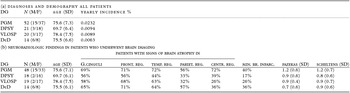
Of these patients, 52 (49%) suffered from PGM, 14 (13%) from DeD, 21 (20%) from DPSY, and 20 (19%) from VLOSP. The average age of all patients was 75 years (SD 7.3) Patients suffering from DPSY were significantly younger than the other (pooled) diagnostic groups (p = 0.003, MWU 510,00, 1 DF) and also as compared to the other diagnostic groups individually; VLOSP (p = 0.004, MWU 98,500 1 DF), DeD (p = 0.046, MWU 206,000, 1 DF), and PGM (p = 0.009, MWU 760,500, 1 DF). Ninety nine (93%) of all patients had either MRI or both MRI and CT scan (74 patients) or only CT scan (25 patients) of the brain during the hospital stay as part of routine clinical examination. A great proportion of these scans revealed atrophy in one or several parts of the brain, but also minor brain infarction were seen in 17–40% of the patients in the different diagnostic groups (see Table 1). Only four patients (one with PGM, one with DPSY, and two with VLOSP) had normal brain scans. The differences between the amount of central brain atrophy between patients with PGM and the other (pooled) patient groups (p = 0.002, MWU 768,00, 1 DF) and with patients with VLOSP (p = 0.008, MWU 572,500, 1 DF) were statistically significant. Otherwise we observed no statistically significant differences between the diagnostic groups. See Table 1.
Demography and imaging findings of elderly first episode psychosis patients

DG = diagnosis, N=number, M/F = male/female, SD = standard deviation, atr = atrophy, G.Cin = Gyrus Cinguli, front=frontal, temp=temporal, centr=central, Fazekas=Fazekas classification of vascular brain pathology, Min. br.inarct =minor brain infarction pariet=parietal, Scheltens = Scheltens classification of Hippocampal atrophy, Yearly incidence % = incidence of people aged 60 years or older PGM (Psycosis due to general medical condition), DPSY (Depression with psychotic features), VLOSP (Schizophrenia), DeD (Delsuional disorder).
Patients with DPSY were significantly younger than patients with PGM, VLOSP and DeD. Patients with PGM had significantly more central atrophy of the brain than the other diagnostic groups
The patient material included no patients with de novo bipolar disorder. Some patients in the PGM group showed symptoms of manic behavior, which, however, were regarded to be related to PGM and not to bipolar disorder.
Discussion
To our knowledge, this is the first time when FEP of the elderly has been studied in an epidemiologically representative patient material. (All patients aged 60 years or more, treated for FEP, living in the Helsinki Region were included in the study) The main findings of this study on FEP in the elderly are as follows:
-
1. First-episode psychosis patients exhibited cortical brain atrophy of the frontal, temporal, and parietal areas to an unexpectedly high degree (clearly more than half of all patients) and with the same frequency in patients with functional psychosis as in patients with psychosis due to a medical condition.
-
2. Psychosis due to a general medical condition was the most common diagnosis consisting nearly half or the patients
-
3. Patients with DPSY features seems to be younger than the other diagnostic groups
-
4. Central brain atrophy seems to be more common in patients with Psychosis due to a general medical condition than in the other categories of FEP of the elderly.
-
5. De novo mania seems to be rare in this age group.
Elderly patients often suffer from more than one medical condition, which can make it difficult to separate Psychosis due to a general medical condition from the functional psychoses. According to the diagnostic criteria in DSM IV (and also to the diagnostic criteria in DSM-5 for the corresponding Psychosis due to another medical condition) this disorder is diagnosed when it is judged that the psychotic symptoms are due to that medical condition. Our present diagnostic guidelines do not provide further advice and thus the diagnosis of PGM is debatable. This may result in gray areas of diagnostics, where it is not easy to decide whether new psychotic symptoms are a result of a psychiatric disorder or another co-existing medical condition. The authors are fully aware of this problem. We tried to overtake this problem by having a working group making consensus diagnoses. However, it can of course always be argued that another working group consisting of different investigators or clinicians could have reached other results.
The present study is a retrospective study based on patient records, and it is uncertain how many of the patients with DPSY had suffered from milder, non-psychotic forms of depression earlier in their lives. Some of them certainly had (according to the available hospital records) as had some patients in the other diagnostic categories. It is therefore, possible that the patients with DPSY features had suffered from depression before the development of psychotic features. This may perhaps explain the younger age of these patients as compared to the other diagnostic groups.
The high frequency of cortical brain atrophy found in all diagnostic groups in the MRI and CT scans, performed as part of clinical routine, in all the diagnostic groups is striking finding, and to our knowledge this has not been reported earlier for elderly patients with FEP. The lack of a control group, nevertheless, limits the interpretations, which can be made from this finding. It is, however, in line with previous reports (Lesser et al., Reference Lesser, Miller, Swartz, Boone, Mehringer and Mena1993; Resnick et al., Reference Resnick, Pham, Kraut, Zonderman and Davatzikos2003; Dotson et al., Reference Dotson, Davatzikos, Kraut and Resnick2009; Benjamin and Steffens, Reference Benjamin and Steffens2011; Sexton et al., Reference Sexton, Mackay and Ebmeier2013; Behdinan et al., Reference Behdinan2015) studying old age psychosis and may implicate that neurodegenerative brain changes may play a role not only in bvFTD and other dementing illnesses with neuropsychiatric symptoms, but also in de novo psychosis of the elderly. It is also possible that degenerative brain diseases and de novo psychoses of the elderly are related and there may be a gray area of patients “falling between” these diagnoses, not presently recognized by our diagnostic system. With no control group, it is presently not possible to estimate the possible impact of the neurodegenerative findings on the psychotic symptoms. Further, controlled and longitudinal studies are needed to clarify this.
Conflict of interest
None.
Description of author's roles
U.-M. Louhija participated in the study design, made a first review of patients collected from the data bases, and wrote the paper together with B. Appelberg, T. Saarela, and K. Juva group categorized patients into diagnostic groups together with U.-M. Louhija and B. Appelberg, and edited and revised the paper. Further analyses of the material were made by U.-M. Louhija and B. Appelberg. B. Appelberg participated in the study design. All authors contributed to and approved the manuscript.