Introduction
The addition of nanofillers improves the properties and performance of polymeric matrices; such materials are referred to as nanocomposites. Organoclays have been used extensively and researched as nanofillers due to their role in strengthening polymeric systems (Bower Reference Bower1949; Pavlidou and Papaspyrides Reference Pavlidou and Papaspyrides2008). Native clay minerals usually must be functionalized in order to increase their compatibility with polymers (Usuki et al. Reference Usuki, Kojima, Kawasumi, Okada, Fukushima, Kurauchi and Kamigaito1993; Reference Usuki, Koiwai, Kojima, Kawasumi, Okada, Kurauchi and Kamigaito1995; Nguyen and Baird Reference Nguyen and Baird2006). The addition of a clay mineral as a filler in polymer matrices has been demonstrated to increase the barrier properties, tensile strength, and temperature sensitivity of the polymer (Marras et al. Reference Marras, Kladi, Tsivintzelis, Zuburtikudis and Panayiotou2008; Rhim et al. Reference Rhim, Hong and Ha2009).
Cloisite 10A (C10A) is a clay derived from montmorillonite (Mnt). Montmorillonite is a 2:1 phyllosilicate with the general formula M x (Al4–x Mg x )Si8O20(OH)4, where M is often a monovalent cation such as Na+, which is exchangeable; x is the degree of isomorphic substitution between 0.5 and 1.3. C10A is produced from Mnt by first exchanging the Na+ with quaternary dimethylbenzyl hydrogenated tallow ammonium ((CH3)2 N+(hydrogenated tallow) (CH2)(C6H6O12)) (Zanetti et al. Reference Zanetti, Lomakin and Camino2000; Manias et al. Reference Manias, Touny, Wu, Strawhecker, Lu and Chung2001) to render it compatible with the polymer (Merah and Mohamed Reference Merah and Mohamed2019), then it is combined with and exfoliated in the selected polymer to form the clay-polymer nanocomposite. Organic cations are known to reduce the surface energy of the nanocomposite and provide better interaction between the clay mineral and the polymer matrix. The long chains of the organic moieties also aid in increasing the interlayer spacing of the Mnt. The increased spacing enhances clay‒polymer interactions (Kornmann et al. Reference Kornmann, Lindberg and Berglund2001).
Clay-polymer nanocomposites are among the most widely used composites, and are preferred because of the ready availability of clay minerals and because they have been studied extensively over many decades (e.g. Pavlidou and Papaspyrides Reference Pavlidou and Papaspyrides2008); the earliest study was by Bower (Reference Bower1949). Biocompatible and environmentally friendly polymers such as poly-L-lactic acid (PLA) and poly-ε-caprolactone (PCL) have been used extensively for packaging and biomedical applications (Elias et al. Reference Elias, Chandran, Souza and Thomas2016; Giannakas et al. Reference Giannakas, Vlacha, Salmas, Leontiou, Katapodis, Stamatis, Barkoula and Ladavos2016; Rodríguez-Tobías et al. Reference Rodríguez-Tobías, Morales and Grande2016; Sun et al. Reference Sun, Fan, Tang, Zhao, Song, Shao and Xu2016; Thomas et al. Reference Thomas, Soumya, Mathew and Radhakrishnan2015). Hence, studies of the interaction between the clay mineral and these polymeric matrices in nanocomposites can determine their suitability for specific applications.
The intercalation of polymer chains into Mnt leading to expanded clay-polymer structures has been reported by several studies (e.g. Mehrotra and Giannelis Reference Mehrotra and Giannelis1989; Vaia et al. Reference Vaia, Ishii and Giannelis1993). The renewal of interest in clay-polymer nanocomposites led to great advancements in clay intercalation chemistry. Interfacial interactions of the polymer and organoclay platelets have been known to influence the ultimate properties of the polymer nanocomposites. Increased interactions have enhanced mechanical properties and fire resistance (Islam et al. Reference Islam, Azmee, Ashaari, AdibAiman, Rasyid and Islam2017; Luo and Daniel Reference Luo and Daniel2003). The interfacial interactions between organoclay and polymer can lead to the formation of polymer crystals on the surface of clay tactoids resulting in a hybrid crystal layer. This hybrid layer promotes enhanced interactions between the clay and polymer (Ning et al. Reference Ning, Fu, Zhang, Chen, Wang, Deng, Zhang and Fu2012). The changes in bioactive properties such as microbicidal efficiency with respect to polymer–filler interactions have not been studied in detail. Microbicidal quaternary ammonium salts within the organosilicate-polymer composites are to be investigated thoroughly. The functional groups in ion-exchanged organoclay confer the bactericidal properties and provide the basis for its increased demand in applications that require antibacterial surfaces. A detailed study into the effect of functional groups of clay minerals on polymer composite properties is important, therefore (Chandran et al. Reference Chandran, Chandran, Maria and Thomas2015; Sun et al. Reference Sun, Fan, Tang, Zhao, Song, Shao and Xu2016).
Staphylococcus aureus is one of the most resilient microorganisms on earth. It is a leading cause of various opportunistic infections such as endocarditis, bacteremia, osteomyelitis, and skin and soft-tissue infections. It is also a leading cause of hospital-mediated and food-borne infections. S. aureus is a very formidable and unpredictable threat to humans due to its global distribution and ability to adapt rapidly to many environments. The persistence of these bacterial strains on every surface makes their eradication highly challenging (Turner et al. Reference Turner, Sharma-Kuinkel, Maskarinec, Eichenberger, Shah, Carugati, Holland and Fowler2019). The distribution of infective MRSA (Methicillin-resistant Staphylococcus aureus) and the associated high mortality rate, despite the introduction of many antibiotics by the Food & Drug Administration (FDA, USA), call for the development of alternative multi-targeted antimicrobial materials (Casapao et al. Reference Casapao, Davis, Barr, Klinker, Gof and Barber2014; Davis et al. Reference Davis, Sud, O'Sullivan, Robinson, Ferguson and Foo2015; Harkins et al. Reference Harkins, Pichon, Doumith, Parkhill, Westh and Tomasz2017).
In a previous study, the current authors worked on differential exhibition of antibacterial properties of the hydrophobic clay cloisite 30B in PCL and PLA (Babu et al. Reference Babu, Kalarikkal, Thomas and Radhakrishnan2018). In continuation of this work, the properties of the hydrophilic cloisite C10A and its interactions with PLA and PCL were studied. The present study was carried out in order to observe and confirm the extent to which the microbicidal activity of C10A was affected by the matrix in which it was enclosed. The present study also aimed to corroborate the findings that were observed in the previous study (Babu et al. Reference Babu, Kalarikkal, Thomas and Radhakrishnan2018) by using techniques such as AFM, FTIR, TEM, SEM, and antimicrobial assay. A further objective was to study the effectiveness of polymer–filler interactions, which, in the current (2020/2021) pandemic scenario, can have a profound effect on the development of antimicrobial materials (such as for use in biomedical and packaging applications) that help to prevent opportunistic bacterial infections.
Materials and Methods
Sample Preparation
The solvent-casting technique was used to prepare nanocomposite thin films: 10 wt.% solutions of PCL (MW 70,000, Sigma Aldrich, St. Louis, Missouri, USA) and PLA (MW 20,000, Sigma Aldrich, St. Louis, Missouri, USA) were prepared in chloroform (Merck, Mumbai, India) and stirred mechanically overnight to ensure uniform mixing and dissolution of polymer chains. The neat PCL and PLA thin films thus prepared were labeled PC0 and PL0, respectively.
Solutions containing 5 wt.% C10A (Rockwood clay additives GmbH, Moosburg, Germany, CEC = 125 meq/100 g) in PCL and PLA were prepared as described above. The solutions containing organoclay were subjected to sonication in a bath sonicator for 30 min, to achieve uniform dispersion of the clay. These were cast uniformly onto glass Petri dishes and allowed to evaporate (Dottori et al. Reference Dottori, Armentano, Fortunati and Kenny2011). The nanocomposite thin films obtained were referred to as PC10 and PL10 for C10A-PCL and C10A-PLA respectively. These prepared membranes were oven dried for 12 h at 40 °C and used for various analyses.
Characterization Studies
Surface topography studies using atomic force microscopy (AFM).
The surface topography of the prepared thin films was evaluated with the help of an atomic force microscope A-100 SPM (APE Research Nanotechnology, Basovizza, Trieste, Italy). The thin films were surface scanned in contact mode at a scan rate of 0.95–1.00 Hz. Mean surface average roughness (Ra) was evaluated using the built-in analysis software of the analytical instrument. The roughness profile was determined from average surface height deviations measured from the mean plane of the AFM images.
Diffraction studies.
X-ray diffraction studies of the thin films (2 cm × 2 cm area; 2 mm thick) and organoclay (C10A) were carried out using a Bruker D8 Advance X-Ray Diffractometer (Hanau, Germany) with nickel-filtered CuKα radiation operating at 45 kV and 44 mA, with minimum step size of 0.001°2θ and Omega: 0.001°. All data were collected in the range 2–40°2θ with a scanning rate of 5°2θ/min. All the samples were examined in the dehydrated state (Babu et al. Reference Babu, Kalarikkal, Thomas and Radhakrishnan2018).
High-resolution Imaging of Thin-film Samples Using HRTEM.
Transmission electron microscopy was performed on the PL10 and PC10 samples. Ultrathin sections (~100 nm) of the thin film were obtained by means of an ultra-microtome fitted with a diamond knife. The PL10 samples were sectioned at room temperature, whereas the PC10 thin films were microtomed at –60°C which is the glass transition temperature of PCL. The micrographs of the samples were taken using a JEOL JEM 2100 transmission electron microscope (JEOL, Tokyo, Japan), with an accelerating voltage of 200 keV. Image J software was used to analyse the micrographs obtained.
Anti-staphylococcal activity of C10A polymer nanocomposites.
Disk diffusion assay on Mueller Hinton Agar (MHA) plates was used to evaluate the antibacterial potential of the thin films (Rhim et al. Reference Rhim, Hong and Ha2009). Staphylococcus aureus MTCC96 strain was used as the test organism and cultured in Nutrient Broth (NB) for 18–24 h at 37ºC. Prepared MHA plates were swabbed with the test organism when their growth showed turbidity equivalent to 0.5 McFarland standard. The clay disks were prepared by immersing Whatman filter paper disks (4 mm in diameter) in a C10A suspension (5% clay in distilled water). These clay control disks were then air-dried and UV-sterilized. Nanocomposite thin-film disks (4 mm in diameter) were UV sterilized and placed on inoculated MHA plates. The plates were then incubated for 24 h at 37°C. The experiment was carried out in triplicate and the zone of inhibition around each disk was measured (mm).
Scanning electron microscopy imaging was also used to confirm the antibacterial activity against S. aureus. For this, the test strain was inoculated into 10 mL of NB in four separate vials. Into these vials, UV-sterilized thin film disks (4 mm in diameter) were introduced and incubated at 37 °C for 18–24 h. The thin-film disks were dehydrated further using increasing concentrations of ethanol (30, 50, 80, and 100% ethanol) for 2 min each. The treated thin-film samples were then dried and coated with gold and examined using a JEOL 6390 SEM JSM at 6 kV (Aggarwal et al. Reference Aggarwal, Surh and Shishodia2007).
Statistical analysis.
Analysis of variance (ANOVA-One way) was performed on all experimental data.
Results
Surface topography studies using AFM revealed thin, uniaxial fibril formations in the 3D topographic images (Fig. 1). The nanocomposite thin films did not exhibit fibrillary formations and clay particles could be detected in the matrix of the nanocomposite (Fig. 1). The Ra values were calculated from the roughness profile determined by AFM. The Ra of unmodified PCL and PLA film surfaces were 52.13 and 25.28 nm, respectively. PC10 showed a surface roughness (Ra) of 63.67 nm, whereas for PL10, Ra was 50.19 nm. Both clay-incorporated membranes showed evidence of increased surface roughness.
Topographic AFM images of thin films of a PC0, b PC10, c PL0, and d PL10

Diffraction Studies
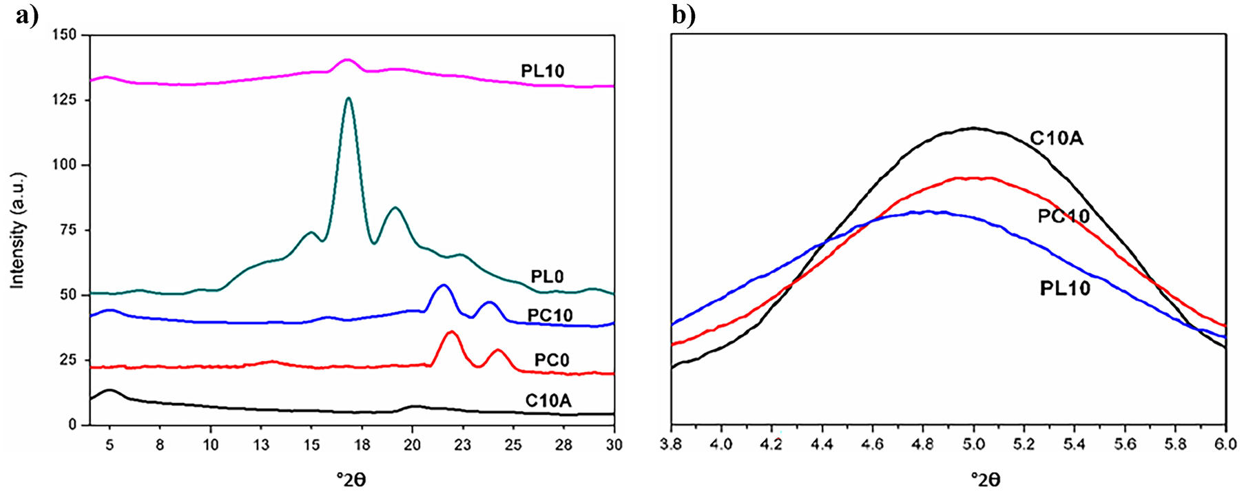
The neat PLA film exhibited a reflection at 16.7°2θ and two other reflections, at 19.3 and 22.6°2θ (Fig. 2). The reflection at 16.7°2θ is ascribed to the (200) and/or (110) planes characteristic of orthorhombic crystals (Das et al. Reference Das, Ray and Banerjee2010). The reflections at 19.3 and 22.6°2θ correspond to the (203) and (105) lattice planes, respectively (Nam et al. Reference Nam, Sinha Ray and Okamoto2003). The XRD profile of PCL had characteristic reflections at 21.4 and 23.8°2θ corresponding to the (110) and (200) planes of the semi-crystalline polymer (JCPDS number: 0–1431). The positions of PCL reflections in the nanocomposite were retained at 21.4 and 23.8°2θ (Fig. 2a). The XRD data of C10A showed a reflection at 5°2θ. The reflection by C10A in the PC10 thin film showed a slight shift to smaller diffraction angle, as it did in PL10 (4.8°2θ). The shift in 2θ values indicated the possible intercalation of the polymer into the interlayer space of the clays.
XRD patterns of a prepared thin films and C10A and b magnified diffraction patterns of C10A, PL10, and PC10 showing a shifting of clay diffraction peaks to lower angles in the polymer matrix, especially in PL10

High-resolution Imaging of Thin-film Samples Using HRTEM
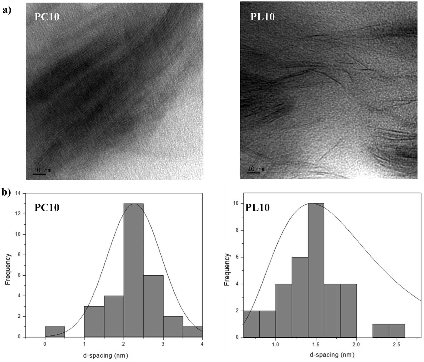
High-resolution TEM images corroborated the XRD data. The d values of the clay in the polymer matrix were measured using Image J software. The measurements were carried out at multiple positions on high-resolution images of the clay-polymer composites. The PL10 micrographs showed a larger degree of polymer intercalation than the PC10 nanocomposite. The PC10 exhibited a greater number of primary structures of C10A tactoids in the polymer matrix than in the PL10 matrix (Fig. 3a). The PL10 composite exhibited an average d value of 2.3 nm, consistent with the d values from XRD. The d values of PC10 averaged around 1.4 nm (Fig. 3b) (Das et al. Reference Das, Ray and Banerjee2010).
TEM images of C10A-PLA nanocomposite showing a high level of intercalation in the C10A-PLA nanocomposite and the presence of primary organoclay structures in the C10A-PCL nanocomposite, and b d-spacing distribution for the polymer nanocomposites

The TEM images also indicated that the organoclay had more exfoliation and slightly more intercalation in the PL10 matrix than in the PC10 matrix. Here, the PC10 matrix was marked by the existence of more primary structures of the organoclay with a d value of 1.4 nm (Fig. 3b). The TEM data provided visual confirmation of the data interpreted from the XRD pattern, therefore.
Anti-Staphylococcal Activity of C10A Polymer Nanocomposites
The antibacterial performance of C10A in PL10 and PC10 matrices was assessed (Fig. 4). The neat polymers had no antibacterial activity. A zone of inhibition of 12 mm was observed for the disks infused with C10A. Interestingly, PL10 and PC10 exhibited clearance zones of 8 and 16 mm, respectively (Fig. 4).
Antimicrobial activity of the prepared thin films (PC0, PL0, PC10, PL10) against Staphylococcus aureus

Upon incubation of the thin films in a nutrient broth culture and studying the SEM images (Fig. 5), the neat polymer thin films showed the presence of intact microbial colonies and the result was comparable to the disk-diffusion assay. The PC10 films showed microbial colonies with extensive cell-wall damage. In the case of PL10 films, isolated, intact microorganisms spread sparingly over the surface were observed, indicating the reduced adherence of the microbial cells to the membrane.
SEM images of polymer thin films and composite thin films treated with Staphylococcus aureus: a PC0 showing an intact microbial colony, b PC10 showing dead bacteria with damaged cell walls, c PL0 showing intact microbial colonies, and d PL10 showing isolated microbes

Discussion
Polymer–filler interactions have been investigated widely (Chen Reference Chen2004; Sorrentino et al. Reference Sorrentino, Tortora and Vittoria2006), and from these, greater levels of interactions are considered to be desirable for enhanced performance of nanocomposites. The influence of these interactions on the expression of bioactive properties of the nanocomposite has not been investigated.
The present study, thus, aimed to investigate polymer–filler interactions and their effect on antibacterial activity of nanocomposite materials.
The morphological analysis of solvent-cast membranes developed in the study indicated the presence of thin, fibrillary structures in both neat thin films. The fibrillary structures probably occurred as a result of chain diffusion induced by crystallization of the polymer chain upon evaporation of the solvent (Cheng and Teoh Reference Cheng and Teoh2004; Das et al. Reference Das, Ray and Banerjee2010). These fibrils were absent from clay-incorporated thin films; in that case, the organoclay may have interfered with the natural tendency of fibrillar crystallization of the polymer chains. The incorporation of clay into the polymer matrix may have induced interaction between the components and thereby enhanced interfacial crystallization, hindering the typical crystallization tendencies of the polymer (Kim et al. Reference Kim, Park, Kim, Park, Ruoff and Hwang2014, Reference Kim, Lee, Hwang, Pei, Jin, Choy and Hwang2016; Ning et al. Reference Ning, Fu, Zhang, Chen, Wang, Deng, Zhang and Fu2012). Several studies have indicated that surface roughness decreases bacterial attachment on material surfaces (e.g. Kinnari et al. Reference Kinnari, Peltonen, Kuusela, Kivilahti, Könönen and Jero2005; Rai et al. Reference Rai, Yadav and Gade2009). The nanoscale surface roughness is associated with high surface energy, resulting in decreased bacterial attachment (Zheng et al. Reference Zheng, Li, Liu and Sun2012). Bacterial contact with the sharp edges of the nanowalls leading to membrane damage has also been demonstrated. In the present study, the addition of nanoclay resulted in the formation of various pointed projections (Fig. 1). In addition, the quaternary ammonium moieties released proved to be responsible for antibacterial activity (Beyth et al. Reference Beyth, Yudovin-Farber, Bahir, Domb and Weiss2006; Yudovin-Farber et al. Reference Yudovin-Farber, Golenser, Beyth, Weiss and Domb2010). The quaternary ammonium products released from the interlayer spaces in the clays also played a significant role in bactericidal action of the nanocomposite thin films (Hong and Rhim Reference Hong and Rhim2008; Maryan and Montazer Reference Maryan and Montazer2015).
The XRD profile provided the structural insights into the polymer and clay‒mineral interactions. The slight shift in organoclay reflections indicated polymer intercalation into the interlayer spaces of the clays. The polymer intercalation into C10A galleries was more pronounced in PLA than in PCL. The decreased intensity of the reflection of C10A in the nanocomposite suggests a highly disordered state of clay platelets in the nanocomposite. This disordered state is predominant in the PLA/C10A nanocomposite.
The XRD and TEM data also indicated better dispersion of C10A in a PLA matrix when compared to that of a PCL matrix. The chaotic arrangement of clay platelets in PLA may, therefore, have formed a barrier to the release of quaternary ammonium products, thereby leading to decreased antibacterial activity. This barrier effect may also have favored the quaternary ammonium moieties following a tortuous path through the nanocomposite matrix, thereby reducing significantly its bactericidal activity in the PLA nanocomposite (Li et al. Reference Li, Yoon and Chen2011). The better dispersion of C10A in PLA was due to increased interaction between the polymer and clay owing to the presence of numerous hydroxyl groups in the organic modifier of C10A. The large surface area of C10A also contributed to the higher levels of interfacial interactions between the PLA and C10A (Chandran et al. Reference Chandran, Chandran, Maria and Thomas2015).
The increased crystallinity of PCL upon C10A addition could be due to the diffraction induced by the polymer layer formed over stacked organoclay induced by increased interfacial interactions between the components (Babu et al. Reference Babu, Augustine, Kalarikkal and Thomas2016a). The decreased PLA crystallinity could be attributed to the highly disordered condition of organoclay in the PLA matrix, which disrupts the native crystallite structure formations characteristic of PLA. This was hampered also by the increased interfacial interactions between PLA and the clay (Fig. 6). The TEM images (Fig. 3) indicated the presence of native clay in their stacked forms in the PC10 films, whereas these highly ordered structures were noticeably absent from the PL10 nanocomposite (Chávez-Montes et al. Reference Chávez-Montes, González-Sánchez and FloresGallardo2016; Das et al. Reference Das, Ray and Banerjee2010). The XRD results presented here are, thus, consistent with the results of TEM analysis.
Schematic representation of clay-polymer interactions in an unmodified semi-crystalline polymer, crystallites forming on the surface of the clay tactoids in PC10, and the highly disordered state of clay tactoids in the PLA matrix and the general decrease in crystallite formation in PL10

In the SEM images, the PC0 membrane showed the typical grape-shaped colonies of S. aureus, whereas the composite film PC10 depicted disrupted cell masses. The PL0 membrane had colonies characteristic of S. aureus, though the PL10 membrane had isolated, intact cells. The primary microbicidal activity observed here was due to alkyl ammonium moieties present in the organoclays. The cell walls of bacteria typically carry a net negative charge as a result of their carboxylate groups (Babu et al. Reference Babu, Kalarikkal, Thomas and Radhakrishnan2018). The organoclay platelets are positively charged, which attracts the bacterial cells through electrostatic interactions onto the surface of nanocomposites. The charge and hydrophobic interactions of the clay attract the bacteria onto the surface of the thin film, where the alkyl ammonium exerts its bactericidal actions (Babu et al. Reference Babu, Mathew, Kalarikkal and Thomas2016b). The three main functional groups of PLA: ester, hydroxyl, and carboxyl groups, interact with the C10A through its hydroxyl groups. The presence of numerous hydroxyl moieties in both PLA and C10A favor greater interaction, thereby improving PLA crystallization (Chávez-Montes et al. Reference Chávez-Montes, González-Sánchez and FloresGallardo2016). The greater levels of interaction of PLA and C10A led to a slower release of the microbicidal moieties, which were manifested as reduced activity of C10A in PLA. The relatively lower levels of interactions between PCL and organoclay facilitated the effective release of the microbicidal moieties, resulting in increased activity (Fig. 6). The enhanced antibacterial activity exhibited by PC10 when compared to pristine organoclay is likely to be a cumulative effect of the diffusion of antibacterial moieties, surface roughness, and the hydrophobic and thermodynamic interactions between the thin film and the test organism (Babu et al. Reference Babu, Mathew, Kalarikkal and Thomas2016b). In the case of PL10, the release of the antibacterial moieties was decreased, which resulted in less potent action compared to the PCL nanocomposite.
Results presented here highlight the significance of nanomaterials and polymer matrix selection in order to bring out the best in both materials, as revealed in a previous study using hydrophobic clay (Babu et al. Reference Babu, Kalarikkal, Thomas and Radhakrishnan2018). The current work sheds light on the interactions between polymer and filler and their direct influence on antibacterial properties. This could aid in engineering materials with greater precision for packaging biomedical commodities where the use of antibiotics is expensive and potentially dangerous.
Conclusions
In the current study, a comparison of the effect of the microbicidal property of an organically modified clay within two polymer matrices was carried out. PLA and PCL were used to study the resultant antibacterial property as a function of their interactions with C10A and surface roughness. The XRD and TEM results revealed greater interactions between PLA and the organoclay when compared with PCL and organoclay. The increased interaction levels between the PLA and C10A resulted in slower release of the alkyl ammonium moieties and thereby decreased microbicidal activity. The reduced interactions between PCL and C10A, increased surface roughness, and physical interaction with the bacteria led to faster release and enhanced microbicidal activity of the nanocomposite. The PC10 composite showed significantly enhanced bactericidal action over the clay-PLA nanocomposite. The present study featured the importance of careful selection of materials in order to cater to the growing market for made-to-order nanocomposites.
ACKNOWLEDGMENTS
The authors are grateful for the facilities provided by the Department of Science and Technology of NANOMISSION (DST-Nanomission) and for the instrument facilities at the International and Inter-University Centre for Nanoscience and Nanotechnology, School of Chemical Sciences, School of Pure and Applied Physics, and School of Biosciences at the Mahatma Gandhi University, India.
Declarations
Conflict of Interest
The authors declare that they have no conflict of interest.