

INTRODUCTION
The Haida Gwaii (Queen Charlotte Islands) archipelago has long intrigued biologists because of its unique flora and fauna, and has prompted speculation and scientific testing of hypotheses that might explain the biological endemism and biogeography of these plants and animals (Calder and Taylor, Reference Calder and Taylor1968; Reimchen and Byun, Reference Reimchen and Byun2005; Shafer et al., Reference Shafer, Cullingham, Côté and Coltman2010). The possible presence of biotic refugia during glacial periods has been central to discussions of Haida Gwaii biological and glacial history, both for the last Pleistocene glaciation of the region (Fraser glaciation, Marine Isotope Stage [MIS] 2; Heusser, Reference Heusser1989) and earlier glaciations, for example that during MIS 4 (Early Wisconsinan; Mathewes et al., Reference Mathewes, Lian, Clague and Huntley2015).
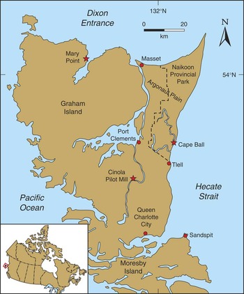
Our understanding of postglacial terrestrial paleoenvironments on Haida Gwaii increased significantly during and after the 1980s (Clague et al., Reference Clague, Mathewes and Warner1982; Warner et al., Reference Warner, Mathewes and Clague1982, Reference Warner, Clague and Mathewes1984; Scudder and Gessler, Reference Scudder and Gessler1989; Barrie et al., Reference Barrie, Bornhold, Conway and Luternauer1991, Reference Barrie, Conway, Mathewes, Josenhans and Johns1993, Reference Barrie, Conway, Josenhans, Clague, Mathewes and Fedje2005; Lacourse et al., Reference Lacourse, Mathewes and Fedje2003; Fedje and Mathewes, Reference Fedje and Mathewes2005; Lacourse and Mathewes, Reference Lacourse and Mathewes2005). The climatic and geological conditions and vegetation on the archipelago during the advance phase of the Fraser glaciation (MIS 2), on the other hand, have not yet been described. This study attempts to fill this knowledge gap. We provide new evidence for MIS 2 glacial conditions from two sea-cliff sections on Graham Island, one at Cape Ball on the east coast, and the second at Mary Point on the north coast (Fig. 1). Both sites were described and radiocarbon dated during the 1980s (Clague et al., Reference Clague, Mathewes and Warner1982; Blaise et al., Reference Blaise, Clague and Mathewes1990), but analyses of pollen, spores, other plant and animal remains, and their paleoecological implications have not previously been reported.
(color online) Locations of the Cape Ball, Mary Point, and Pilot Mill study sites on Graham Island (Haida Gwaii).

GEOLOGIC CONTEXT
During most Pleistocene glaciations, Haida Gwaii supported mountain ice caps and a network of valley glaciers that were independent of the Cordilleran ice sheet (Clague, Reference Clague1989). At the Fraser glaciation maximum, however, large lobes of the Cordilleran ice sheet extended west across northern Hecate Strait and Dixon Entrance and coalesced with local glaciers on eastern and northern Graham Island (Sutherland Brown, Reference Sutherland Brown1968; Clague, Reference Clague1989). Sediments exposed in sea cliffs at Cape Ball and Mary Point provide evidence for the advance of the Hecate and Dixon lobes of the Cordilleran ice sheet during the early part of the Fraser glaciation. The Quaternary succession in these sea cliffs includes till and outwash of the Fraser glaciation, glaciomarine sediments of Early Wisconsin age (MIS 4), and nonglacial beds above and below the Fraser glaciation deposits (Clague et al., Reference Clague, Mathewes and Warner1982; Warner et al., Reference Warner, Mathewes and Clague1982). In this paper, we briefly describe early Fraser glaciation sediments at the Cape Ball and Mary Point sea-cliffs to provide context for our discussion of ice extent, vegetation, and climate on the northwest coast during the advance phase of the Fraser glaciation.
METHODS
Geology
Clague logged the stratigraphy of the two sea-cliff sections that are the subject of this paper in 1980 (Cape Ball; Clague et al., Reference Clague, Mathewes and Warner1982, section 80-37) and 1988 (Mary Point). Observations upon which the two sections were compiled include lithology, Munsell color, mineralogy and clast composition, clast roundness, sorting, stratal thickness, sedimentary structures, visible fossils, unit thicknesses, and unit contact relations. Elevations, referenced to mean sea level, were determined with an altimetric barometer and a tape measure. Plant fossil samples were radiocarbon dated at the former Radio-Isotope Direct Detection Laboratory (RIDDL) at McMaster University. We calibrated the radiocarbon ages using the OxCAL4.2 online calibration program with the IntCal 13 dataset. Two-sigma calibrated age ranges are rounded off to the nearest 100 yr.
Paleoecology
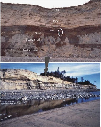
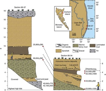
We collected sediment samples for pollen and macrofossil analysis at Cape Ball section 80-37 (Clague et al., Reference Clague, Mathewes and Warner1982) in August 1986 (Figs. 1, 2, and 3). They comprise 10 samples spaced 20 cm vertically in the upper laminated silt unit (Figs. 2 and 3; 8.2–10 m above sea level [asl]), and three samples from organic stringers in the sand directly below the silt unit (7–8.2 m asl). After cleaning the section, we extracted approximately 100 mL of sediment per sample and placed the samples in labeled plastic bags. We returned the samples from the field to Simon Fraser University and stored them in a cold room at 4°C until analyzed. Subsamples of 2 mL were separated for pollen analysis; the remainder of each sample was wet sieved through nested 1 mm and 180 μm screens to concentrate macrofossils. We also collected macrofossils of mosses and other plants from peaty stringers exposed in a low coastal bluff (Cape Ball section 80-34, Fig. 3), about 1 km south of section 80-37. Mathewes examined macrofossil residues from both sections under a dissecting microscope and picked fossils for identification and potential radiocarbon dating. He performed pollen analysis using standard methods on the 2 mL subsamples, after adding a tablet of exotic Eucalyptus pollen containing 16,180 ± 1460 grains (batch 903722) to calculate pollen concentrations. Pollen subsamples were first treated with 10% hot HCl and 10% KOH, then sieved through a 250 µm mesh screen, and finally treated with hot 48% HF to remove silicates. Following this treatment, subsamples were dehydrated in glacial acetic acid and acetolysed to remove cellulose and then stained red with safranin O. The residues were mounted in glycerin jelly for microscopic identification.
(color online) View of the high Cape Ball sea cliffs (bottom), looking west, with annotations at the location of site 80-37, shown in detail (above). The photograph above shows Mathewes (circled) sampling for the pollen diagram (Figure 8).

(color online) Stratigraphy of Late Pleistocene sediments exposed in the sea cliff at Cape Ball sections 80-34 and 80-37. Details of radiocarbon ages are provided in Table 1.

Identification and counting of palynomorphs were done at 400x magnification using Zeiss and Nikon eclipse research microscopes. Selected taxa were photographed under oil immersion (1000x magnification) using a Nikon Eclipse 80i with Nikon DS camera control. Identifications were made using a pollen and spore reference collection at Simon Fraser University and standard keys (Faegri et al., Reference Faegri, Kaland and Krzywinski1989; Moore et al., Reference Moore, Webb and Collinson1991). Pollen sums used for percentage calculations are based on total terrestrial pollen. For spores and other palynomorphs, we use total pollen plus sums of non-pollen taxa. Sums range from 300 to 437 pollen at Cape Ball, and 190 to 330 pollen at Mary Point. We generated the Cape Ball pollen diagram using TILIA software (Grimm, Reference Grimm2011).
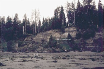
Mathewes and several of his colleagues collected the paleoecological samples at the Mary Point section (Fig. 4) in July 1988 using similar methods. One difference was that they collected and processed larger quantities of sediment to check for insect remains and plant macrofossils. J.V. Matthews Jr. and Alice Telka field-sieved large sediment samples for macrofossils and recovered beetle fossils. The beetle fossils have not been published and are only briefly mentioned in this paper. Six samples were collected at Mary Point for pollen analysis from silty beds with organic stringers, previously sampled for accelerator mass spectrometry (AMS) radiocarbon dating (fig. 10 in Blaise et al., Reference Blaise, Clague and Mathewes1990). We chose these samples for comparison with the six upper dated levels at Cape Ball. Two mL sediment subsamples were processed using the same protocols as for the Cape Ball samples. Finally, we generated a pollen diagram for Mary Point in the same form as for Cape Ball.
(color online) Photograph of the Mary Point sea cliffs in the vicinity of site 80-130 (see Fig. 5 for location). Slumps and slides obscure much of the Mary Point exposures, but the light-colored outwash sand in the upper part of the section and darker laminated mud below are clear in this photograph. A man on the beach (circled) provides scale.

Selected macrofossils were imaged under a scanning electron microscope (ETEC autoscan system) after attachment to an aluminum stub with double-sided tape, vacuum drying, and coating with evaporated gold. The ecology of the fossil mosses is taken from Vitt et al. (Reference Vitt, Marsh and Bovey1988), Klinka et al. (Reference Klinka, Krajina, Ceska and Scagel1989), and Schofield (Reference Schofield1992).
STRATIGRAPHY AND RADIOCARBON AGES
Cape Ball
The northeast corner of Graham Island, which includes Cape Ball, is a roughly triangular-shaped, low-lying plain (Argonaut Plain) underlain by thin patchy till and thick sand. The till, which is generally less than 2 m thick, is massive to stratified diamicton consisting of stones up to small boulder size set in a matrix of silt and sand. Till stones are dominantly volcanic and sedimentary rocks of local provenance; many of the stones are striated and faceted. Where till is present, the ground surface is characterized by long, low ridges oriented in a northwest to north-northwest direction. Sutherland Brown (Reference Sutherland Brown1968) interpreted these landforms to be glacial flutings oriented parallel to the direction of ice flow. They formed when local ice flowing from the mountains on Haida Gwaii coalesced with, and was deflected northward by, the Hecate lobe of the Cordilleran ice sheet. The Cape Ball area thus was an interlobate area between two glacier complexes at the Fraser glaciation maximum.
At the Cape Ball sample site, the till is underlain across a sharp to gradational contact, by up to 7 m of well sorted, horizontally and cross-bedded sand (Figs. 2 and 3). Cross-beds indicate deposition by southerly and southwesterly flows. The sand consists mainly of quartz, feldspar, and lithic grains derived from granitic and gneissic rocks that dominate the Coast Mountains on the British Columbia mainland to the east. We interpret the sand to be outwash deposited in an eastward-sloping, glacio-isostatically formed depression in front of the Hecate lobe as it flowed west and southwest across the northern Hecate Strait from the British Columbia mainland. It is similar to Esperance Sand and Quadra Sand, widespread units of Fraser glaciation advance outwash in northwestern Washington and south-coastal British Columbia (Mullineaux et al., Reference Mullineaux, Waldron and Rubin1965; Clague, Reference Clague1976, Reference Clague1977).
At Cape Ball, laminated and thin-bedded clayey silt and sand conformably underlie the sand described above (Figs. 2 and 3). These sediments contain pollen and plant macrofossils. They probably accumulated in shallow ponds or perhaps along the coastline in a brackish marine environment (see Discussion); we interpret them to record the initial approach of the Hecate lobe to Cape Ball during the Fraser glaciation. These sediments fill shallow basins or channels cut in glaciomarine sediments of probable MIS 4 age (Clague et al., Reference Clague, Mathewes and Warner1982; Mathewes et al., Reference Mathewes, Lian, Clague and Huntley2015).
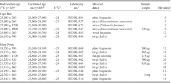
Plant fossils collected from the laminated clayey silt-sand unit yielded AMS radiocarbon ages ranging from 23,200±280 14C yr BP to 25,100±1000 14C yr BP (Fig. 3, Table 1). There is no significant trend in radiocarbon ages through the unit. The most reliable ages, that is those with uncertainties of less than 1000 14C yr, range from 23,200±280 14C yr BP to 26,650±390 14C yr BP (27,000–31,400 cal yr BP). We interpret this suite of radiocarbon ages to indicate that the Hecate lobe approached Cape Ball sometime between 30,000 and 31,000 cal yr BP.
Radiocarbon ages from Cape Ball and Mary Point.

a Error term is±1σ.
b Age range at±2σ determined using OxCAL4.2 online calibration program with the IntCal 13 dataset. Values are rounded off to the nearest 100 yr.
c Delta 13C values are assumed values based on Stuiver and Polach (Reference Stuiver and Polach1977).
d RIDDL, Radio-Isotope Direct Detection Laboratory, McMaster University.
e Datum is mean sea level.
Mary Point
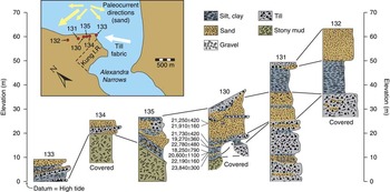
The Mary Point section is located on the north coast of Graham Island about 60 km northwest of Cape Ball (Figs. 1 and 4). As at Cape Ball, patchy thin till unconformably overlies sand and clayey silt in the sea-cliff at Mary Point (Fig. 5). The till is massive to stratified and consists of stones of local provenance in a matrix of silt and sand. Many of the stones are striated and faceted. Elongate, rod-shaped stones are preferentially oriented in a northwest-southeast direction, parallel to flutings, drumlins, and drumlinoid ridges, which are prominent landforms on this part of northern Graham Island. These landforms are continuous with more northerly-oriented flutings on the lowland west of Cape Ball, indicating continuity of flow of the Hecate and Dixon lobes, and coalescence with local ice flowing from the mountains of Haida Gwaii.
(color online) Stratigraphy of Pleistocene sediments in the sea cliff at Mary Point. The inferred ice-flow direction based on till fabric data and paleocurrent directions derived from the sand unit are summarized on the map at the upper left. Till fabric – lower hemisphere projection; contours 4%, 6%, 8%. Details of radiocarbon ages are provided in Table 1.

The thick sand-mud unit below the till is horizontally stratified and consists of beds of fine to medium sand, laminated clayey silt, and organic-rich silt and sand (Figs. 4 and 5). This unit correlates with the thick sub-till outwash sand unit at Cape Ball site 80-37. Thin layers of peat and lenses of diamicton are also present in the unit. Laminated clayey silt is common in the lower part of the unit, whereas sand dominates the upper part. Structures in the sand include cross-beds, ripples, and some graded beds. Some strata are warped and folded, probably due to loading and gravitational movement of the sediment pile during deposition. The uppermost few meters of the unit contain shears that we interpret to be glaciotectonic in origin. The laminated mud beds clearly were deposited in standing water, probably a pond or lagoon.
Sedimentary structures in the sand are consistent with subaqueous deposition. Our interpretation is that the sand-silt unit was deposited in shallow freshwater or the sea on glacio-isostatically depressed ground in front of the Dixon lobe as it advanced on Mary Point. This interpretation is supported by measurements of orientations of ripples and cross-beds, which indicate flow mainly to the south and west. The paleoflow data are incompatible with the alternative explanation that streams flowing northward off Graham Island deposited the sediments. The sand at Mary Point, like that at Cape Ball, is dominated by quartz, feldspar, and lithic particles probably derived from granitic rocks on the British Columbia mainland. The sand-silt unit fills a shallow basin cut in glaciomarine gravelly mud, which may correlate with the similar unit at Cape Ball.
The sub-till sand-mud unit at Mary Point was deposited between 18,250±790 and 23,840±300 14C yr BP (Fig. 5, Table 1). Most of the nine AMS ages from this unit are statistically equivalent at the 95% confidence level, and there is no apparent trend in age through the unit. We take the oldest Mary Point radiocarbon age, which, when calibrated, yields a calendric age range of 27,500–28,600 cal yr BP, as the approximate time the sediments were deposited and glacier ice approached the site. Shortly after this time, the Dixon lobe overrode Mary Point. The youngest of the Mary Point ages (18,250±790 14C yr BP) could, if valid, be taken as a maximum for the time of glacier overriding. However, this age was obtained on a very small sample (200 μg) of unidentified plant fragments and has a large analytical uncertainty. We thus are reluctant to use it to constrain the time of glacier overriding. Five ages higher in the sequence, all on identified plant macrofossils (Fig. 5, Table 1), are older than 18,250±790 14C yr BP, lending support to our cautious interpretation.
PALEOECOLOGY
Macrofossils
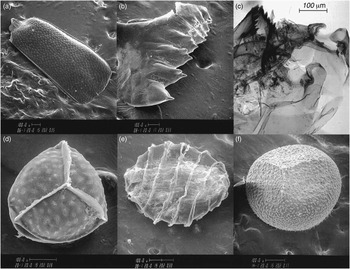
Invertebrates recorded at Cape Ball and Mary Point include beetles (Coleoptera, Fig. 6a), tadpole shrimp (Lepidurus cf. arcticus, Fig. 6b), and chironomid head capsules, typified by the midge Pseudodiamesa (Fig. 6c).
Invertebrate macrofossils and selected plant and algal spores from Cape Ball. (a) Right elytron of a staphylinid beetle. (b) Mandible of the fresh-water tadpole shrimp (Lepidurus cf. L. arcticus). (c) Head capsule of the cold water chironomid Pseudodiamesa. (d) Trilete megaspore of aquatic quillwort (Isoetes). (e) Oospore of aquatic fresh-water alga (Characeae). (f) Trilete megaspore of the boreal spikemoss Selaginella selaginoides.

Plant remains at both study sites are dominated by twigs and leaf fragments, some of which are from small willows (Salix), as well as fragmented and whole mosses (bryophytes). Megaspores of aquatic quillworts (Isoetes, Fig. 6d) and algal oospores of Characeae (Fig. 6e) were found at Cape Ball and indicate a mostly freshwater environment. Megaspores of the boreal spikemoss, Selaginella selaginoides (Fig. 6f), are present at both Cape Ball and Mary Point. The presence of the cold stenotherm chironomid Pseudodiamesa, the tadpole shrimp, and the boreal spikemoss are evidence for colder temperatures than today.
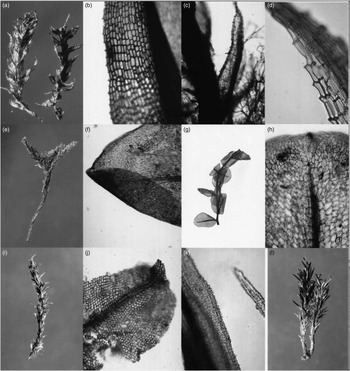
The well-preserved macroscopic moss remains at both study sites were examined by Schofield, who identified 18 genera or species (Fig. 7, Table 2). These mosses indicate a range of local site conditions, including wet areas, seepage sites, mesic soils, and well-drained sandy or gravelly areas.
Bryophyte macrofossils from Mary Point (a–b, e– j, and l) and Cape Ball (c, d, and k). (a) Meesia triquetra habit with three-ranked leaves. (b) Detail of M. triquetra leaf margin with small teeth. (c) Portion of stem and leaves of Philonotis fontana with thread-like paraphyllia. (d) Detail of toothed leaf tip of Philonotis fontana. (e) “Tree-shaped” whole plant of Climacium dendroides, (f) Leaf tip of Climacium dendroides with strong midrib and elongated cells. (g) Stem of Cyrtomnium hymenophylloides with well–preserved ovate leaves. (h) Detail of leaf of Cyrtomnium hymenophylloides with strong midrib and small apiculus (apex). (i) Gametophyte of Tortula ruralis with worn leaf tips. (j) Detail of Tortula ruralis leaf with strong midrib, square-shaped cells. Leaf hair at the tip is missing. (k) Microscopic detail of Rhacomitrium canescens complex showing the rough papillate leaf surfaces. (l) Whole gametophyte of Polytrichum juniperinum with two aerial shoots.

Fossil mosses recovered from sediments at Cape Ball and Mary Point.

a Identified by W.B. Schofield.
Pollen and spores
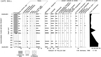
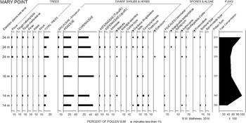
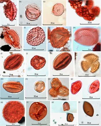
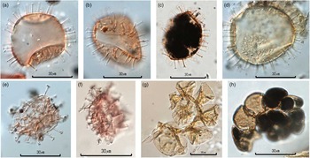
The dominant pollen and spore types and selected less common taxa from the Cape Ball section are summarized as percentage frequencies in a pollen diagram (Fig. 8). Mary Point pollen and spore frequencies are plotted in a separate diagram (Fig. 9) for comparison with Cape Ball. Rare pollen, spore, and other microfossils (≤1%, and many as single occurrences) at both Cape Ball and Mary Point are listed in Table 3. Although rare, some of these taxa are significant paleoenvironmental indicators, as noted in following sections. Palynomorphs discussed in this paper include taxa that are not commonly illustrated in publications, including cryptogam spores, algal cells, fungal spores, stomates, and marine dinoflagellate cysts and foraminiferan linings. Accordingly, we have included a color plate (Fig. 10) illustrating terrestrial and fresh water taxa that are key to our paleoenvironmental reconstruction. Figure 11 illustrates the marine indicators from the two sites.
Percentage pollen diagram for the Cape Ball section showing dominant pollen and spores and dinoflagellate cysts (dots indicate values of 1% or less). Radiocarbon ages and a total pollen concentration curve are also shown. Rare taxa not shown here are listed in Table 3 or shown in Figures 10 or 11.

Percentage pollen diagram for the Mary Point section, showing dominant pollen types and rare taxa of paleoecological interest (dots indicate values of 1% or less). Analyzed samples from silty beds are ordered by elevation in meters above sea level (asl), with three closely spaced samples (10–20 cm apart) at 24 m asl and two at 14 m asl. Radiocarbon ages are given in Table 1.

(color online) Selected pollen, spores, and other microfossils from Cape Ball (a–b, d–e, g, and i) and Mary Point (c, f, h, and j–t). All images photographed under oil immersion at 1,000x magnification. (a) Echinate Nuphar pollen. (b) Leaf-hair base of Nymphaeaceae. (c) Callitriche pollen. (d) Botryococcus algal colony. (e) Picea (spruce) stomata. (f) Proximal view of Anthoceros spore. (g) Microspore of Selaginella selaginoides. (h) Test of rhizopod Assulina. (i) Gentiana douglasiana pollen. (j) Koenigia islandica pollen. (k) Bistorta (Polygonum) pollen. (l) Large verrucate Anenome pollen. (m) Caltha type pollen, tricolpate with small verrucae. (n) Thalictrum periporate pollen. (o) Three joined Caryophyllaceae grains. (p) Ligusticum calderi pollen grains showing the distinctive shape and H-shaped pore. (q) Polemonium caeruleum type pollen. (r) Campanula pollen with large annulate pores and spines. (s) Sporormiella type bullet-shaped terminal spore and rectangular spore showing germinal apertures. (t) Fungal spore of Sordaria type with terminal pore.

(color online) Marine microfossils from Cape Ball and Mary Point photographed at 1,000x magnification. (a–c) Operculodinium centrocarpum cysts from Cape Ball. (d) Operculodinium centrocarpum cyst from Mary Point. (e and f) Spiniferites cysts from Cape Ball. (g and h) Microforaminiferan chitinous linings from Mary Point.


Pollen and spore taxa present at Cape Ball and Mary Point are similar, although some qualitative and quantitative differences are apparent, as might be expected from sites separated by 60 km and differing by several thousand years in age. Tree pollen (arboreal pollen, AP) is moderately abundant at Cape Ball, with lodgepole pine (Pinus contorta type) reaching up to 20% of terrestrial pollen and mountain hemlock (Tsuga mertensiana) ranging from 5% to 10%; values for other trees, including spruce (Picea) and true fir (Abies), are less than 5% (Fig. 8).
Megaspores (Fig. 6d) and microspores of the submerged aquatic quillwort Isoetes indicate shallow fresh waters, as do rare pollen of Nuphar and leaf hair bases of water lily, found at Cape Ball (Fig. 10a and b). Water starwort (Callitriche) pollen (Fig. 10c) is recorded only at Mary Point. Algal colonies of the benthic Botryococcus (Fig. 10d) and planktonic Pediastrum were found at both localities. Oospores of submerged Characeae algae (Fig. 6e) are present at Cape Ball; they also are among the earliest postglacial botanical remains identified by Warner et al. (Reference Warner, Mathewes and Clague1982) at that locality.
Monolete ferns (Filicales) range from a few percent up to 20%, and spores of Sphagnum (peat moss) are consistently present, although at less than 5%. Also present are spores of the liverwort Anthoceros (Fig. 10f). Rare occurrences of the spikemoss Selaginella selaginoides microspores (Fig. 10g) confirm the local presence of this boreal bog species, consistent with the presence of megaspores at Mary Point and Cape Ball (Fig. 6f).
As seen in Figure 8, pollen of graminoids (Poaceae and Cyperaceae) and the sunflower family Asteraceae are dominant at both localities. They are accompanied by small frequencies of pollen of alpine-subalpine herbs: Koenigia islandica (Fig. 10j) and Bistorta (Fig. 10k) are present at <1% in most samples. Other herbs include three members of the Ranunculaceae family, Anenome (Fig. 10l), Caltha type (Fig. 10m), and Thalictrum (Fig. 10n), with the last genus being more common at Mary Point. Pollen of the parsley family (Apiaceae) is registered by rare occurrences of Heracleum, Angelica type, Oenanthe, and Calder’s lovage (Ligusticum calderi, Fig. 10p and q) at Cape Ball. The distinctive large periporate pollen of Polemonium caeruleum (Fig. 10r) is rare but also environmentally significant.
Marine dinoflagellate cysts are present at both study sites and reach values of up to 9.6% in the uppermost Cape Ball samples (Fig. 8). The more common taxon is the marine dinoflagellate cyst Operculodinium centrocarpum (Figs. 11a–d); the less common taxon is Spiniferites sp. (Figs. 11e and f).
A surprise was the discovery of two marine micro-foraminiferan tests in the uppermost Mary Point sample. The complete chitinous test (Fig. 11g) shows the characteristic coiling of a foraminiferan, which is also apparent in a second specimen (Fig. 11h).
DISCUSSION
Ice limits and chronology of the early Fraser glaciation
Radiocarbon-dated sediments at Cape Ball and Mary Point constrain the age of the Late Wisconsin advance of the Hecate and Dixon lobes of the Cordilleran ice sheet. The sediments, along with glacier flow indicators, also provide information on the interaction of the ice sheet with independent local glaciers on Haida Gwaii.
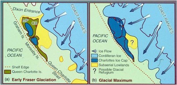
Our data indicate that the Hecate lobe, which was fed chiefly by ice flowing down the Skeena River valley from sources in the Coast Mountains and the British Columbia interior, advanced across the northern Hecate Strait about 30,000–31,000 cal yr BP, early during the Fraser glaciation (Fig. 12a). Fans of sandy and silty outwash were deposited in the vicinity of the present east coast of Graham Island near the margin of the advancing ice. The extent of Haida Gwaii glaciers at this time is uncertain, although the advance outwash at Cape Ball does not have a local provenance. In Figure 12a, we show local glaciers approaching the lowland plains of Haida Gwaii at this time.
(color online) Diagrammatic maps showing inferred extent of glaciers on Haida Gwaii and adjacent areas. (a) About 30,000–31,000 yr ago during the early phase of the Fraser glaciation and (b) 17,000–19,000 yr ago at the local last glacial maximum. The extent of the exposed continental shelf at these times is uncertain; the approximate location of an inferred “Hecate Refugium” is indicated by the question mark. CB, Cape Ball; MP, Mary Point.

At the Fraser glaciation maximum, local ice coalesced with the Hecate lobe and flowed northwestward, producing the flutings west of Cape Ball. Peaks in ice-rafted detritus in a deep-sea core in the eastern North Pacific Ocean south of Haida Gwaii (Blaise et al., Reference Blaise, Clague and Mathewes1990) suggest that ice cover in Dixon Entrance and in Dixon Entrance south of Moresby Island was greatest between about 17,000 and 19,000 calyrBP.
The Hecate lobe coalesced with the Dixon lobe to the north (Fig. 12a). The latter, nourished by ice flowing down Nass River valley and Observatory Inlet, flowed west along Dixon Entrance. About 27,000–29,000 cal yr BP, the advancing Dixon lobe approached Mary Point, and meltwater streams flowing off the ice deposited an outwash fan similar to that deposited several thousand years earlier at Cape Ball. The Dixon lobe reached the edge of the continental shelf at the mouth of the Dixon Entrance at the Fraser glaciation maximum (Fig. 12b). At that time, Mary Point was covered by hundreds of meters of ice.
The difference in age of correlative outwash at our two study sites is consistent with the more distant location of Mary Point from source areas of the Cordilleran ice sheet on the British Columbia mainland. Furthermore, Dixon Entrance is deeper than northern Hecate Strait – whereas the Hecate lobe may have crossed Hecate Strait when it was subaerially exposed due to low eustatic sea levels prevailing at the time (Barrie et al., Reference Barrie, Conway, Mathewes, Josenhans and Johns1993; Josenhans et al., Reference Josenhans, Fedje, Conway and Barrie1995), the Dixon lobe probably advanced in contact with the sea, perhaps more slowly than the Hecate lobe.
Local Haida Gwaii glaciers eventually coalesced with the Dixon lobe and were deflected northwestward along the north coast of Graham Island. Glacial flutings, drumlins, and drumlinoid ridges oriented in this direction record the pattern of ice flow at the Fraser glaciation maximum when most of the present land area of Haida Gwaii was covered by ice (Fig. 12b).
Paleoenvironmental interpretation
The environments recorded by both macrofossils and microfossils at both study sites are primarily freshwater ponds, lakes, or slow streams on a coastal plain, although the presence of marine dinoflagellate cysts and foraminiferan linings suggests a possible marine influence (see Other environmental indicators). Isoetes, Callitriche heterophylla, and Nuphar polysepalum are present today on Haida Gwaii as aquatic macrophytes found in shallow ponds and pools, and at the margins of lakes (Calder and Taylor, Reference Calder and Taylor1968). Characeae oospores (Fig. 6e) are recorded at our Cape Ball section, along with mandible fragments of tadpole shrimp (Lepidurus arcticus, Fig. 6b). Chara and Nitella oospores and Lepidurus remains have also been recorded as fossils in subarctic lakes in Greenland (Fredskild, Reference Fredskild1983). Head capsules of the cold-stenotherm midge Pseudodiamesa (Fig. 6c) indicate cold freshwater conditions at Cape Ball consistent with glacial runoff. Walker et al. (Reference Walker, Levesque, Cwynar and Lotter1997) have documented the narrow cold temperature preference of this midge genus from its modern distributions in the Canadian Arctic.
Tree pollen types recorded at Cape Ball are also dominant in the pollen spectra at the mid-Wisconsin Pilot Mill site on Graham Island (Warner et al. Reference Warner, Clague and Mathewes1984), and may have persisted, at least locally, into the early part of the Fraser glaciation. The radiocarbon age of the top of the Pilot Mill section of 27,500 ± 400 14C yr BP (GSC-3530) defines a time of declining, but still high, tree percentages. Given that the oldest Cape Ball radiocarbon age (26,650 ± 390 14C yr BP; ca. 30,000–31,400 cal yr BP) is only slightly younger, it seems likely that some trees were still present on Graham Island during the earliest stage of the Fraser glaciation. It is unclear whether the pollen concentrations at Cape Ball (10,000 to 30,000/cm3, Fig. 8) indicate local tree presence 30,000–31,400 yr ago, but this possibility is supported by generally good pollen preservation and by the ubiquitous presence of tree pollen in all samples from Cape Ball. Rare conifer leaf stomata (Fig. 10e), identified as Picea using the identification criteria of Hansen (Reference Hansen1995), support this interpretation. The recovery of fossil stomata suggests that some trees were locally present on Haida Gwaii, given that modern analogues at treeless alpine tundra sites in northwestern Canada lack stomates, whereas almost all treed sites have stomate subfossils (Pisaric et al., Reference Pisaric, Szeicz, Karst and Smol2000).
The differences between Cape Ball and Mary Point AP percentages are notable and support cooling of climate and tree losses over the period separating deposition of the sampled sequence at the two localities. This trend is supported by the lower pollen concentrations at Mary Point (4,000 to 10,000/cm3, Fig. 9) compared to Cape Ball. Arboreal pollen frequencies in the uppermost six samples at Cape Ball range from 20.4% to 34%, with a mean value of 26.5%. In contrast, the uppermost six samples at Mary Point have a range in AP of 5.4–21%, with a mean of only 11.3%. Further work is needed to confirm this difference, but the data so far indicate a significant reduction of tree pollen input as glaciation proceeded, with corresponding increases of non-arboreal pollen. The mean value of sedge (Cyperaceae) pollen, for example, is 15% at Cape Ball and 46% at Mary Point. The other major difference that we note is the greater abundance of willow (Salix) pollen at Mary Point compared to Cape Ball—the peak value at the former site is 6.7%, whereas all 13 Cape Ball samples have values less than 2%. The sparse presence of Ericales pollen at Mary Point (<4.7%) contrasts with values of 12–28% at Cape Ball. Sunflower family (Asteraceae) pollen types, including sage (Artemisia), are also common at Cape Ball, with values up to 10%.
The presence of peat moss and S. selaginoides spores suggests local boggy or peaty environments, which fits with the stratigraphic presence of peaty organic stringers. A fossil test of the sphagnicolous rhizopod Assulina (Fig. 10h) at Mary Point confirms the presence of Sphagnum peat there. Additional support for boggy or peaty environments is provided by the presence of Gentiana douglasiana pollen grains (Fig. 10i).
Moss macrofossils from both study sites (Table 2) help to reconstruct local microhabitats. Meesia triquetra (Figs. 7a and b), Philonotis fontana (Figs. 7c and d), Dichodontium pellucidum, Calliergon stramineum, and Amblystegium cf. riparium indicate very wet conditions. Intermediate moist to wet conditions are favored by “tree moss” (Climacium dendroides, Figs. 7e and f) and the Arctic-alpine moss Cyrtomnium hymenophylloides (Figs. 7g and h). This last species occurs in moist fens, heaths, seepage areas, and in fine-grained alluvium in the tundra of North America and Greenland (Miller Reference Miller1996).
In contrast, mosses typical of well-drained, dry, sandy, or rocky habitats are also present, including Tortula ruralis (Figs. 7i and j), Rhacomitrium canescens species complex (Fig. 7k), Polytrichum juniperinum (Fig. 7l), and Bryum capillare. Other taxa in Table 2 have a range of microhabitats, including disturbed open soils. Overall, the bryophytes support an open lowland environment with a mosaic of microhabitats, disturbed soils, and ponds.
Other environmental indicators
Less abundant pollen and spores are commonly useful in paleoecological reconstructions. Koenigia islandica is a small herb of moist to wet open soils in subalpine-alpine sites in British Columbia and in similar habitats throughout the circumpolar Arctic (Douglas et al., Reference Douglas, Meidinger and Pojar1999). Pollen of Bistorta bistortoides (syn. Polygonum bistortoides) and B. vivipara (syn. P. viviparum) have similar pollen grains, but both species are indicative of subalpine-alpine distributions and environments (Douglas et al., Reference Douglas, Meidinger and Pojar1999). A single pollen grain of Anenome at Mary Point may also support an interpretation of cool Arctic-alpine conditions. Although 12 species of Anenome are present in British Columbia today (Douglas et al., Reference Douglas, Meidinger and Pojar1999), only three species currently are found on Haida Gwaii. Anenome multifida and A. parviflora are rare on limestone, and A. narcissiflora is a more common plant in subalpine-alpine settings (Calder and Taylor, Reference Calder and Taylor1968). The fossil Anenome grain from Mary Point (Fig. 10l) best matches the arctic-alpine species A. narcissiflora based on comparison with modern reference pollen, but species identification remains uncertain since comparison reference material for other species of the genus is incomplete, precluding a positive identification. Pollen of Caltha type (Fig. 10m) is present at both localities. It is similar to Anenome type pollen, but is smaller and may also represent some other ranunculaceous genera such as Aquilegia. Caltha also fits with moist soils and cool climatic conditions. A plant cover of subalpine-alpine or Arctic affinity with abundant graminoids is supported by the presence of Thalictrum pollen (Fig. 10n) at Mary Point, with values up to 2.9%. The only meadow rue species that occurs today on Haida Gwaii is T. alpinum, found as a single colony on open alpine heath (Calder and Taylor, Reference Calder and Taylor1968). Another taxon at Mary Point with unusually high frequencies is the chickweed family (Caryophyllaceae, Fig. 10o). Its local presence at Mary Point is indicated by percentages averaging 3.3% (range 2.5–4.8%). In contrast, values in all 13 Cape Ball samples are <1%, and most samples have only one or two pollen grains.
Hebda (Reference Hebda1985) reviewed Apiaceae pollen morphologies, including Ligusticum calderi, a former Queen Charlotte endemic (Calder and Taylor, Reference Calder and Taylor1968), which is also present today on Banks Island east of Haida Gwaii, Kodiak Island, and other islands as far south as Vancouver Island. He argued that pollen of L. calderi can be identified to species by its H-shaped pore, subrhomboidal outline in equatorial view, and its larger size compared to L. canbyi. Hebda’s criteria confirm the presence of L. calderi in six samples from Cape Ball (Fig. 10p). The taxon is significant in that it is a possible indicator of refugia in coastal British Columbia during the Pleistocene (Hebda, Reference Hebda1985). Ligusticum calderi pollen was found in MIS 4 sediments at Cape Ball (Mathewes et al., Reference Mathewes, Lian, Clague and Huntley2015), suggesting that it persisted through both MIS 4 and MIS 2 glaciations on Haida Gwaii. Another species that has been recorded both in MIS 4 (Mathewes et al., Reference Mathewes, Lian, Clague and Huntley2015) and early MIS 2 sediments at our study sites is tall Jacob’s-ladder, Polemonium caeruleum (Fig. 10q), a circumboreal taxon found on moist soils in meadows, marshes, and stream edges in montane and northern locales (Mathewes, Reference Mathewes1979a). This species is no longer present on Haida Gwaii (Mathewes, Reference Mathewes1980), but is found in early Fraser glaciation sediments at Vancouver (Mathewes, Reference Mathewes1979b) and at Late Glacial sites on Haida Gwaii.
Potential refugial taxa in addition to Ligusticum and Polemonium include Koenigia islandica and species of genera such as Bistorta, Thalictrum, Empetrum, Artemisia, Campanula, Caltha type, Huperzia, and many cryptogams and mosses. The bluebell genus Campanula thrives in open, dry, sandy, and rocky disturbed environments, based on personal observations and herbarium records. Its pollen is distinctive (Fig. 10r), but rarely encountered, and may represent one of the two species currently on Haida Gwaii – C. alaskana and alpine C. lasiocarpa, which Heusser (Reference Heusser1989) identified as a species of present-day nunataks in the Juneau Ice Field of southeast Alaska.
Further study of pollen and macrofossils will clarify the composition of likely refugial flora on Haida Gwaii. The question of glacial refugia on Haida Gwaii and adjacent areas in southeast Alaska has a long controversial history, but is converging on an interpretation favoring the existence of ice-free areas in this region during the Fraser glaciation (Heusser, Reference Heusser1989; Mathewes, Reference Mathewes1989; Heaton et al., Reference Heaton, Talbot and Shields1996; Barrie et al., Reference Barrie, Conway, Josenhans, Clague, Mathewes and Fedje2005; Carrara et al., Reference Carrara, Ager and Baichtal2007; Shafer et al., Reference Shafer, Cullingham, Côté and Coltman2010; Pruett et al., Reference Pruett, Topp, Maley, McCracken, Rohwer, Birks, Sealy and Winker2013). In addition to widely accepted refugia on nunataks at high elevations in the Queen Charlotte Range, a large lowland “Hecate Refugium” probably existed on the now-submerged continental shelf off the coast of Moresby Island (Barrie et al., Reference Barrie, Conway, Josenhans, Clague, Mathewes and Fedje2005; and Fig. 12).
The presence at Mary Point of up to 1.2% coprophilous fungal spores of Sporormiella type (Fig. 10s) and up to 1.9% Sordaria type spores (Fig. 10t) was unexpected because these palynomorphs were not found in the MIS 2 samples at Cape Ball or seen at other postglacial localities. Spores of Sporormiella and Sordaria type were previously detected on Haida Gwaii only in 57,000-year-old (MIS 4) sediments at Cape Ball (Mathewes et al., Reference Mathewes, Lian, Clague and Huntley2015), and indicate the presence of mammal dung. A follow-up study is planned to increase the number of sampling sites and pollen counts to better assess the significance of these occurrences.
The possibility that the marine dinoflagellate cysts at both our study sites were reworked from older glaciomarine sediments was considered but rejected because these marine fossils were not seen in the older sediments and are particularly abundant in some Cape Ball samples (Fig. 8). It is conceivable that the dinoflagellates were deposited by sea spray, but their high frequencies in some Cape Ball samples and the presence of foraminifera tests at Mary Point favor a primary marine influence.
Two taxa were identified based in part on descriptions in Zonneveld and Pospelova (Reference Zonneveld and Pospelova2015) and confirmed by Pospelova. Operculodinium centrocarpum is a cosmopolitan species, but is most abundant in temperate to subpolar North Atlantic waters. As noted by Zonneveld and Pospelova (Reference Zonneveld and Pospelova2015), this species can be abundant in areas where upper water temperatures are <0°C throughout the year, and where salinities are reduced as a result of meltwater inputs during summer. These ecological observations accord well with an interpretation of glacier buildup on Haida Gwaii early during the Fraser glaciation.
Mathison and Chmura (Reference Mathison and Chmura1995) discuss marine foraminiferal linings and conclude that there is no relationship between salinity and fossil concentration. Their presence at Mary Point supports an interpretation of crustal depression and attendant marine influence during the early Fraser glacial advance.
CONCLUSIONS
At the maximum of the Fraser glaciation, lobes of the Cordilleran ice sheet flowed westward across Hecate Strait and down Dixon Entrance, where they briefly coalesced with glaciers sourced on Graham Island. Sediments exposed in the sea cliffs at Cape Ball and Mary Point provide evidence for the advance of these glacier lobes during the early phase of the Fraser glaciation. We analyzed macrofossils and microfossils recovered from sandy and silty strata beneath till and outwash at Cape Ball. These strata date to between about 27,000 and 31,400 cal yr BP. Sand, silt, and peat stringers in a similar stratigraphic position at Mary Point, 60 km to the north, appear to be several thousand years younger.
Vegetation at Cape Ball during the advance phase of the Fraser glaciation was tundra-like, with grasses, sedges, composites, and abundant Ericales pollen. The presence of Arctic-alpine indicator plants at both Cape Ball and Mary Point indicates a seasonally cold climate. Some trees may have persisted in protected microenvironments based on moderate pollen percentages of spruce, mountain hemlock, and lodgepole pine type. These tree types appear to be relicts of the interstade that preceded the Fraser glaciation, when the same three trees were dominant. The Mary Point site records a treeless tundra with much lower tree pollen than at Cape Ball; peak values of willow, chickweeds, and meadow-rue pollen; and a dominance of grasses and sedges. The presence of abundant marine dinoflagellate cysts at both Cape Ball and Mary Point, and foraminiferal remains at Mary Point support an interpretation of significant crustal depression during the Fraser glaciation ice advance in this area.
The presence of coprophilous fungus spores at Mary Point suggests that mammals were present on northern Haida Gwaii during the buildup of glacier ice. Although no mammal fossils have yet been recovered, caribou is a likely candidate based on a find of a fossil antler fragment in pre-Fraser glaciation gravel on Graham Island (Wigen, Reference Wigen2005).
The paleoecological evidence presented here ties together previous palynological evidence spanning the Wisconsin Stage (MIS 2, 3, and 4). The presence of Ligusticum calderi and Polemonium caeruleum pollen in Early Wisconsin sediments (Mathewes et al., Reference Mathewes, Lian, Clague and Huntley2015), early Fraser glaciation sediments (this study), and Late-Glacial sediments at Cape Ball (Mathewes, Reference Mathewes1979b) suggests survival of these taxa and others in glacial refugia. The most likely refugial area is the coastal shelf, now drowned in western Hecate Strait, and referred to here as the “Hecate Refugium”. This large refugium may have supported a variety of plants and animals.
ACKNOWLEDGMENTS
We thank Calvin J. Heusser (deceased), John V. Matthews Jr., Alice Telka, and Erle Nelson for field assistance at Mary Point. We also thank W.B. Schofield (deceased) for fossil moss identifications, Ian R. Walker for chironomid identification, and Vera Pospelova for confirming our dinoflagellate identifications. Erle Nelson provided the McMaster University (RIDDL) radiocarbon ages. Marlow Pellatt and Matt Huntley helped prepare some of the figures. Two anonymous reviewers helped to improve the manuscript. Funding was provided by the Natural Sciences and Engineering Research Council of Canada through Discovery Grants A3835 to RM and A24595 to JC, and from Simon Fraser University through Associate Dean research funds to RM.