Takotsubo cardiomyopathy, also known as transient apical ballooning syndrome or colloquially as Broken Heart syndrome, often presents with congestive heart failure and electrocardiographic changes that mimic acute coronary syndrome. The vast majority of these cases occur in women over 50 years of age. It is estimated that ~3.5–5% of these patients will have a recurrence of takotsubo.Reference Sharkey, Lesser and Maron 1 , Reference Gianni, Dentali, Grandi, Sumner, Hiralal and Lonn 2 Limited data exist describing takotsubo in children, and to our knowledge there are no reports describing recurrent takotsubo in children. In this study, we describe the case of a 14-year-old boy who was diagnosed with two episodes of takotsubo cardiomyopathy triggered by status epilepticus.
Case
The patient was a 14-year-old boy with asthma and medically refractory epilepsy who was one year status post anterotemporal lobectomy. He presented to the emergency department following a prolonged generalised tonic–clonic seizure. His review of systems was negative for any recent illnesses or fevers.
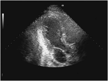
The boy was admitted directly to the paediatric ICU. The next day, he was noted to be hypotensive and was placed on a dopamine drip to maintain his blood pressure. An echocardiogram revealed decreased systolic function and hypokinesis of the apex, with dilation of the apical portion of the left ventricle, findings suggestive of takotsubo cardiomyopathy (Fig 1). He was supported on milrinone after the diagnosis was made.
Echocardiographic apical three-chamber view obtained during initial takotsubo admission. The image was obtained at end systole. Note that the apex balloons out as the base contracts.

Laboratory studies showed an elevated troponin level of 3.37 ng/ml (normal <0.2 ng/ml) and a B-type natriuretic peptide level of 1173 pg/ml (normal <100 pg/ml). The patient became febrile during his hospitalisation. Blood, urine, cerebrospinal fluid studies, and viral studies for potentially causative organisms of myocarditis, including enterovirus, herpes simplex virus, adenovirus, and parvovirus, were negative.
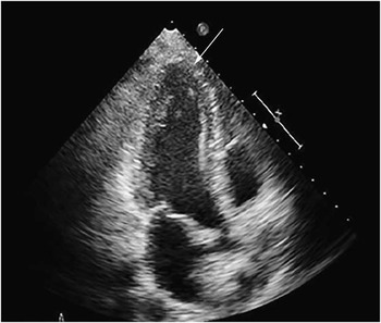
His pressors were gradually weaned off, and he was transitioned to lisinopril. His cardiac function and morphology normalised before hospital discharge (Fig 2).
He remained seizure free for ~6 months, at which time he had another generalised tonic–clonic seizure and was brought to the emergency department. Again he was found to have hypotension requiring an ICU admission with inotropic support. An echocardiogram demonstrated recurrent severe left ventricular apical dysfunction. During this second presentation, his initial troponin level was 5.76 ng/ml. An electrocardiogram showed T-wave inversion in the inferolateral leads. Angiography revealed normal coronary arteries.
The hospital course during this admission mirrored the first, with gradual recovery of cardiac function and a transition back to oral lisinopril. Given the recurrence of this rare cardiomyopathy, now with a certain inciting event of prolonged generalised seizures, it was believed that catecholaminergic surge was playing a role. For this reason, metoprolol was added specifically to help prevent or at least mitigate future occurrences if he were to have another profound seizure.
Echocardiographic apical three-chamber view obtained after recovery from takotsubo cardiomyopathy. The image was obtained at end systole, similar to Figure 1. Note that the apex contracts similarly to the base.

The child has been subsequently admitted for status epilepticus and did not have recurrent episodes of takotsubo. He remains on treatment for seizures and on metoprolol. Lisinopril was weaned off as function normalised.
Discussion
Takotsubo cardiomyopathy was first recognised in Japan in 1990. The condition is named for the appearance of the akinetic left ventricular apex, which balloons out during the acute phase and resembles a Japanese pot used to trap the octopus – the tako-tsubo. It is characterised by both left ventricular dysfunction and electrocardiographic changes.Reference Gianni, Dentali, Grandi, Sumner, Hiralal and Lonn 2
Most patients have a sudden onset of symptoms including chest pain and shortness of breath, which are often mistaken for an acute coronary syndrome; however, on angiography, the coronary arteries are not found to have a significant amount of blockage, and typical markers for myocardial injury, such as troponins, are only mildly elevated. Electrocardiographic findings include ST-segment elevations and T-wave inversion, particularly in pre-cordial leads.Reference Gianni, Dentali, Grandi, Sumner, Hiralal and Lonn 2
It is estimated that takotsubo is diagnosed in ~1.7–2.2% of adult patients who present for evaluation of acute coronary syndrome;Reference Gianni, Dentali, Grandi, Sumner, Hiralal and Lonn 2 85% of patients are able to identify an emotionally or physically stressful event.Reference Sharkey, Lesser and Maron 1 Nevertheless, the exact pathophysiology of takotsubo remains unclear. Current evidence suggests a role of an exaggerated sympathetic nervous system response to catecholamines.Reference Hernandez 3 Patients with takotsubo cardiomyopathy have a myocardium with decreased glucose uptake.Reference Hernandez 3 There may also be factors related to microvascular impairment and low oestrogen levels, which may explain this condition’s aetiology and explain why older women are disproportionately affected.Reference Hernandez 3
In a retrospective literature review, Hernandez found only 12 cases published of children or young adults with takotsubo cardiomyopathy. In our literature search, reported triggers included physical and emotional stress, withdrawal of medicine, and co-existent diseases such as coeliac disease and haemophagocytic lymphohistiocytosis.Reference Hernandez 3 – Reference Firdouse, Garwal, Lace De Almeida and Mondal 7 Takotsubo-like cardiomyopathies have also been described in the presence of intra-cranial haemorrhage and pheochromocytoma.Reference Von Berge, Lyon and Edens 8 – Reference Wittekind, Yanay, Johnson and Gibbons 9
In this patient, catecholamine surge secondary to status epilepticus was thought to trigger the takotsubo cardiomyopathy during both presentations. Despite his extensive seizure history, it does not appear that this patient had any previous clinically evident cardiomyopathy before the first presentation described in this case. It was suspected that perhaps the patient also had an underlying viral illness, which in combination with the status epilepticus led to sufficient catecholamine surge. A work-up for pheochromocytoma was also performed and was negative.
In general, 95% of adult patients do not have a recurrence, even in light of a subsequent stressful event.Reference Sharkey, Lesser and Maron 1 The remaining 5% of patients may have recurrence for a second or even a third time, again triggered by a stressor. Based on the limited paediatric data available, it does not appear that a child or young adult has ever been reported to have had a recurrence of takotsubo.
Patients usually require supportive care for 3–7 days. Patients who have had long-term follow-up have not shown lasting loss of strength or function as a result of this diagnosis.Reference Sharkey, Lesser and Maron 1 In almost all patients, left ventricular wall function returns to normal over the course of a few weeks.Reference Lee, Lee, Choi, Choi, Chun and Choi 10 As of yet, no data show that multiple episodes of takotsubo cardiomyopathy cause cumulative cardiac damage. To our knowledge, no cases of mortality secondary to takotsubo have been reported in children. In adults, in-hospital mortality is ~1.1%, and the most commonly reported clinical complication is heart failure with or without pulmonary oedema in ~18%;Reference Gianni, Dentali, Grandi, Sumner, Hiralal and Lonn 2 however, other clinical complications can include ventricular arrhythmias and ventricular rupture.
Acknowledgement
None.
Financial Support
This research received no specific grant from any funding agency, commercial, or not-for-profit sectors.
Conflicts of Interest
None.