Introduction
Post-operative maxillary mucocele, also known as surgical ciliated maxillary cystReference Gardner and Gullane1, Reference Sugar, Walker and Bounds2 or as a post-operative maxillary cyst in Japan,Reference Tamura and Higaki3, Reference Kaneshiro, Nakajima, Yoshikawa, Iwasaki and Tokiwa4 is a late complication following radical maxillary sinus surgery, particularly using the Caldwell–Luc procedure.Reference Kaneshiro, Nakajima, Yoshikawa, Iwasaki and Tokiwa4 Although relatively common in Japan, post-operative maxillary cyst is rare in Europe and the USA.Reference Nishioka, Pittella, Hamagaki, Okada and Takagi5 Consequently, fewer studies have investigated post-operative maxillary cyst treatment using endonasal functional endoscopic sinus surgery (FESS). Conventional post-operative maxillary cyst treatment is comprised of complete excision of the mucocele lining using the canine fossa approach; FESS has become the preferred post-operative maxillary cyst treatment in Japan in recent years.Reference Ikeda, Takahashi, Oshima, Suzuki, Satake and Hidaka6 The present study investigates indications for FESS in treating post-operative maxillary cyst using a retrospective analysis.
Patients and methods
The records of 118 patients treated for post-operative maxillary cyst between 2003 and 2012 at the ENT Surgery Centre, Yuai-Kai Oda Hospital, Saga, Japan and the Otolaryngology Kyushu Central Hospital, Fukuoka, Japan were reviewed. The population comprised 66 men and 52 women aged 30–87 years (mean, 60 years). Post-operative maxillary cyst was diagnosed based on the history of Caldwell–Luc surgery, clinical symptoms of buccal swelling and pain and computed tomography (CT) examination. All patients previously underwent Caldwell–Luc surgery and pre-operative CT. Functional endoscopic sinus surgery was performed under general or local anaesthesia. All patients underwent endonasal marsupialisation of the cystic cavity with middle- and/or inferior meatus antrostomy using a 4-mm rigid endoscope. When the use of a transnasal endoscopic approach alone was not possible, a combined endonasal endoscopic and canine fossa approach was performed.
If adequate marsupialisation could not be performed, a T-shaped ventilation stent was placed spanning the cyst and nasal cavities to avoid post-operative meatal antrostomy stenosis. In many cases, nasal packing was not required. In cases where patients required nasal packing, it was removed within 1–2 days after surgery.
Patients were followed for 6–67 months (mean 23 months) post-operatively. Endoscopic or CT examinations were performed three or four months after surgery.
Clinical outcome measures, including the length of stay at the hospital, and surgical duration, were determined based on the medical records and compared according to multiple demographic factors using a non-parametric analysis.
Statistical analysis was performed using the Mann–Whitney U test or the Fisher's exact test. A p-value of <0.05 was considered to indicate statistical significance.
Results and analysis
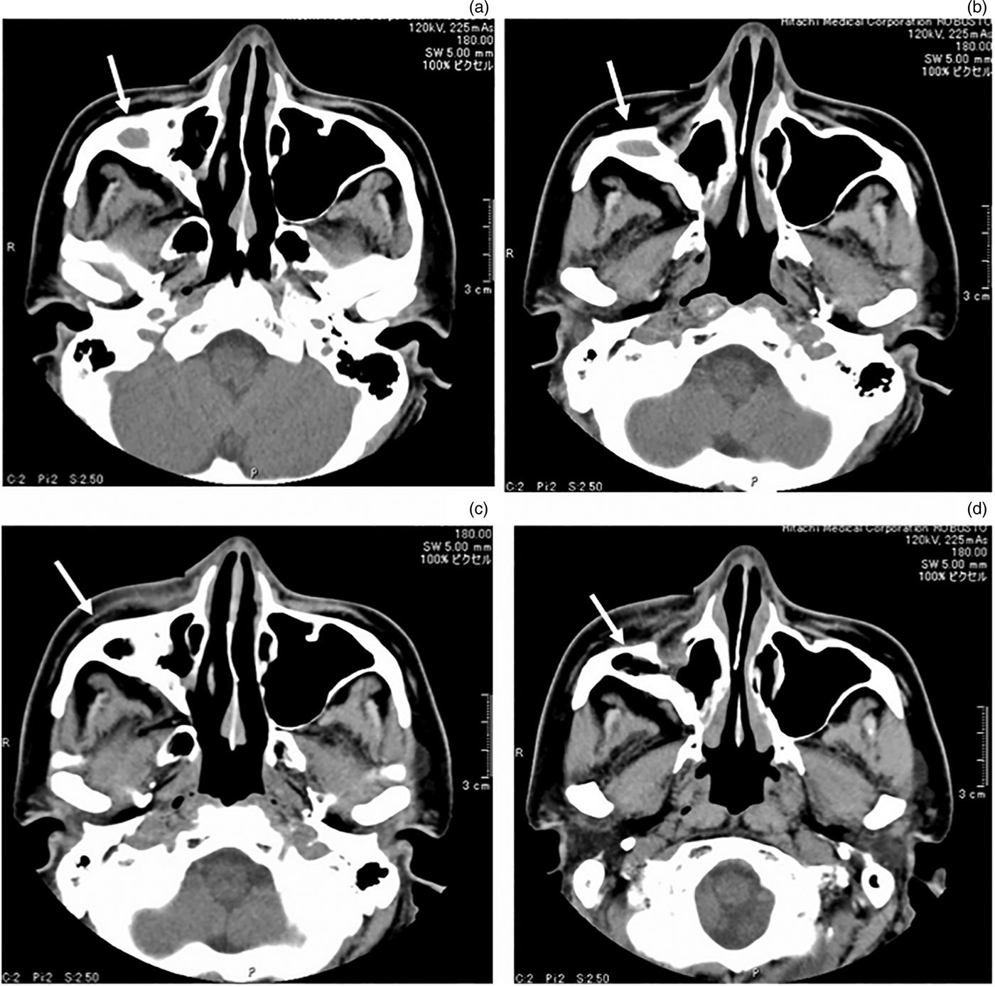
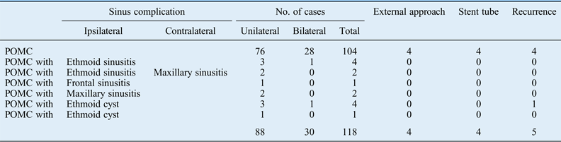
In total, 148 procedures were performed on post-operative maxillary cysts which comprised 43 left lateral, 45 right lateral and 30 bilateral lesions. Significant cranial extension towards the orbit with osteolysis at the orbital floor was identified in nine patients (7.6 per cent). Five patients (4.2 per cent) had multiple cysts, and nine patients (7.6 per cent) had post-operative maxillary cysts with ethmoid sinusitis. Post-operative maxillary cyst with subcutaneous abscessation was present in five patients (4.2 per cent). A transnasal endoscopic approach was performed in 144 cysts (97.3 per cent). For the remaining cysts (4 of 148 cysts), a combined endonasal endoscopic and canine fossa (external) approach was performed because the cysts were located in areas that were too distant from the nasal cavity and had a thick bony wall (Table I, Figures 1 and 2). A ventilation stent was placed in four patients (four cysts), two of whom underwent the transnasal procedure and the other two the combined procedure, to avoid post-operative meatal antrostomy stenosis (Figures 1 and 2). Recurrence was observed in five patients (4.2 per cent), all of whom underwent additional transnasal endoscopic surgery (5 of 148 lesions, 3.4 per cent). No further recurrence was noted in these five patients after revision surgery.
Axial non-contrast computed tomography (CT) image of a case treated with canine fossa approach. (a, b) Pre-operative CT demonstrates a cyst (arrow) in the right buccal region, in areas too distant from the nasal cavity and has a thick bony wall. (c,d) Post-operative CT images at one year after surgery.

Image analysis of a case in which a ventilation stent was placed. (a,b) Pre-operative computed tomography (CT) images. The soft tissue CT window demonstrates a cyst in the left buccal region, which has a bony and thick wall (white arrow). (c) A ventilation stent (black arrow) was placed spanning the cyst cavity and inferior meatus. (d) Cyst cavity with the T-shaped ventilation stent (white arrow).
Clinical characteristics of post-operative maxillary cyst (POMC) repair
POMC = post-operative maxillary cyst
Early post-operative complications occurred in two cases: buccal haematoma and hard palate fistula. These complications resolved within one month. Post-operative epistaxis was not observed.
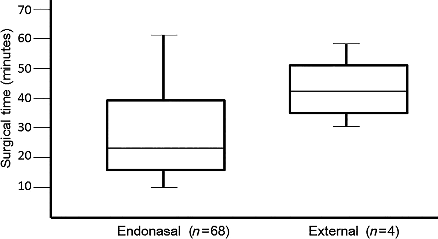
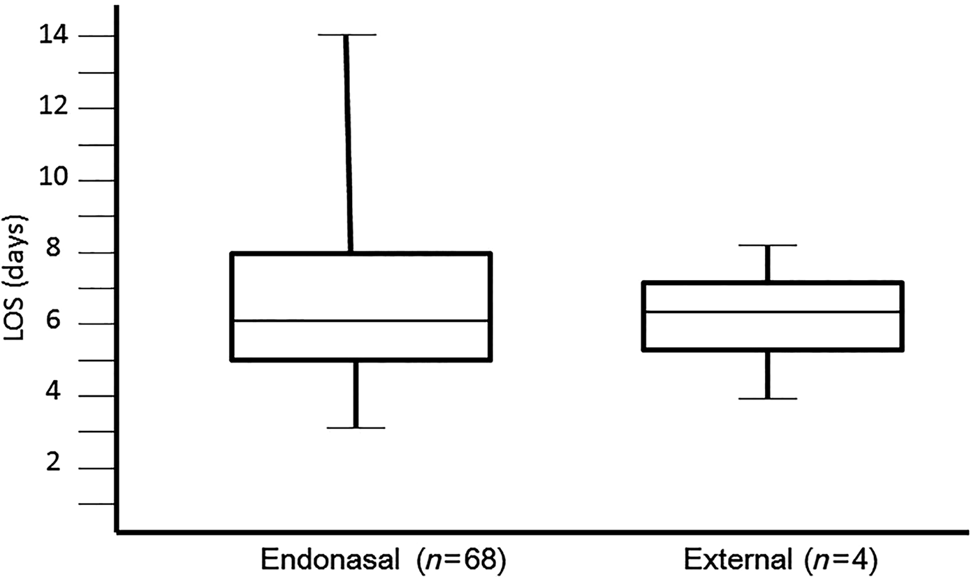
Patients stayed at the hospital for an average of 6 days. The length of stay at the hospital ranged from 3 to 18 days, and the mean length of stay was 6 days. Hospital discharge was delayed due to post-operative complications, including buccal haematoma and hard plate fistula, personal or family reasons, as well as insurance and medical complications involving diabetes mellitus and heart angina. The mean FESS surgical duration was 28 minutes (range, 9–63 minutes; median, 24 minutes). In contrast, the mean surgical duration for the external approach was 42 minutes (range, 29–56 minutes; median, 41 minutes). Although the surgical duration was significantly shorter for FESS compared with the external approach (p < 0.05, Figure 3), there was no significant difference in the length of stay between the FESS and external approaches (Figure 4).
Relationships between the surgical duration and surgical approaches. Box plot comparing the surgical duration (minutes) in endonasal (left) and external (right) procedures.
Relationships between length of stay (LOS) at the hospital and surgical approaches. Box plot comparing the LOS (days) at the hospital for endonasal (left) and external (right) procedures.
Discussion
Post-operative maxillary cyst was first described by Kubo in 19277 in a 26-year-old man diagnosed with a left lateral buccal cyst who previously underwent radical bilateral maxillary sinus surgery (the Caldwell–Luc procedure). Post-operative maxillary cyst was considered a late complication of radical maxillary sinus surgery, and the buccal cyst was attributed to residual maxillary sinus mucosa trapped within the cheek, or fluid or blood retention creating a tissue space post-operatively.Reference Kubo7
In Japan, post-operative maxillary cyst is the most common cause of sinus mucocele.Reference Kaneshiro, Nakajima, Yoshikawa, Iwasaki and Tokiwa4–Reference Ikeda, Takahashi, Oshima, Suzuki, Satake and Hidaka6 Tsuzuki et al. reported post-operative maxillary cysts in 173 of 218 (79.4 per cent) patients with paranasal sinus cysts.Reference Tsuzuki, Fukazawa, Takebayashi, Oka and Sakagami8 Post-operative maxillary cyst is diagnosed based on clinical symptoms, previous maxillary surgery, radiography and histology.Reference Nishioka, Pittella, Hamagaki, Okada and Takagi5, Reference Huang, Chen, Lee, Chang, Chen and Chen9 Computed tomography examination and magnetic resonance imaging are also useful for the diagnosis of post-operative maxillary cyst.Reference Chindasombatjaroen, Uchiyama, Kakimoto, Murakami, Furukawa and Kishino10, Reference Ishihara and Muraoka11
Post-operative maxillary cyst occurs 10–30 years following maxillary surgery, especially following the Caldwell–Luc procedure to treat chronic maxillary sinusitis. The Caldwell–Luc procedure had been widely used to treat chronic sinusitis, before FESS with the middle meatal approach, which is now the standard procedure, was developed. Functional endoscopic sinus surgery was devised based on studies demonstrating microciliary flow through the natural maxillary ostium. Therefore the prevalence of post-operative maxillary cyst in Japan should decrease in the near future.
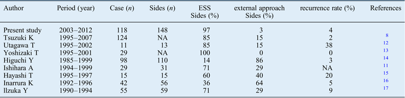
Previously, there were two approaches for post-operative maxillary cyst surgery. One was an open approach with the Caldwell–Luc procedure, which entailed complete excision of the mucocele lining through the inferior nasoantral window. The other was an intranasal approach involving complete marsupialisation. In recent Japanese reports, the use of complete mucocele lining excision (the canine fossa approach) has decreased, and FESS has become the widely accepted treatment for post-operative maxillary cyst due to the development of new instruments or power toolsReference Tsuzuki, Fukazawa, Takebayashi, Oka and Sakagami8, Reference Utagawa, Ynanagi, Ishii and Imai12–Reference Murata, Tomoda, Kitani and Yamashita18 (Table II). The present study showed that 97.3 per cent of cysts (144 cysts) were treated by the transnasal endoscopic approach. When cysts were located distant from the nasal cavity and had a thick bony wall, an external approach (the canine fossa approach) was required. Although post-operative maxillary cyst surgery was performed without a navigation system, it should be possible to excise thick-walled cysts by FESS using a navigation system, avoiding the external approach entirely. Murata et al. showed that indications for the use of a navigation system during post-operative maxillary cyst surgery were as follows: a thick bony cyst wall, multiple cysts, anterior, lateral, or infra-orbital lesion and large distance between the cyst and infra- or middle meatus.Reference Ikeda, Hirano, Oshima, Shimomura, Suzuki and Sunose19
Summary of the previous reports on post-operative maxillary cysts in japan
ESS = endoscopic sinus surgery; External approach = canine fossa approach
Although there was no significant difference in the length of hospital stay between FESS and external approaches, the surgical duration was shorter for FESS. The FESS procedure offers additional benefits, including decreased haemorrhage and complications.Reference Ikeda, Hirano, Oshima, Shimomura, Suzuki and Sunose19 The primary post-operative complication following FESS to treat post-operative maxillary cyst was meatal antrostomy stenosis.Reference Utagawa, Ynanagi, Ishii and Imai12, Reference Inamura, Kawai and Aoyagi16, Reference IIzuka, Fukami, Yanagi, Asai, Ohtori and Moriyama17 Inserting a T-shaped ventilation stent between the cyst and the nasal cavity proved to be an effective option for avoiding this complication. In the present study, the ventilation stent was placed in four patients (four cysts) to avoid post-operative meatal antrostomy stenosis, and recurrence was not observed in these patients.
• Transnasal endoscopic surgery is an effective treatment for post-operative maxillary cyst with the exception of cysts located distant from the nasal cavity
• Furthermore, 97.3 per cent of post-operative maxillary cysts are treatable using the transnasal endoscopic approach alone
Recently, 3D CT examination has provided a powerful tool in evaluating post-operative maxillary cyst before FESS. Using a plastic bone model created using 3D printer technology, FESS can be simulated pre-operatively. Combining these powerful tools and technologies, including the navigation system, will enable FESS without an external approach.
In conclusion, the present study showed that the use of the transnasal endoscopic approach alone was possible to treat 97.3 per cent of cysts. Transnasal endoscopic surgery is an effective treatment for post-operative maxillary cyst, with the exception of cysts located distant from the nasal cavity.
Acknowledgement
We thank Dr Masaharu Washizaki (Washizaki ENT clinic) for his excellent technical assistance.
Financial support
This research received no specific grant from any funding agency, commercial or not-for-profit entity.
Ethical standards
The authors assert that all procedures contributing to this work comply with the ethical standards of the relevant national and institutional guidelines on human experimentation (Oda Hospital Institutional Review Board 2009) and with the Helsinki Declaration of 1975, as revised in 2008.