Despite excellent long-term survival rates,Reference Murphy, Gersh and Mair 1 , Reference Nollert, Fischlein, Bouterwek, Böhmer, Klinner and Reichart 2 most patients with tetralogy of Fallot are left with substantial pulmonary regurgitation following surgical repair with its consequent long-term deleterious effects.Reference Bouzas, Kilner and Gatzoulis 3 , Reference Gatzoulis, Balaji and Webber 4 Cardiac MRI is the gold standard for evaluating anatomy, ventricular function, right ventricular mass, and valvar insufficiency following surgical repair for tetralogy of Fallot and has been used mostly during long-term follow-up to guide the decision to intervene on the pulmonary valve.Reference Geva, Sandweiss, Gauvreau, Lock and Powell 5 – Reference Rebergen, Chin, Ottenkamp, Van der Wall and De Roos 10 Although perioperative outcomes have been described in tetralogy of Fallot, there is little information about the early ventricular adaptations, including pulmonary regurgitation, which occur immediately following surgical repair; these may be important to understanding the changes that occur later in life and put the magnitude of these alterations into perspective.Reference Pigula, Khalil, Mayer, Pedro and Jonas 11 – Reference Mercer-Rosa, Pinto, Yang, Tanel and Goldmuntz 13 The feed and sleep technique is an anaesthesia-free method for performing cardiac MRI, and has been used in select patients with proven utility in the assessment of intra- and extra-cardiac anatomy in certain CHDs.Reference Fogel, Pawlowski and Harris 14 – Reference Windram, Grosse-Wortmann, Shariat, Greer, Crawford and Yoo 16
The objectives of our study were to investigate right ventricle remodelling in terms of right ventricular function, pulmonary regurgitation, pulmonary artery anatomy, and residual lesions in the early postoperative period in patients operated for tetralogy of Fallot and to investigate the feasibility of the feed and sleep technique to obtain this information in postoperative neonates and infants.
Materials and method
Subjects were recruited sequentially following complete surgical repair for tetralogy of Fallot between March, 2013 and January, 2016. Institutional Review Board approval and parental informed consent were obtained before cardiac MRI exams. The medical history was reviewed to assess patient safety before undergoing unsedated cardiac MRI. Inclusion criteria were the diagnosis of tetralogy of Fallot and under 1 year of age at the time of enrolment. Patients requiring mechanical ventilation, medically unstable at scheduled time of cardiac MRI, or those with contraindications to cardiac MRI, such as pacemakers, were excluded. Complete surgical repair was defined as ventricular septal defect closure with or without right ventricular outflow tract intervention – transannular patch or right ventricle to pulmonary artery conduit. Protocol-based cardiac MRIs were performed with the feed and sleep technique just before hospital discharge, at a time when the patient was deemed stable to undergo the study without sedation. If the patient was discharged on a weekend before scheduling the cardiac MRI, the test was performed as soon as possible after hospital discharge.
The cardiac MRI protocol used has been previously described.Reference Mercer-Rosa, Pinto, Yang, Tanel and Goldmuntz 13 Briefly, subjects were fed 3–4 hours before the planned initiation of the procedure and fasted up until their cardiac MRI. Immediately before the procedure, electrocardiographic leads were placed, the patient was fed, swaddled in an infant blanket, and placed in a vacuum immobiliser (MedVac bag; CFI Medical Solutions/Contour Fabricators, Fenton, Michigan, United States of America). After a pulse oximeter probe (Invivo Corporation, Gainesville, Florida, United States of America) and ear muffs (Natus MiniMuffs; Natus Medical Inc, San Carlos, CA, United States of America) were applied, the lights were dimmed in the scanner to minimise stimuli. The study proceeded once the patient fell asleep or remained motionless. Cardiac MRI sequences were prioritised to assess anatomy using static steady state free precession axial imaging; ventricular function using cine steady state free precession four-chamber and short axis; and flow assessments using phase-contrast cardiac MRI across the aortic root and main and branch pulmonary arteries. An exam was considered complete if the intra-cardiac anatomy, right and left ventricular ejection fraction, and pulmonary regurgitant fraction could be assessed. Contrast was not used as part of the study protocol as subjects were preparing for hospital discharge and therefore did not have intravenous lines. Pulmonary regurgitation was considered moderate if the regurgitant fraction was >20% and severe if the regurgitant fraction was >40%. Right ventricular mass z score was calculated based on normative data in children and main, right, and left pulmonary artery dimensions were indexed to (body surface area)0.5 based on the linear relationship between vascular size and this indexing method.Reference Buechel, Kaiser, Jackson, Schmitz and Kellenberger 17 , Reference Sluysmans 18
Statistical analysis
Continuous variables were described using mean with standard deviation or median with interquartile range, if not normally distributed. Categorical variables were described with counts and percentages. For all analyses, statistical significance was indicated by a two-sided p value <0.05. Pearson’s or Spearman’s correlation coefficients were determined to assess the association between cardiac MRI variables.
Results
Baseline data
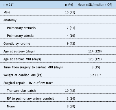
Cardiac MRI was attempted in 21 subjects. Baseline demographics for these patients are listed in Table 1. Five patients did not complete the study owing to inability to remain motionless for the entire exam. in total, 16 patients (76%) completed the cardiac MRI protocol using the feed and sleep technique. Of those, nine (60%) were asleep for the entire exam, two (13%) were asleep for part of the exam, and four (27%) were awake throughout the exam. One patient received sedation for another procedure before cardiac MRI, which was then performed during recovery. The median age at cardiac MRI for the entire group (n=21) was 123 days, with an interquartile range of 121 and a mean weight of 5.2±1.7 kg. Those who could not complete the study were older at the time of attempted cardiac MRI as compared with those who could complete the study – 174 days, with an interquartile range of 23, versus 77, with an interquartile range of 114, respectively, p=0.03.
Baseline demographic data.

IQR=interquartile range; RV=right ventricle
* Includes one patient recovering from sedation during the start of the cardiac MRI
Right ventricular remodelling
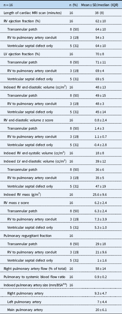
Among the 16 patients who completed the study, there was overall normal ventricular function, indexed right ventricular volumes, and right ventricular volume z scores. There was on average moderate pulmonary regurgitation among the 11 subjects requiring right ventricular outflow tract intervention – transannular patch or right ventricle to pulmonary artery conduit; the regurgitant fraction was 27±16%, as shown in Table 2. Of this group, four (36%) had moderate regurgitation and two (11%) had severe regurgitation. Subjects requiring only ventricular septal defect closure had mild regurgitation. There was overall significant right ventricular hypertrophy, with a right ventricular mass z score of 6.2±2.4, as shown in Table 2. Subjects had a well-balanced circulation with a pulmonary to systemic blood flow ratio of 0.9±0.2. A small residual ventricular septal defect was present in three of 10 subjects. There was no association between pulmonary regurgitant fraction and indexed right ventricular end-diastolic volume (r= 0.19, p=0.5) or right ventricular mass z score (r=−0.19, p=0.5).
Cardiac MRI data.

BSA=body surface area; IQR=interquartile range; LV=left ventricle; RV=right ventricle
Discussion
Pulmonary regurgitation resulting from surgical repair is the main determinant of outcome in tetralogy of Fallot, in particular with regard to its deleterious effect on the right ventricle. Characterisation of right ventricular remodelling by cardiac MRI early after surgical repair for tetralogy of Fallot has not been described. In this study, we sought to investigate the early right ventricular remodelling in patients operated for tetralogy of Fallot and to test the feasibility of unsedated cardiac MRI to obtain this information. The study found that neonates and infants have moderate pulmonary regurgitation and increased right ventricular mass with normal volumes and function. The feed and sleep technique allows for complete cardiac assessment in tetralogy of Fallot patients less than 5 months of age in the early postoperative period.
Right ventricular remodelling
There was overall moderate pulmonary regurgitation among those requiring a transannular patch or right ventricle to pulmonary artery conduit. Other reports have also demonstrated moderate pulmonary regurgitation in children and adolescents with tetralogy of Fallot.Reference Geva, Sandweiss, Gauvreau, Lock and Powell 5 , Reference Niezen, Helbing, Van der Wall, Van der Geest, Rebergen and De Roos 8 , Reference Lee, Lee and Kwak 19 , Reference Mercer-Rosa, Paridon and Fogel 20 The determinants of pulmonary regurgitation are multifactorial,Reference Chaturvedi and Redington 21 , Reference Redington 22 and include right ventricular compliance, pulmonary vascular resistance, heart rate, and orifice size. Although we cannot comment on the longitudinal changes in pulmonary regurgitation based on these comparisons, our findings would suggest that the degree of pulmonary regurgitation might be established early postoperatively. Longitudinal assessment of pulmonary regurgitation is thus necessary to further elaborate changes in the long term.
Right ventricular mass was significantly increased in the early postoperative period, which is a reflection of insufficient time for right ventricular remodelling in response to relief of the outflow tract obstruction; right ventricular hypertrophy is thus most likely a reflection of the preoperative physiology. Right ventricular mass could be an important parameter to monitor in this patient population, and follow-up studies to determine the longitudinal changes in right ventricular mass, and physiologic drivers of these changes, are necessary.
Right ventricular volumes were within normal limits in our cohort, most likely owing to the early interval between surgery and cardiac MRI. As such, there was no association between right ventricular end-diastolic volume and pulmonary regurgitant fraction. This may suggest that the right ventricle does not dilate immediately, but rather remodels over the course of a number of years.
Even though on average mild residual disease was present early postoperatively, some patients demonstrated significant pulmonary regurgitation, and they may be at risk for accelerated right ventricular dilation and early dysfunction and should therefore be carefully monitored.
This study determined a number of parameters that can be used as a reference. Pulmonary artery size and architecture was obtained, which can be used as a baseline for future assessments; branch pulmonary artery stenosis is a potentially manageable residual lesion that has deleterious effects if not addressed.Reference Redington 22 , Reference Harris, Whitehead and Gillespie 23 Right ventricular function was also obtained early after surgery. Better understanding of the early right ventricular remodelling after tetralogy of Fallot repair may provide insight into postoperative management strategies and guide follow-up. Early postoperative cardiac MRI could be of potential clinical use, as it can establish the burden of residual lesions, define the branch pulmonary artery anatomy, and help identify patients who could benefit from early follow-up and interventional procedures.
Feed and sleep technique
The feed and sleep technique provides a complete cardiac assessment in postoperative neonates and infants with tetralogy of Fallot. This technique was previously shown to be feasible in brain and extra-cardiac MRIs, as well as in cardiac MRIs that had specific anatomic questions.Reference Fogel, Pawlowski and Harris 14 – Reference Windram, Grosse-Wortmann, Shariat, Greer, Crawford and Yoo 16 , Reference Rangamani, Varghese and Li 24 To our knowledge, our study is the first attempt to extend the use of the feed and sleep technique to perform a comprehensive cardiac MRI evaluation in tetralogy of Fallot. A complete data set was obtained in ~75% of cardiac MRIs in this study. The failure rate is multifactorial, and includes greater time to obtain a full protocol, attempting feed and sleep in an older age group, and having all of our patients scanned before discharge. Indeed, three of the infants who failed feed and sleep had an attempted cardiac MRI within 5 days of surgery, and thus it is possible that postoperative pain may have contributed to an inability to fall asleep or remain motionless during the scan. Because our study protocol sought to perform cardiac MRI before discharge, the window for performing a study for research purposes was quite narrow and dependent on both the patient’s stability and schedule availability.
This study demonstrates that the feed and sleep technique is feasible in postoperative patients who are younger than 5 months of age. One advantage of this technique is that sedation is not required, and although sedation is generally safe it does carry some risks. In addition, the feed and sleep allows for normal physiology to be present, which could be distorted by sedation with or without intubation and mechanical ventilation, is relatively fast, and complete data sets can be obtained using the duration of the child’s nap.Reference Rangamani, Varghese and Li 24 , Reference Dorfman, Odegard, Powell, Laussen and Geva 25 Although we did not investigate the cost-effectiveness of this technique, a non-sedated protocol is probably cost-effective, given that the required support staff is limited to the bedside nurse and cardiac MRI personnel, and post-procedure recovery is unnecessary.Reference Windram, Grosse-Wortmann, Shariat, Greer, Crawford and Yoo 16
Limitations
This was a prospective pilot study; therefore, we are limited by a small sample size. Thus, we cannot comment on the statistical significance of differences in pulmonary regurgitation or right ventricular volume based on anatomy or type of surgical repair owing to a lack of power. However, the goal of the study was to demonstrate the right ventricular changes that occur early after tetralogy of Fallot repair and to test the feasibility of the feed and sleep technique. Further studies with larger sample sizes are necessary to address these questions. Because this analysis was restricted to patients who were clinically stable and ready for discharge, our sample may not have included those with more significant disease who may not have been stable for examination, or those who had less significant disease and early hospital discharge.
Conclusions
Early right ventricular remodelling after surgical repair for tetralogy of Fallot is characterised by significant pulmonary regurgitation, right ventricular hypertrophy, and lack of dilation. Longitudinal examination in this cohort could provide further insight into right ventricular remodelling and help identify a subset of children at particular risk for deleterious consequences. Unsedated cardiac MRI is a feasible approach for infants less than 5 months of age and could be considered in other forms of CHDs.
Acknowledgements
None.
Financial Support
This work was supported by the National Institutes of Health [M.P.D, NIH grants T32 HL007915; L.M.R, K01HL125521], and Pulmonary Hypertension Association supplement to K01HL125521 (L.M.R).
Conflicts of Interest
None.
Ethical Standards
The authors assert that all procedures contributing to this work comply with the ethical standards of Ethical Advisory Board and the National Institute of Health, and with the Helsinki Declaration of 1975, as revised in 2008, and has been approved by the institutional committee (Institutional Review Board, Children’s Hospital of Philadelphia).