


Introduction
The genus Helicotylenchus Steiner, 1945 (Nematoda: Hoplolaimidae) represents one of the most diverse and widely distributed groups of plant-parasitic nematodes, with around 227 nominal species described worldwide (Mokhtari et al. Reference Mokhtari, Bazeghi, Ruiz-Cuenca, Palomares-Rius, Eskandari and Pedram2026; Sher Reference Sher1966; Uzma et al. Reference Uzma, Nasira, Firoza and Shahina2015). Species of this genus are commonly associated with agricultural and natural ecosystems, where they parasitise roots of a wide range of host plants, occasionally causing economic damage (Maina et al. Reference Maina, Sulaiman and Musa2023; O’Bannon and Inserra Reference O’Bannon and Inserra1989; Shokoohi Reference Shokoohi2025). Despite their ecological and agricultural importance, species delimitation within Helicotylenchus remains challenging due to high morphological conservatism, overlapping morphometric characters, and intraspecific variability, particularly in tail morphology and stylet features (Fortuner and Wong Reference Fortuner and Wong1984; Mwamula et al. Reference Mwamula, Kwon, Kwon, Kim, Kim and Lee2024; Subbotin et al. Reference Subbotin, Inserra, Marais, Mullin, Powers, Roberts, van den Berg, Yeates and Baldwin2011; Reference Subbotin, Vovlas, Yeates, Hallmann, Kiewnick, Chizhov, Manzanilla-López, Inserra and Castillo2015).
Traditionally, species identification in Helicotylenchus has relied primarily on morphological and morphometric characters, such as lip region configuration, stylet length and shape, lateral field incisures, position of phasmids, tail shape, and the presence or absence of a mucro (Sher Reference Sher1966; Siddiqi Reference Siddiqi2000; Uzma et al. Reference Uzma, Nasira, Firoza and Shahina2015). However, in some cases, some species exhibit close morphological/morphometric features, leading to taxonomic ambiguities and frequent misidentifications. In recent decades, the integration of molecular data, especially ribosomal DNA markers such as 18S rRNA gene, D2–D3 expansion segments of 28S rRNA gene, and ITS rDNA regions, has greatly improved species delimitation and phylogenetic inference within the genus (Pour Ehtesham et al. Reference Pour Ehtesham, Azimi and Pedram2021; Subbotin et al. Reference Subbotin, Sturhan, Chizhov, Vovlas and Baldwin2006, Reference Subbotin, Inserra, Marais, Mullin, Powers, Roberts, van den Berg, Yeates and Baldwin2011, Reference Subbotin, Vovlas, Yeates, Hallmann, Kiewnick, Chizhov, Manzanilla-López, Inserra and Castillo2015).
To date, 34 species of Helicotylenchus have been recorded from Vietnam (Nguyen et al. Reference Nguyen, Trinh, Nguyen, Le and Bert2024a). Several species are recognised as economically important plant-parasitic nematodes, capable of causing severe root damage and exhibiting high pathogenic potential on a wide range of cultivated crops in Vietnam (Nguyen et al. Reference Nguyen, Nguyen and Trinh2024b). Lai Chau ginseng (Panax vietnamensis var. fuscidiscus) is an endemic medicinal plant of high economic and conservation value, cultivated mainly in mountainous regions of northern Vietnam (Le et al. Reference Le, Nguyen, Pham, Le, Luong, Huynh, Nguyen, Nong, Teixidor-Toneu, de Boer and Manzanilla2022). In particular, information on the diversity and composition of nematode species inhabiting the rhizosphere of ginseng is still scarce. In Vietnam, research on nematodes associated with ginseng has been especially limited, and published reports are largely lacking. Investigating nematode diversity in ginseng-growing areas is therefore essential for improving our understanding of the nematode community structure associated with this economically important crop and for identifying potential plant-parasitic species that may affect ginseng production. During a survey of plant-parasitic nematodes associated with Lai Chau ginseng in Lai Chau Province, a population of Helicotylenchus was recovered whose morphological characteristics did not match any known species key or original description. From a molecular perspective, it should be noted that unless all species within the genus have been sequenced, molecular data alone cannot definitively confirm a species as new.
The objectives of the present study are to provide a comprehensive description of this new species, Helicotylenchus laichauensis sp. n., using an integrative taxonomic approach based on morphology, morphometrics, and molecular data from three ribosomal RNA markers.
Materials and methods
Sampling and nematode extraction
Soil and root samples from Lai Chau ginseng (Panax vietnamensis var. fuscidiscus) growing areas in Lai Chau Province were collected randomly, placed in separate nylon bags, and transported to the laboratory. The decanting and sieving process was used to recover nematodes from soil samples (Nguyen Reference Nguyen2003). For extracting nematodes from the roots, root samples were divided into 0.5-cm pieces and put on a tray using the modified Baermann tray method described by Whitehead and Hemming (Reference Whitehead and Hemming1965).
Morphological characterisation
Light microscopy: Nematodes were killed by immersing them in hot water (60–70 °C) for 30 seconds. They were then fixed in TAF solution (8% formalin and 2% triethanolamine in distilled water) for 4–5 days, following the procedure of Courtney et al. (Reference Courtney, Polley and Miller1955). Subsequently, the specimens were dehydrated using the method described by Seinhorst (Reference Seinhorst1959). Finally, a Carl Zeiss Axio Lab.A1 microscope was used to measure and photograph the nematodes.
Molecular techniques and polymerase chain reaction (PCR)
Live nematodes were cut into small fragments and put into PCR tubes for DNA extraction. Subsequently, 20 μL of worm lysis buffer (WLB) solution, which contained 50 mM KCl, 10 mM Tris pH 8.3, 2.5 mM MgCl2, 0.45% NP-40, and 0.45% Tween-20, was added. After that, the samples were incubated for at least 10 min at –20°C. Following the addition of 2 μL of proteinase K (1.2 mg mL−1), the samples were incubated for 1 h at 65°C and then for 10 min at 95°C in a PCR machine.
For amplification of the 18S, 28S and ITS rRNA were used as follow: for 18S rRNA the forward primer MN 18F (5′-CGCGAATRGCTCATTACAACAGC-3′) and reverse primer Nem_18S_R (5′-GGGCGGTATCTGATC GCC-3′) (Floyd et al. Reference Floyd, Rogers, Lambshead and Smith2005); for D2–D3, the forward primer D2A (5′–ACAAGTACCGTGGGGAAAGTTG–3′) and reverse primer D3B (5′–TCGG AAGGAACCAGCTACTA–3′) (Subbotin et al. Reference Subbotin, Sturhan, Chizhov, Vovlas and Baldwin2006); and for ITS rRNA the forward primer Vrain2F (5′-CTTTGTACACACCGCCCGTCGCT-3’) and reverse primer Vrain2R (5′-TTTCACTCGCCGTTACTAAGGGAATC-3’) (Vrain et al. Reference Vrain, Wakarchuk, Lévesque and Hamilton1992) were used. All PCR reactions contained 12.5 μL Hot start green PCR Master Mix (2x) (Promega, USA), 1 μL of each of the forward and reverse primer (10 μM each), 3 μL DNA template, and sterile Milli-Q water to a total volume of 25 μL. All PCR reactions were performed in SimpliAmp Thermal cycler (Thermo Fisher Scientific) as follows: an initial denaturation step at 95°C for 4 min, followed by 40 cycles at 95°C for 30 sec, 54°C for 30 sec, and 72°C for 60 sec with a final incubation for 5 min at 72°C. After amplification, the PCR products were visualised by running 1% agarose gel and purified by ExoSAP-IT™ (Thermo Fisher Scientific). The purified PCR products were sent to Apical Scientific Company in Selangor, Malaysia, for sequencing. The resulting sequences were deposited in the NCBI database under the accession numbers presented in the corresponding tree of each locus.
Sequence assembly of the forward and reverse reads was performed using ChromasPro. In the next step, sequences closely related to the query sequences were identified by performing a search using BLAST in the National Center for Biotechnology Information (NCBI) database. Separate datasets were constructed for D2–D3 expansion segments of 28S rRNA, nearly full-length 18S rRNA, and ITS rRNA sequences. Newly generated sequences of Helicotylenchus laichauensis sp. n. were aligned with selected sequences. Sequences were aligned using ClustalW implemented in BioEdit and the resulting alignments were manually edited to remove ambiguously aligned regions. Phylogenetic analyses were performed separately for the 18S rRNA, D2–D3 expansion domains of the 28S rRNA, and ITS rRNA datasets using the maximum likelihood (ML) method implemented in MEGA 12 (Kumar et al. Reference Kumar, Stecher, Suleski, Sanderford, Sharma and Tamura2024). The best-fitting nucleotide substitution model for each dataset was selected using the model selection function implemented in the software. Node support was assessed using 1,000 bootstrap replicates, and only bootstrap values greater than 50% are shown on the trees.
Results
Helicotylenchus laichauensis sp. n.
(Figures 1–3; measurements in Table 1)
Drawings of Helicotylenchus laichauensis sp. n. A: Entire body of male; B: Entire body of female; C: Pharynx; D: Anterior body region; E: Vulva region; F, G: Tail of female; H: Tail tip variation of female; I, J: Tail region of the male. (Scale bars: 10 μm).

Figure 1. Long description
The drawing consists of ten panels labeled A through J, each with a 10 mu m scale bar.
Panel A shows the entire body of a male nematode in a C-shaped curve.
Panel B shows the entire body of a female nematode in a spiral shape.
Panel C illustrates the pharynx region, showing the stylet, median bulb, and esophageal glands.
Panel D provides a close-up of the anterior body region, highlighting the heavily sclerotized cephalic framework and the dark, needle-like stylet.
Panel E displays the vulva region in a cross-section, showing the vaginal opening and surrounding cellular structures.
Panels F and G show detailed views of the female tail, featuring the anus and the pointed terminus.
Panel H presents six variations of the female tail tip, ranging from rounded to sharply pointed with irregular notches.
Panels I and J focus on the male tail region. Panel I shows the spicule and gubernaculum, while Panel J highlights the bursa, a fin-like extension of the cuticle used during mating, with a shaded area indicating internal musculature.
Micrographs of Helicotylenchus laichauensis sp. n. A: Entire body of female; B–D: Anterior end; E, F: Vulva region; G: Lateral field at midbody. H, I: Tail region of male; J, K: Tail region of the females. (Scale bars: 10 μm).

Figure 2. Long description
A collection of 11 light micrographs labeled A through K. Each panel includes a black vertical scale bar representing 10 micrometers.
* Panel A: A full-body view of a female nematode curved into a C-shape, showing the internal digestive and reproductive tracts.
* Panels B, C, and D: High-magnification views of the anterior end. Panel B shows the esophagus and nerve ring. Panels C and D provide close-ups of the cephalic region, highlighting the robust stylet and rounded head shape.
* Panels E and F: Detailed views of the mid-body vulva region, showing the transverse slit and internal uterine structures.
* Panel G: A close-up of the mid-body lateral field, displaying longitudinal lines or incisures along the cuticle.
* Panels H and I: The posterior end of a male nematode, showing the spicules and a pointed tail tip.
* Panels J and K: The posterior end of female nematodes, showing a shorter, more rounded tail shape compared to the male.
Female tail variation of Helicotylenchus laichauensis sp. n. (Scale bars: 10 μm).

Figure 3. Long description
A multi-panel figure containing twelve light micrographs labeled A through L arranged in three rows of four. Each panel displays a lateral view of the posterior end of a female nematode.
Row 1 (A to D):
- Panel A shows a tail with a rounded terminus and a small ventral projection.
- Panel B displays a slightly more tapered tail with a distinct mucro.
- Panel C and D show variations in the curvature of the dorsal side and the sharpness of the terminal point.
Row 2 (E to H):
- Panels E through H illustrate tails with prominent cuticular annulations along the ventral and dorsal margins. The tail shapes vary from hemispherical to slightly conoid, each ending in a small, variable process or mucro at the terminus.
Row 3 (I to L):
- Panels I through L show further variations in the tail tip morphology, including differences in the length and thickness of the terminal projection. Panel I and J highlight the internal granular structure of the body cavity proximal to the tail.
Each panel includes a vertical black scale bar in the bottom-left corner representing 10 micrometers. The cuticle is clearly visible as a translucent outer layer with rhythmic indentations (annules) along the edges of the tail.
Morphometric data of Helicotylenchus laichauensis sp. n. from fixed specimens. All measurements are in μm (except for ratio) and in the form: mean ± s.d. (range)

Table 1. Long description
The table presents morphometric data for fixed specimens of Helicotylenchus laichauensis sp. n. with measurements in micrometers. The columns are divided into Characters, Holotype (Female), and Paratypes (Female and Male). The sample size n is 29 for female paratypes and 28 for male paratypes.
Key measurements include:
* L (Body length): Holotype 612.1; Female paratypes 624 plus or minus 32.5; Male paratypes 519 plus or minus 21.9.
* V (or T): Holotype 62.8; Female paratypes 62.7 plus or minus 1.1; Male paratypes 39.6 plus or minus 3.
* Stylet length: Holotype 25.8; Female paratypes 25.6 plus or minus 0.6; Male paratypes 21.6 plus or minus 0.7.
* Tail length: Holotype 17.4; Female paratypes 17.4 plus or minus 1.1; Male paratypes 18.5 plus or minus 1.5.
* Spicule length: Male paratypes 21.2 plus or minus 0.9.
Other characters listed include ratios a, b, b prime, c, and c prime; O and m values; genital branch lengths (G 1 and G 2); lip dimensions; conus length; stylet knob dimensions; D G O; distances from the anterior end to the median bulb, nerve ring, secretory/excretory pore, cardia, and pharyngeal gland; cuticle annuli width; body diameters; spermatheca dimensions; mucro length; and number of tail annuli. Male-specific data includes testis length, spicule width, and gubernaculum length.
Female. Body shape generally C-shaped to open spiral when heat-killed. Lip region hemispherical, not set off from the rest of the body contour, with 4–5 lip annuli. Body annuli distinct, their width 1.7 (1.4–2.0) μm at midbody. Lateral field with four incisures, occupying about 20–30% of the corresponding body diameter and extending to terminal of tail. Inner lateral-field incisures fused distally at the tail end into an m shape. Stylet well developed, stylet knobs flattened to slightly indented anteriorly, 2.1 (1.7–2.5) μm high and 4.2 (3.2–4.8) μm wide, conus forming 51.5 (50.1–53.5)% of entire stylet length. Dorsal pharyngeal gland opening 11.0 (10.1–12.2) μm from the base of stylet knobs. Median bulb oval to round-shaped. Hemizonid distinct, usually about 1.5–2 annuli wide and situated 0–2 annuli anterior to the excretory pore. Excretory pore located anterior to the esophago-intestinal junction. Pharyngeal glands overlap the intestine ventrally or ventrolaterally, ventral overlap longer than dorsal overlap. Reproductive system with two functional genital branches of equal length, well developed and outstretched, oocytes in a single row, spermatheca rounded and filled with sperm. Vulva a transverse slit. Vagina well developed, occupying approximately 40–50% of the corresponding body width at the vulval level. Phasmids distinct, situated 3–7 annuli anterior to the anus. Tail short, conical, dorsally convex, terminus ventrally tapering to an irregularly annulated projection, smooth in 7 specimens, and mucronate in 22 specimens with a variably shaped mucro 1.1 (0.5–1.9) μm long. Ventral surface of the tail features 10 (8–12) annuli.
Males. Habitus C-shaped. Lip region hemispherical, bearing 4–5 annuli. Stylet knobs generally flattened to slightly indented anteriorly, conus 51.9 (50.3–53.5)% of entire stylet length. Dorsal pharyngeal gland opening 9.5 (8.1–10.6) μm from the base of stylet knobs. Median bulb small, oval to round-shaped. Hemizonid distinct, usually about 1.0–1.5 annuli wide and situated 0–2 annuli anterior to the excretory pore. Excretory pore located anterior to the pharyngo-intestinal junction. Pharyngeal glands overlap the intestine ventrally or ventrolaterally, ventral overlap longer than dorsal overlap. Testis extending anteriorly. Spicules paired, slightly arcuate, ventrally curved, 21.2 (19.0–22.5) μm long. Gubernaculum wedge-shaped, slightly curved, short, and slim. Bursa extending from proximal end opposite the retracted spicules to tail terminus. Phasmids pre-cloacal. Tail short, conical, with a curved, pointed terminus, bearing a variable mucro in all specimens, with a length of 2.2 (1.2–3.0) μm.
Diagnosis and relationships
Helicotylenchus laichauensis sp. n. is characterised by a hemispherical lip region not set off from the body contour and bearing 4–5 lip annuli; well-developed stylet with knobs flattened to slightly indented anteriorly, length ranges from 24.5 to 26.7 μm; the lateral lines in the tail region distally fused into an m-shaped pattern; a round spermatheca filled with sperm; males present; phasmids located 3–7 annuli anterior to the anus; c′ value 1.3 (1.2–1.4); tail short, conical, dorsally convex, tapering to a prominent ventral projection bearing a variable mucro.
Based on the overall morphology, particularly the lip region structure, stylet morphology, lateral field pattern, phasmid position, and tail configuration, Helicotylenchus laichauensis sp. n. most closely resembles H. microcephalus Sher, Reference Sher1966 , H. craigi Knobloch and Laughlin, Reference Knobloch and Laughlin1973 , H. oleae Inserra, Vovlas and Golden, Reference Inserra, Vovlas and Golden1979, and H. amplius Anderson and Eveleigh, Reference Anderson and Eveleigh1982.
Helicotylenchus laichauensis sp. n. is most closely related to H. microcephalus Sher, Reference Sher1966, morphologically and molecularly. However, the new species differs from H. microcephalus by having a hemispherical lip region with 4–5 annuli (vs truncated lip region), stylet knobs flattened to slightly indented anteriorly (vs rounded posteriorly and mostly flattened anteriorly), a spermatheca filled with sperm (vs indistinct or round, thick-walled and empty), males present (vs unknown), and a tail terminating in a prominent ventral projection bearing a variable mucro (vs small ventral projection, some of the specimens have a small spike on the tip of the projection).
Helicotylenchus laichauensis sp. n. resembles H. craigi Knobloch and Laughlin, Reference Knobloch and Laughlin1973, in having a hemispherical lip region with four annuli, stylet knobs with slightly indented anterior surfaces and a tail tapering to a pronounced ventral projection. However, H. laichauensis sp. n. differs in having a spermatheca filled with sperm (vs empty), males present (vs unknown), phasmids situated 3–7 annuli anterior to the anus (vs from two annuli posterior to six annuli anterior to anus), and the lateral field terminating in a characteristic m-shaped fusion pattern that has not been reported for H. craigi.
Helicotylenchus laichauensis sp. n. is also similar to H. oleae Inserra, Vovlas and Golden, Reference Inserra, Vovlas and Golden1979 in having a lip region with 4–5 annuli, stylet knobs flattened and indented anteriorly, and phasmids located anterior to the anus. However, the new species differs in its hemispherical lip region (vs bluntly conical and anteriorly flattened), spermatheca filled with sperm (vs empty), males present (vs unknown), and a prominent ventral tail projection bearing a variable mucro (vs tail gradually tapering to a dorsally curved terminus without a distinct ventral projection).
Helicotylenchus laichauensis sp. n. differs from H. amplius Anderson and Eveleigh, Reference Anderson and Eveleigh1982, in having a conical tail, tapering to a prominent ventral projection bearing a variable mucro (vs the tail bears a broadly rounded ventral projection with a terminal mucro), a spermatheca filled with sperm (vs not observed), males present (vs unknown), a C-shaped to open spiral-shaped (vs tight spiral when relaxed).
In addition to the morphologically closest species discussed above, Helicotylenchus laichauensis sp. n. also shares certain morphological features with H. neopaxilli Inserra, Vovlas and Golden, Reference Inserra, Vovlas and Golden1979, H. paxilli Yuen, Reference Yuen1964, H. persiaensis Kashi and Karegar, Reference Kashi and Karegar2014, H. wilmae Marais, Gaidashova and Tiedt, Reference Marais, Gaidashova and Tiedt2008, H. valdeclarus (Orton Williams Reference Orton Williams1983) Ebsary, 1991, and H. verecundus Zarina and Maqbool, Reference Zarina and Maqbool1991. These species possess one or more characters in common with the new species, such as a modified tail terminus with a ventral or terminal projection, phasmids situated anterior to the anus, a well-developed stylet, and a lateral field with four incisures. Nevertheless, they differ from H. laichauensis sp. n. in important qualitative characters, including the shape and annulation of the lip region, stylet knob morphology, spermathecal condition, occurrence of males, the termination pattern of the lateral field, and the morphology of the tail projection or mucro.
Helicotylenchus neopaxilli Inserra, Vovlas and Golden, Reference Inserra, Vovlas and Golden1979 differs from H. laichauensis sp. n. by having a conical, truncate lip region (vs hemispherical), rounded stylet knobs (vs flattened to slightly indented anteriorly), a spermatheca without sperm (vs filled with sperm), males unknown (vs present), and a tail with a ventral extension only occasionally bearing a small mucro (vs a prominent ventral projection bearing a distinct, variable mucro).
Helicotylenchus paxilli Yuen, Reference Yuen1964 differs from H. laichauensis sp. n. in having a bulbous lip region with six annuli (vs hemispherical with 4–5 annuli), massive stylet knobs (vs flattened to slightly indented), lateral fields areolated anteriorly (vs not areolated), a digitate tail projection with only an occasional terminal mucro (vs a conical tail terminating in a prominent ventral projection bearing a variable mucro), and males unknown (vs present).
Helicotylenchus persiaensis Kashi and Karegar, Reference Kashi and Karegar2014 differs from H. laichauensis sp. n. by having an almost conical, truncate lip region (vs hemispherical), an empty spermatheca (vs filled with sperm), males unknown (vs present), the inner lateral-field incisures terminating in a V- or U-shaped pattern (vs m-shaped), phasmids situated only 1–3 annuli anterior to the anus (vs 3–7 annuli), and a broadly rounded tail terminus with only a slight projection (vs a prominent ventral projection bearing a variable mucro).
Helicotylenchus wilmae Marais, Gaidashova and Tiedt, Reference Marais, Gaidashova and Tiedt2008 differs from H. laichauensis sp. n. by having 10–11 lip annuli (vs 4–5), a spermatheca usually without sperm (vs filled with sperm), males extremely rare (vs consistently present), phasmids located 6–11 annuli anterior to the anus (vs 3–7 annuli), and a centrally positioned annulated tail projection, occasionally bearing a mucro (vs a ventral projection bearing a variable mucro).
Helicotylenchus valdeclarus (Orton Williams Reference Orton Williams1983) Ebsary, 1991 differs from H. laichauensis sp. n. by having a lip region lacking distinct annuli (vs 4–5 annuli), a reduced, non-functional posterior genital branch (vs two functional genital branches), lateral-field incisures that do not fuse on the tail (vs forming a distinct m-shaped pattern), and a tail terminating in a slender posterior process that may bear one or more fine spines (vs a ventral projection bearing a variable mucro). Males are unknown in H. valdeclarus but present in the new species.
Helicotylenchus verecundus Zarina and Maqbool, Reference Zarina and Maqbool1991 differs from H. laichauensis sp. n. by having 5–7 lip annuli (vs 4–5), the lateral field areolated on the anterior part of the body (vs not areolated), inner lines of lateral field fused in the tail region (vs forming a distinct m-shaped pattern), and a tail with a variable but non-mucronate terminus (vs bearing a distinct variable mucro).
Molecular characterisation
18S rRNA
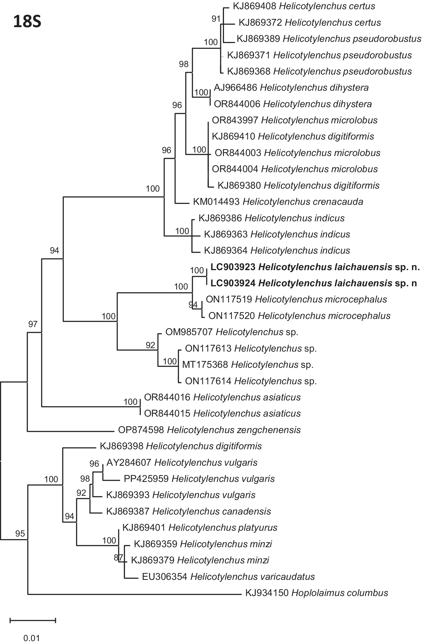
Two nearly full-length 18S rRNA gene sequences of Helicotylenchus laichauensis sp. n. were obtained and found to be identical. Both sequences were 1,663 bp long and deposited in GenBank under accession numbers LC903923 and LC903924. BLAST analysis showed that the 18S rRNA sequences of the new species had the highest identity (99.31%) to those of Helicotylenchus microcephalus from China (ON117520; ON117519). In the inferred phylogenetic tree, the 18S sequences of H. laichauensis sp. n. formed a distinct lineage clustered with the corresponding sequences of H. microcephalus with maximal bootstrap support (100%) (Figure 4).
Maximum Likelihood (ML) phylogenetic tree inferred from 18S rRNA gene sequences showing the phylogenetic position of Helicotylenchus laichauensis sp. n. among selected species of the genus Helicotylenchus. The tree was reconstructed under the TN93+ G nucleotide substitution model. Numbers to the left of the branches are bootstrap values for 1,000 replications. The sequences from this study are shown in bold.

Figure 4. Long description
A rectangular cladogram labeled 18 S at the top-left. The tree branches from a common root on the left toward specific species labels on the right.
* The top major clade includes species such as H. certus, H. pseudorobustus, H. dihystera, H. microlobus, H. digitiformis, H. crenacauda, and H. indicus, with bootstrap values ranging from 91 to 100.
* The middle clade contains the study’s focus species in bold: L C 9 0 3 9 2 3 Helicotylenchus laichauensis sp. n. and L C 9 0 3 9 2 4 Helicotylenchus laichauensis sp. n., which cluster with H. microcephalus (bootstrap 100) and other Helicotylenchus species (bootstrap 92 to 100).
* A separate branch below this includes H. asiaticus and H. zengchenensis.
* The bottom major clade includes H. digitiformis, H. vulgaris, H. canadensis, H. platyurus, H. minzi, and H. varicaudatus.
* The outgroup at the very bottom is K J 9 3 4 1 5 0 Hoplolaimus columbus.
* A scale bar at the bottom-left indicates 0.01 substitutions per site.
D2–D3 expansion domains of 28S rRNA region
Two D2–D3 28S rRNA gene sequences were obtained from two individuals of Helicotylenchus laichauensis sp. n. from Vietnam. Both sequences were 706 bp in length and differed from each other by only a single nucleotide over the aligned region. The sequences were deposited in GenBank under accession numbers LC903925 and LC903926.
BLAST searches revealed their highest identity (98.0–99.1%) with sequences of Helicotylenchus microcephalus from China (PP425970; MW716315; ON117539; MW716317). Pairwise sequence divergence between D2–D3 sequences of H. laichauensis sp. n. and H. microcephalus ranged from 0.7% to 2.0%. In contrast, sequence divergence between the new species and H. brevis (HM14299; HM14300) was considerably higher, ranging from 8.1% to 8.6%. The divergence value with sequences of other Helicotylenchus species available in GenBank was 8.8–15.2%.
The final alignment of the D2–D3 dataset comprised 53 sequences and 829 nucleotide positions. In the resulting phylogenetic tree, the two Vietnamese sequences of Helicotylenchus laichauensis sp. n. clustered together and formed a highly supported clade (bootstrap support = 100%) with the corresponding sequences of H. microcephalus from China. This lineage was recovered as a sister clade to a clade containing sequences of H. brevis (Figure 5).
Maximum Likelihood (ML) phylogenetic tree inferred from D2–D3 of 28S rRNA gene sequences showing the phylogenetic position of Helicotylenchus laichauensis sp. n. among selected species of the genus Helicotylenchus. The tree was reconstructed under the K2 + I + G nucleotide substitution model. Numbers to the left of the clades are bootstrap values for 1,000 replications. The newly generated sequences in this study are shown in bold.

Figure 5. Long description
A maximum likelihood phylogenetic tree based on D 2-D 3 expansion-segment sequences of 28 S r R N A. The tree begins with a root on the left and branches into several major clades.
* The top major clade includes species such as Helicotylenchus microlobus, H. paxilli, H. broadbalkiensis, H. platyurus, H. digonicus, and H. dihystera, with bootstrap values ranging from 88 to 100.
* The middle section contains clades for H. indicus, H. multicinctus, H. crenacauda, H. caudatus, H. labiodiscinus, and H. erythrinae.
* The lower section features a clade containing H. oleae and H. cuspicaudatus, followed by a group including H. sp., H. brevis, and the target species.
* The target species, Helicotylenchus laichauensis sp. n. (sequences L C 9 0 3 9 2 5 and L C 9 0 3 9 2 6), is shown in bold. It forms a sister group with a clade containing H. microcephalus (M W 7 1 6 3 1 5, O N 1 1 7 5 3 9, M W 7 1 6 3 1 7, and P P 4 2 5 9 7 0) with a bootstrap value of 98.
* Below this are clades for H. zengchenensis and H. asiaticus.
* The tree is outgrouped at the bottom by Scutellonema clavicaudatum and S. brachyurum.
* A scale bar at the bottom left indicates 0.05 substitutions per site. The label 28 S is positioned at the top left.
ITS rRNA
Two ITS rRNA gene sequences were obtained from two individuals of Helicotylenchus laichauensis sp. n. from Vietnam. Both sequences were 1,074 bp long. Pairwise comparison showed that the two ITS rRNA gene sequences were identical (100% sequence identity). The sequences were deposited in GenBank under accession numbers LC903927 and LC903928.
BLAST searches revealed that the ITS sequences showed 94.0–94.9% identity with sequences of Helicotylenchus microcephalus from China (GQ906355; MW714361; MW714362; OP329220).
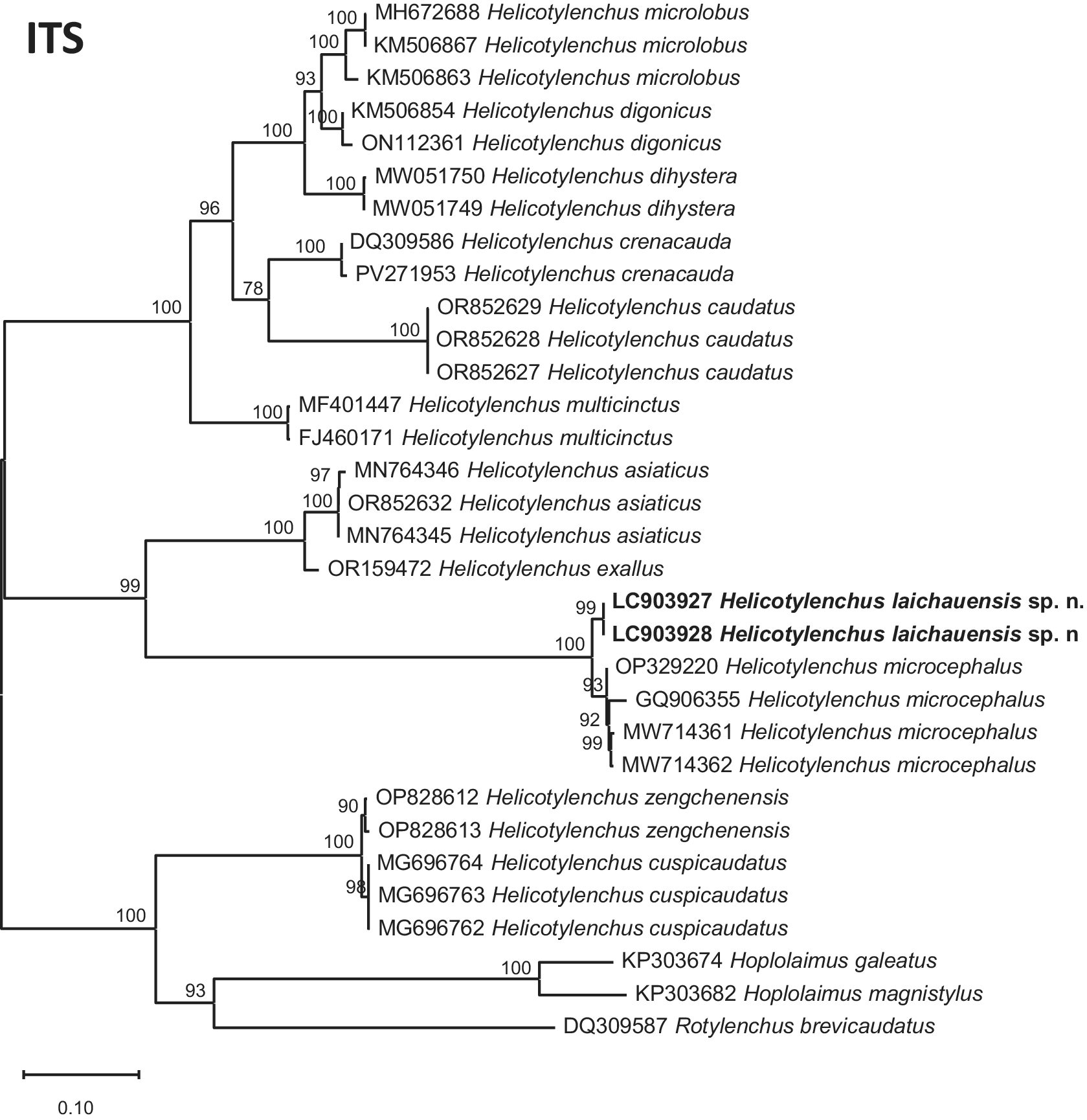
In the resulting tree, the two Vietnamese ITS sequences clustered together and formed a strongly supported clade with sequences of H. microcephalus (bootstrap support = 100%) that was clearly separated from the remaining Helicotylenchus sequences (Figure 6).
Maximum Likelihood (ML) phylogenetic tree inferred from ITS rRNA gene sequences showing the phylogenetic position of Helicotylenchus laichauensis sp. n. among selected species of the genus Helicotylenchus. The tree was reconstructed under the K2 + I + G nucleotide substitution model selected using the Akaike Information Criterion (AIC). Numbers to the left of the branches are bootstrap values for 1,000 replications. The sequences from this study are shown in bold.

Figure 6. Long description
The phylogenetic tree begins at a root on the left and branches rightward. Bootstrap values are indicated at each node.
Top Clade: Includes Helicotylenchus microlobus, Helicotylenchus digonicus, Helicotylenchus dihystera, Helicotylenchus crenacauda, and Helicotylenchus caudatus.
Middle Clades:
- A branch for Helicotylenchus multicinctus.
- A cluster containing Helicotylenchus asiaticus and Helicotylenchus exallus.
- A significant cluster containing the study sequences in bold: L C 9 0 3 9 2 7 Helicotylenchus laichauensis sp. n. and L C 9 0 3 9 2 8 Helicotylenchus laichauensis sp. n., which are sister to a group of Helicotylenchus microcephalus sequences.
Bottom Clades:
- A cluster containing Helicotylenchus zengchenensis and Helicotylenchus cuspicaudatus.
- An outgroup at the bottom includes Hoplolaimus galeatus, Hoplolaimus magnistylus, and Rotylenchus brevicaudatus.
A scale bar at the bottom left indicates 0.10 substitutions per site. The label I T S is positioned at the top left.
Type host and locality
The new species was extracted from the rhizosphere soil of Laichau ginseng (Panax vietnamensis var. fuscidiscus) in Lai Chau Province, Vietnam, at GPS coordinates N: 22°40′41′′, E: 102°34′42′′, altitude 1,891 m.
Type material
The holotype female (slide no. DL45-7703-1) and paratypes (slides nos. DL45-7703-2 to DL45-7703-4) are deposited in the Nematode Collection, Institute of Biology, Vietnam Academy of Science and Technology, 18 Hoang Quoc Viet, Nghia Do, Hanoi, Vietnam. ZooBank ID: urn:lsid:zoobank.org:pub:D080AF88-C9A3-48BA-B521-56F663D08105.
Etymology
The specific epithet, the Latin term laichauensis refers to the type locality of Lai Chau Province in Vietnam, where the species was found.
Discussion
The present study describes Helicotylenchus laichauensis sp. n., a new species associated with Lai Chau ginseng in northern Vietnam. This discovery increases the known diversity of Helicotylenchus in Vietnam and highlights the still insufficiently explored diversity of plant-parasitic nematodes associated with economically important and medicinal plants in the country. Although more than 30 species of Helicotylenchus have been reported from Vietnam, the nematode fauna associated with many native and cultivated hosts remains poorly documented. The discovery of a new species from a medicinal plant emphasises the need for further nematological surveys in both agricultural and natural ecosystems, which are likely to reveal additional undescribed taxa.
The taxonomy of Helicotylenchus remains challenging because many species share overlapping morphometric characters and exhibit considerable intraspecific variation, making species identification based solely on measurements unreliable (Fortuner and Wong Reference Fortuner and Wong1984; Rybarczyk-Mydłowska et al. Reference Rybarczyk-Mydłowska, Dmowska and Kowalewska2019; Subbotin et al. Reference Subbotin, Vovlas, Yeates, Hallmann, Kiewnick, Chizhov, Manzanilla-López, Inserra and Castillo2015). Consequently, qualitative morphological characters have long been regarded as more stable and taxonomically informative than morphometric traits for species delimitation within the genus (Fortuner and Wong Reference Fortuner and Wong1984; Subbotin et al. Reference Subbotin, Vovlas, Yeates, Hallmann, Kiewnick, Chizhov, Manzanilla-López, Inserra and Castillo2015; Uzma et al. Reference Uzma, Nasira, Firoza and Shahina2015). In the present study, the separation of H. laichauensis sp. n. from related species was supported primarily by qualitative morphological characters rather than by morphometric differences alone. The most informative diagnostic features include the hemispherical lip region with 4–5 annuli, stylet knobs flattened to slightly indented anteriorly, the characteristic m-shaped fusion of the inner lateral-field incisures on the tail, the presence of a spermatheca filled with sperm, the occurrence of males, and the conical tail terminating in a prominent ventral projection bearing a variable mucro. These characters, considered together, provide a reliable basis for distinguishing the new species from morphologically similar taxa such as H. microcephalus, H. craigi, H. oleae, H. amplius, H. neopaxilli, H. paxilli, H. persiaensis, H. wilmae, H. valdeclarus, and H. verecundus. Similar conclusions have been reached in previous taxonomic studies, which demonstrated that characters such as lip region morphology, stylet knob configuration, lateral-field pattern, phasmid position, spermathecal condition, tail configuration, and the occurrence of males are among the most reliable morphological traits for distinguishing closely related Helicotylenchus species (Fortuner and Wong Reference Fortuner and Wong1984; Sher Reference Sher1966; Uzma et al. Reference Uzma, Nasira, Firoza and Shahina2015; Subbotin et al. Reference Subbotin, Vovlas, Yeates, Hallmann, Kiewnick, Chizhov, Manzanilla-López, Inserra and Castillo2015). Therefore, the present study further confirms the continued importance of detailed morphological examination, particularly of qualitative characters, in the taxonomy of Helicotylenchus.
The molecular data generated in this study also contribute to improving the taxonomic framework of the genus. Despite the large number of described species, molecular information remains unavailable for many taxa, limiting the resolution of phylogenetic relationships and the accurate identification of morphologically similar or cryptic species (Subbotin et al. Reference Subbotin, Vovlas, Yeates, Hallmann, Kiewnick, Chizhov, Manzanilla-López, Inserra and Castillo2015; Rybarczyk-Mydłowska et al. Reference Rybarczyk-Mydłowska, Dmowska and Kowalewska2019). The addition of new 18S, D2–D3 expansion domains of 28S, and ITS rRNA sequences for H. laichauensis sp. n. expands the molecular database for the genus and provides valuable data for future phylogenetic, taxonomic, and DNA barcoding studies. Furthermore, the congruent placement of the Vietnamese sequences across all three gene trees demonstrates the usefulness of combining multiple ribosomal markers to assess phylogenetic relationships and delimit closely related species within Helicotylenchus. Similar integrative approaches have been recommended as the most reliable strategy for resolving species boundaries in plant-parasitic nematodes, particularly in taxonomically difficult genera characterised by high morphological similarity and intraspecific variability (Mwamula et al. Reference Mwamula, Kwon, Kwon, Kim, Kim and Lee2024; Rybarczyk-Mydłowska et al. Reference Rybarczyk-Mydłowska, Dmowska and Kowalewska2019; Subbotin et al. Reference Subbotin, Sturhan, Chizhov, Vovlas and Baldwin2006, Reference Subbotin, Inserra, Marais, Mullin, Powers, Roberts, van den Berg, Yeates and Baldwin2011, Reference Subbotin, Vovlas, Yeates, Hallmann, Kiewnick, Chizhov, Manzanilla-López, Inserra and Castillo2015).
In conclusion, the combined morphological and molecular evidence supports the recognition of Helicotylenchus laichauensis sp. n. as a distinct species. The discovery of this species and the generation of new molecular data contribute to the knowledge of nematode biodiversity in Vietnam and improve the taxonomic resolution of one of the most species-rich genera of plant-parasitic nematodes.
Acknowledgements
This research was supported by a funding from the Vietnam Academy of Science and Technology (project code: VAST04.06/25-26).
Competing interests
The authors declare no conflicts of interest.
Ethical standard
The result of this work has not been published previously and is not under consideration elsewhere.
Ethical approval
The research did not involve human participants or vertebrate animals. Therefore, institutional ethical approval was not required.




 Open access

